1029
A Rapid Deep Learning Approach to Parcellate 280 Anatomical Regions to Cover the Whole Brain1The Russell H. Morgan Department of Radiology and Radiological Science, The Johns Hopkins University School of Medicine, Baltimore, MD, United States, 2Department of Applied Informatics, Hosei University Graduate School of Science and Engineering, Tokyo, Japan, 3The Richman Family Precision Medicine Center of Excellence in Alzheimer’s Disease, Johns Hopkins University School of Medicine, Baltimore, MD, United States
Synopsis
Keywords: Segmentation, Segmentation
Motivation: Whole-brain MRI parcellation serves as a feature extraction technique, allowing for the condensation of over a million pixels of information into a few hundred neuroanatomically defined elements.
Goal(s): The multi-atlas label-fusion (MALF) method is known for accurate parcellation but typically necessitates several hours to process a single image. Our goal was to develop a faster parcellation tool with an accuracy comparable to that of MALF.
Approach: We introduce open-source multiple anatomical parcellation T1 (OpenMAP-T1), based on deep learning and multi-processing.
Results: The OpenMAP achieves an equivalent parcellation performance to MALF and is 40 times faster.
Impact: OpenMAP significantly accelerates processing speed, allowing for large-scale data analysis using volumetric information derived from detailed parcellation of the whole brain, including both gray and white matter regions.
Introduction
Brain image parcellation constitutes a pivotal aspect of neuroscientific and clinical research, delineating a repertoire of parcels corresponding to biologically or functionally pertinent brain units. The electronic version of a brain atlas1-3 commonly serves as a reference for demarcation of anatomical or functional areas.To adeptly segment an array of brains, including those pathologically affected, the multi-atlas label-fusion technique4-7 has garnered significant traction since the 2010s. In the multi-atlas approach, typically, 10-30 atlases are curated and subsequently transformed into the target brain. These atlases are judiciously chosen to encompass a diverse range of morphological features, ensuring accommodation for inter-individual variations in cerebral morphology. However, the multi-atlas technique is characteristically and computationally intensive, necessitating intricate mathematical transformations across multiple atlases followed by label fusion.
In recent years, there has been a growing inclination to employ deep learning models8-13 to expedite the parcellation of brain MRI while simultaneously enhancing accuracy. However, models capable of detailed structural parcellation across the whole brain, including gray and white matter areas, and those offering results comparable to the multi-atlas label-fusion (MALF) method, remain to be developed. In this paper, we propose open-source multiple anatomical parcellation T1 (OpenMAP-T1). The OpenMAP-T1 is a deep learning-based approach that parcellates a whole-brain T1-weighted image into 280 anatomical regions in 90 seconds.
Methods
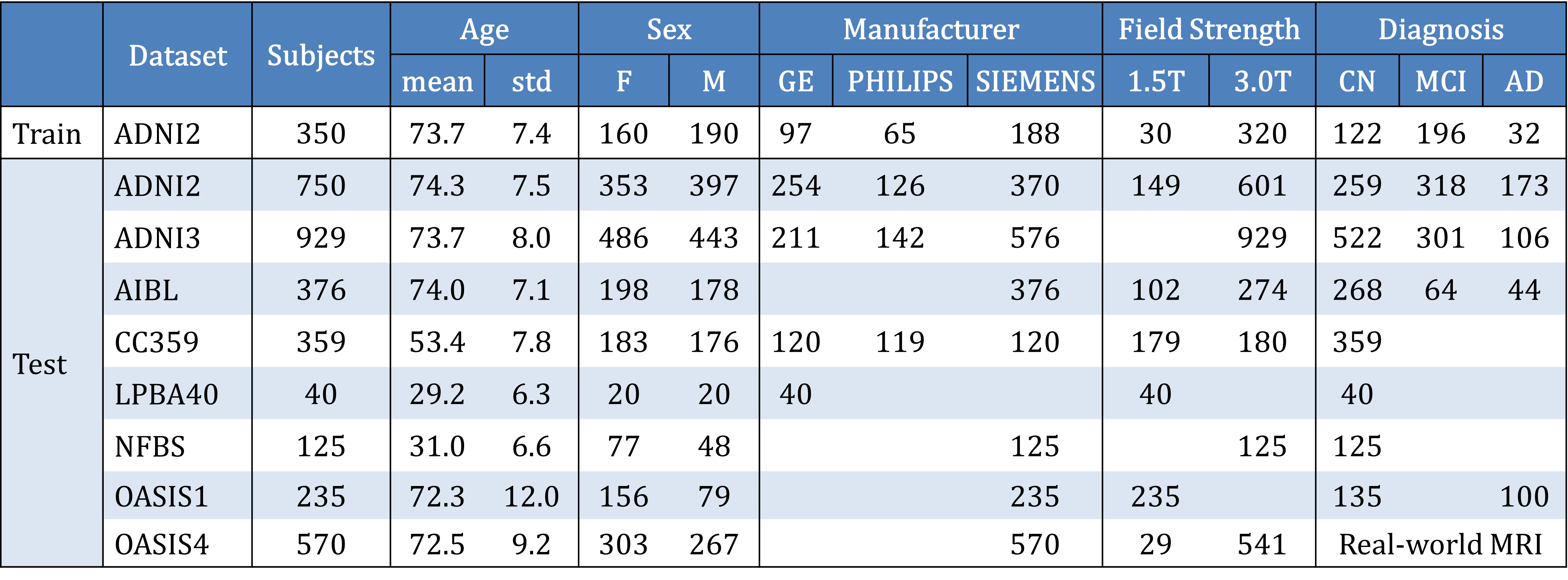
ParticipantsThis study utilized eight open MRI data resources14-20, as outlined in Figure 1. A total of 350 T1-weighted brain MRIs from ADNI2 served as the training dataset, while the remaining images were used as the test dataset.
Ground Truth (GT) Anatomical Labeling
The MALF algorithm21,22 developed at the Johns Hopkins University, was applied to the MRI to generate a parcellation map, a set of anatomical labels comprising 280 anatomical regions based on the JHU-MNI atlas3.
Model Design
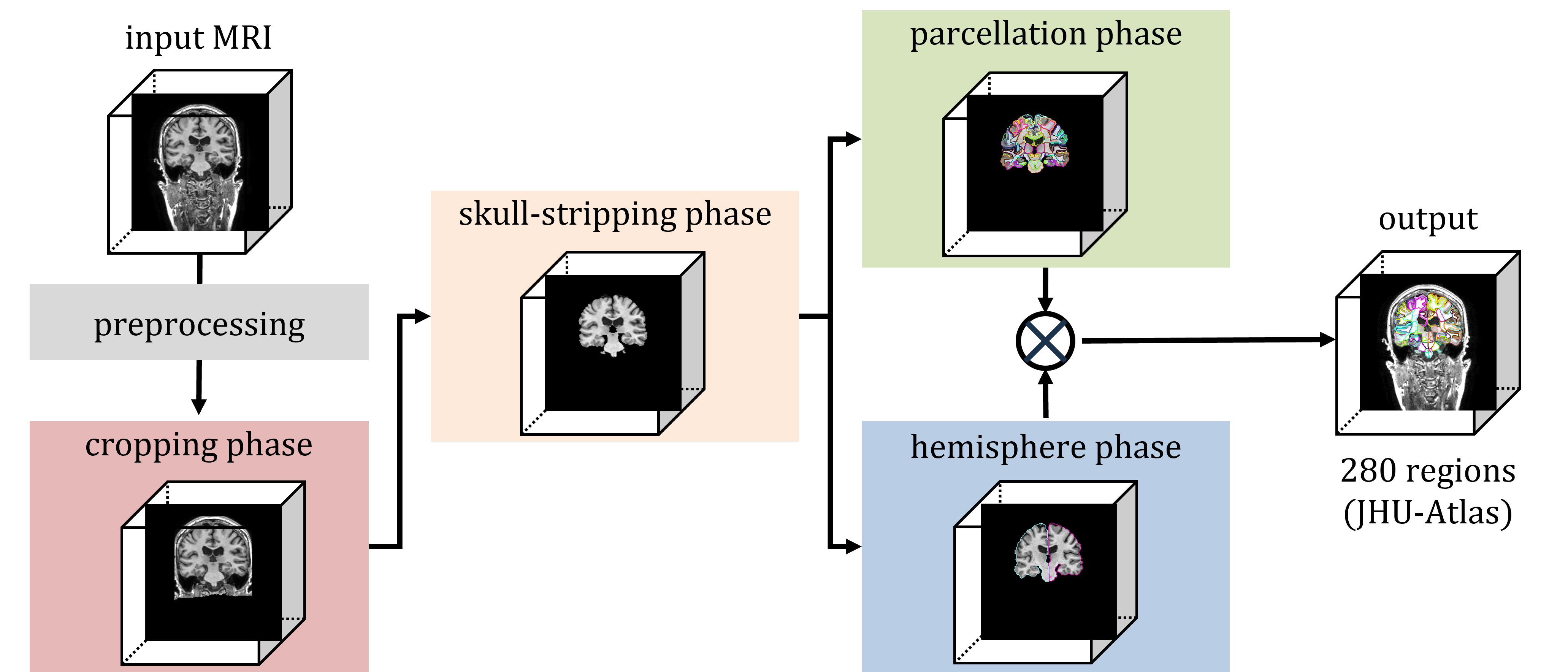
The OpenMAP-T1 operates through six steps (Figure 2): (1) preprocessing; (2) application of a 2D U-Net23 to crop the area surrounding the head in the input MRI; (3) application of a 2D U-Net to extract the brain; (4) application of a 2.5D U-Net to parcellate the whole brain into 141 anatomical areas; (5) application of a 2D U-Net to segment the whole brain into the right and left hemisphere; and (6) separation of 139 regions of the 141 from the parcellation map created in step 4 into right and left sides based on the hemisphere map from step 5, with the exception of the 3rd and 4th ventricles. Consequently, OpenMAP -T1 produces a parcellation map consisting of 280 neuroanatomically defined regions.
Model Evaluation
To assess the parcellation performance of OpenMAP-T1, four metrics were utilized: (1) the Dice score obtained between the parcellation maps generated by MALF and OpenMAP-T1; (2) correlation analysis of the predicted regional volumes between MALF and OpenMAP-T1; (3) calculation of the percentage difference in predicted volumes between MALF and OpenMAP-T1; and (4) examination of the relationship between the volume predicted by MALF and the Dice score. These metrics were computed for each individual region.
Result
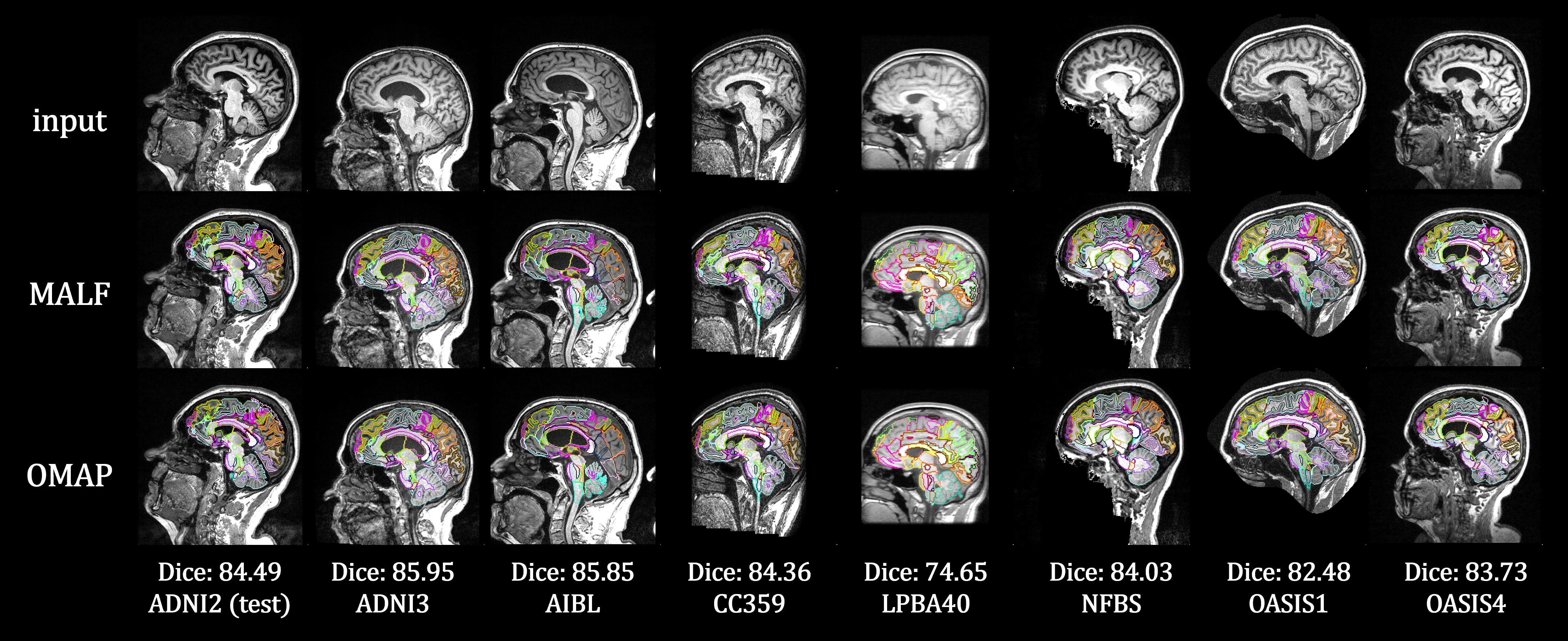
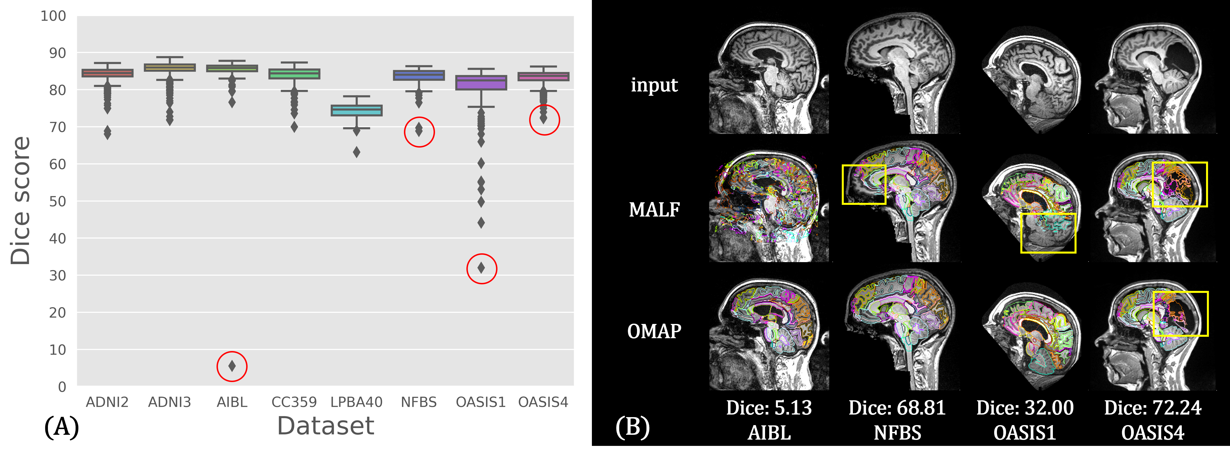
Figure 3 presents a comparison of a parcellation result between MALF and OpenMAP-T1. Despite the considerable variation in head appearance across the datasets, OpenMAP-T1 was visually confirmed to perform adequately for all eight datasets.Figure 4A shows the average Dice score of each region of parcellation labels between MALF and OpenMAP-T1, with scores exceeding 0.75, indicative of substantial agreement. The red circles in the boxplot highlight four cases depicted in Figure 4B, highlighting instances of disagreement between the two methods. The disagreements were predominantly attributed to mislabeling on the part of MALF, except for the rightmost image, which features a large lesion (arachnoidal cyst) that neither method successfully labeled.
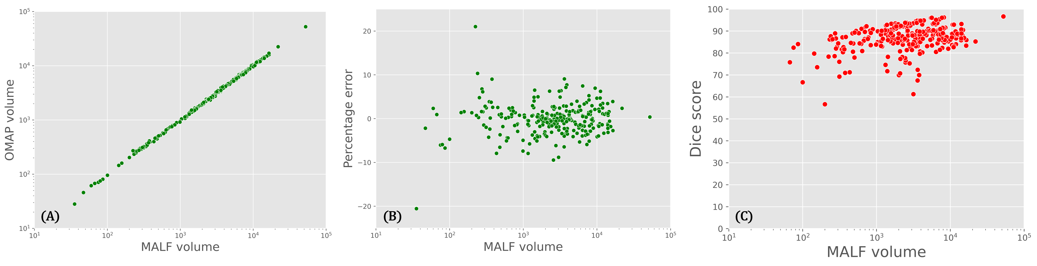
Figure 5A demonstrates a strong correlation between the predicted volumes of MALF and OpenMAP-T1. Figure 5B shows that the predicted volumes by OpenMAP-T1 are within 10% of those predicted by MALF for most regions, regardless of the region’s volume. Figure 5C shows that the Dice score exceeded 0.7 for the majority of the regions. This suggested that the variance tends to be slightly larger in smaller regions.
Discussion
The accuracy of OpenMAP-T1 is comparable to that of MALF, while it offering 40 times faster processing speed. Despite being trained solely on the ADNI2 dataset, OpenMAP-T1 demonstrated high parcellation performance on test datasets, highlighting its potential for clinical applications. Moreover, there is room for further improvement in its performance, particularly by refining the region labels of the GT.Conclusion
We have developed OpenMAP-T1, a fast, robust, and accurate whole brain parcellation tool. The OpenMAP-T1 holds great potential to accelerate the analysis of large clinical datasets.Acknowledgements
Part of the MRI data used in the preparation of this article was obtained from the Australian Imaging Biomarkers and Lifestyle flagship study of ageing (AIBL) and the Alzheimer's Disease Neuroimaging Initiative (ADNI). See www.aibl.csiro.au for further details. Data were provided in part by OASIS Cross-Sectional: Principal Investigators: D. Marcus, R, Buckner, J, Csernansky J. Morris; P50 AG05681, P01 AG03991, P01 AG026276, R01 AG021910, P20 MH071616, U24 RR021382 and Clinical Cohort: Principal Investigators: T. Benzinger, L. Koenig, P. LaMontagne. This work was supported by the Richman Family Precision Medicine Center of Excellence in Alzheimer's Disease, including significant contributions from the Richman Family Foundation, the Rick Sharp Alzheimer’s Foundation, the Sharp Family Foundation, and others. KOi is a consultant for “AnatomyWorks” and “Corporate-M.” This arrangement is being managed by the Johns Hopkins University in accordance with its conflict-of-interest policies. The authors thank the Brain Resource Center for providing the brain specimens and Mary McAllister for English-language editing.References
1. Lancaster, Jack L., et al. "Automated Talairach Atlas Labels For Functional Brain Mapping." Human brain mapping 10.3 (2000): 120-131.
2. Desikan, Rahul S., et al. "An automated labeling system for subdividing the human cerebral cortex on MRI scans into gyral based regions of interest." Neuroimage 31.3 (2006): 968-980.
3. Oishi, Kenichi, et al. "Atlas-based whole brain white matter analysis using large deformation diffeomorphic metric mapping: application to normal elderly and Alzheimer's disease participants." Neuroimage 46.2 (2009): 486-499.
4. Wu, Guorong, et al. "A generative probability model of joint label fusion for multi-atlas based brain segmentation." Medical image analysis 18.6 (2014): 881-890.
5. Asman, Andrew J., et al. "Multi-atlas learner fusion: An efficient segmentation approach for large-scale data." Medical image analysis 26.1 (2015): 82-91.
6. Bai, Wenjia, et al. "Multi-atlas segmentation with augmented features for cardiac MR images." Medical image analysis 19.1 (2015): 98-109.
7. Mori, Susumu, et al. "MRICloud: Delivering High-Throughput MRI Neuroinformatics as Cloud-Based Software as a Service." Computing in Science & Engineering 18.5 (2016): 21-35.
8. Huo, Yuankai, et al. "3D whole brain segmentation using spatially localized atlas network tiles." NeuroImage 194 (2019): 105-119.
9. Zhao, Yuan-Xing, et al. "Multi-view Semi-supervised 3D Whole Brain Segmentation With a Self-ensemble Network." Medical Image Computing and Computer Assisted Intervention–MICCAI 2019: 22nd International Conference, Shenzhen, China, October 13–17, 2019, Proceedings, Part III 22. Springer International Publishing, 2019.
10. Henschel, Leonie, et al. "Fastsurfer-a fast and accurate deep learning based neuroimaging pipeline." NeuroImage 219 (2020): 117012.
11. Coupé, Pierrick, et al. "AssemblyNet: A large ensemble of CNNs for 3D whole brain MRI segmentation." NeuroImage 219 (2020): 117026.
12. Yee, Evangeline, et al. "3D hemisphere-based convolutional neural network for whole-brain MRI segmentation." Computerized Medical Imaging and Graphics 95 (2022): 102000.
13. Lim, Eun-Cheon, et al. "DeepParcellation: a novel deep learning method for robust brain magnetic resonance imaging parcellation in older East Asians." Frontiers in Aging Neuroscience 14 (2022): 1027857.
14. Weiner, Michael W., et al. "The Alzheimer's Disease Neuroimaging Initiative: Progress report and future plans." Alzheimer's & Dementia 6.3 (2010): 202-211.
15. Ellis, Kathryn A., et al. "The Australian Imaging, Biomarkers and Lifestyle (AIBL) study of aging: methodology and baseline characteristics of 1112 individuals recruited for a longitudinal study of Alzheimer's disease." International psychogeriatrics 21.4 (2009): 672-687.
16. Souza, Roberto, et al. "An open, multi-vendor, multi-field-strength brain MR dataset and analysis of publicly available skull stripping methods agreement." NeuroImage 170 (2018): 482-494.
17. Shattuck, David W., et al. "Construction of a 3D probabilistic atlas of human cortical structures." Neuroimage 39.3 (2008): 1064-1080.
18. Puccio, Benjamin, et al. "The preprocessed connectomes project repository of manually corrected skull-stripped T1-weighted anatomical MRI data." Gigascience 5.1 (2016): s13742-016.
19. Marcus, Daniel S., et al. "Open Access Series of Imaging Studies (OASIS): Cross-sectional MRI Data in Young, Middle Aged, Nondemented, and Demented Older Adults." Journal of cognitive neuroscience 19.9 (2007): 1498-1507.
20. Koenig, Lauren N., et al. "Select Atrophied Regions in Alzheimer disease (SARA): An improved volumetric model for identifying Alzheimer disease dementia." NeuroImage: Clinical 26 (2020): 102248.
21. Mori, Susumu, et al. "Brain atrophy in middle age using magnetic resonance imaging scans from Japan’s health screening programme." Brain Communications 4.4 (2022): fcac211.
22. Fujita, Shohei, et al. "Characterization of Brain Volume Changes in Aging Individuals With Normal Cognition Using Serial Magnetic Resonance Imaging." JAMA Network Open 6.6 (2023): e2318153-e2318153.
23. Ronneberger, Olaf, Philipp Fischer, and Thomas Brox. "U-Net: Convolutional networks for biomedical image segmentation." Medical Image Computing and Computer-Assisted Intervention–MICCAI 2015: 18th International Conference, Munich, Germany, October 5-9, 2015, Proceedings, Part III 18. Springer International Publishing, 2015.
Figures