1028
Active Gradient Guidance Based Susceptibility and Magnitude Information Complete Network for Basal Ganglia Segmentation1Department of Electronic Science, Xiamen University, Xiamen, China
Synopsis
Keywords: Segmentation, Segmentation, Susceptibility Imaging, Basal Ganglia, Segmentation Network, Magnitude Information Complete, Active Gradient Guidance
Motivation: Accurate segmentation of basal ganglia is a crucial prerequisite for subsequent clinical practice and research. The boundaries of BG remain challenging to segment especially when dealing with data affected by severe artifacts.
Goal(s): This work aims to propose an automatic BG segmentation method with radiologist comparable accuracy and high inference speed.
Approach: An active gradient guidance-based susceptibility and magnitude information complete network(AGNet). With newly designed modules, AGNet can efficiently capture the inter-slice information and exploit it as attention guidance to facilitate the segmentation process.
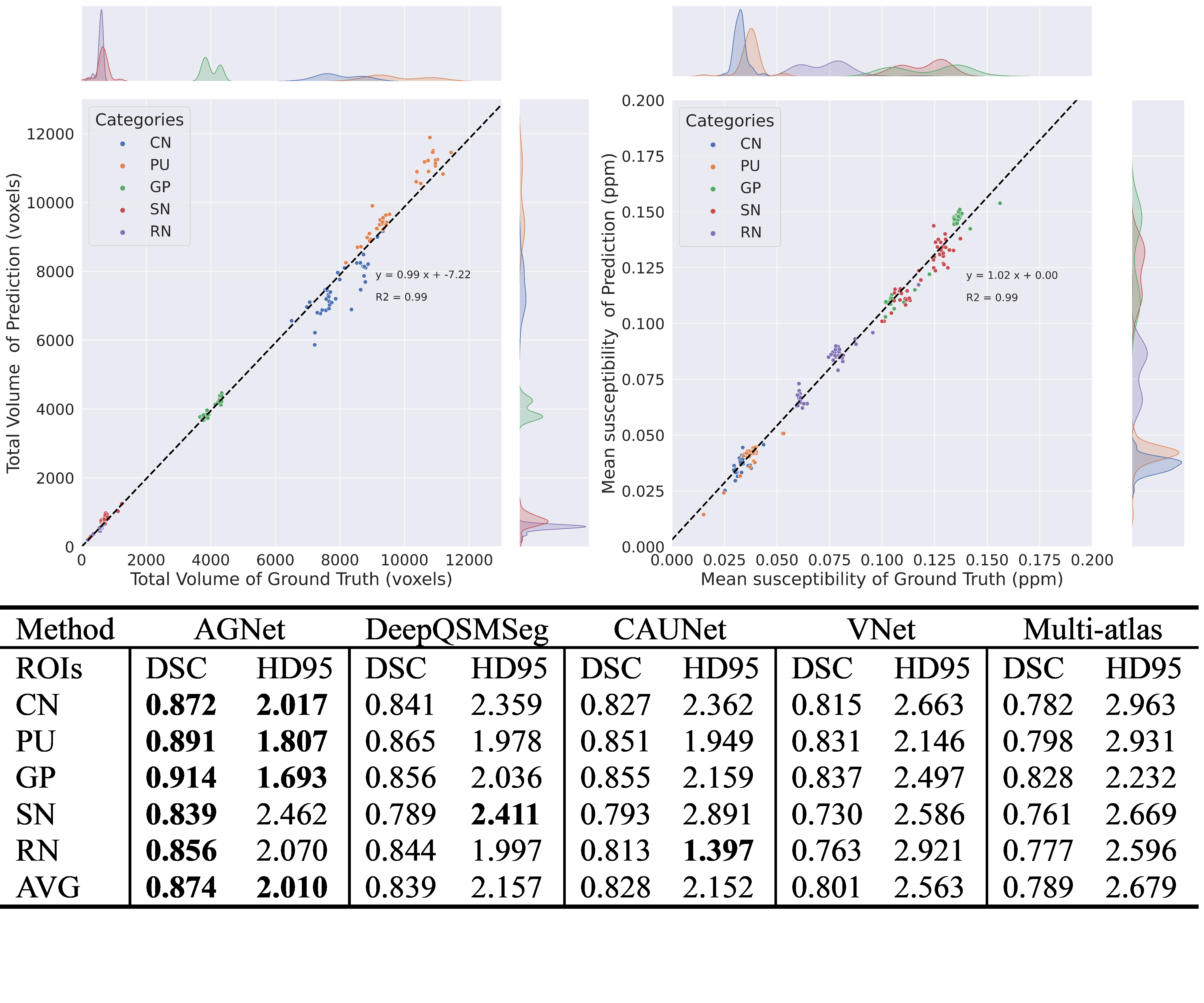
Results: AGNet has superior segment accuracy over existing methods with ADSC=0.874 and AHD=2.010, especially near boundaries of target VOI.
Impact: The proposed model achieves more accurate segmentation at the boundary contour. Automatic and precise segmentation of basal ganglia is a prerequisite for the quantification of tissue magnetic susceptibility analysis and can serve as a fundamental tool for neurodegenerative disease research.
Introduction
Accurate segmentation of Basial Ganglia (BG) is a crucial prerequisite for subsequent disease diagnosis, surgical planning, and quantitative research1. The conventional method of localizing and segmenting BG heavily relies on layer-by-layer manual annotation by experts, resulting in a tedious amount of workload. Deep learning-based approaches have been applied to address this task. However, the specific characteristics of the input image were not adequately considered into the model design2. For these DL-based models, the boundaries of BG remain challenging especially when dealing with data acquired from clinical practice and data affected by severe artifacts. In this study, we investigated a gradient guidance based susceptibility and magnitude information complete network for BG segmentation, named AGNet. It exhibits the capability to achieve more accurate segmentation and enhanced robustness on both healthy human data and clinical data.Methods
The detailed architecture of AGNet is presented in Fig.1. It utilizes a dual-branch architecture, with the Seg-branch aiming to generate a proper segmentation map and the Grad-branch to reconstruct the gradient map of ROI. Additionally, the Grad-map provides attention guidance for the Seg-branch, facilitating it to focus on the boundary of target nuclei. AGNet takes QSM maps and Magnitude maps as two seperated inputs, whose features are selectively enhanced in the proposed Magnitude Information Complete (MIC) module. The distinction between the foreground and the background across different channels is enlarged through learning inside MIC module. The enhanced maps are then delivered to the dual-branch as contrast-shifted inputs for specific downstream tasks. Like inter-frame information acquired between continuous frames in video segmentation tasks, inter-layer information in medical images contains valuable continuity and steep change information, often reflecting rich boundary structural details. The newly designed Active Gradient Module (AGM) can adaptively capture inter-slice information of input feature maps according to the given sample interval. To reflect edge information properly without the generated gradient map being a coarser edge band or vanishing at the boundary contour, the sample intervals should be set properly. Rather than setting constant sample intervals, they are determined according to the resolution and receptive field level of input feature maps inside AGM. The captured information can be utilized as gradient guidance through the Gradient Guiding Module (GGM), which essentially serves as the information exchange path between the Seg-branch and Grad-branch. Feature maps from Seg-branch ($$$I_{skip}$$$ and $$$I_{seg}$$$) are delivered into AGM inside GGM to generate corresponding gradient maps, which can facilitate the Grad-branch to filter out non-related gradient information. Reversely, feature maps from Grad-branch ($$$I_{grad}$$$) are activated into gradient guidance through sigmoid in GGM. The detailed architecture compositions of proposed AGM and GGM are shown in Fig.3.Results
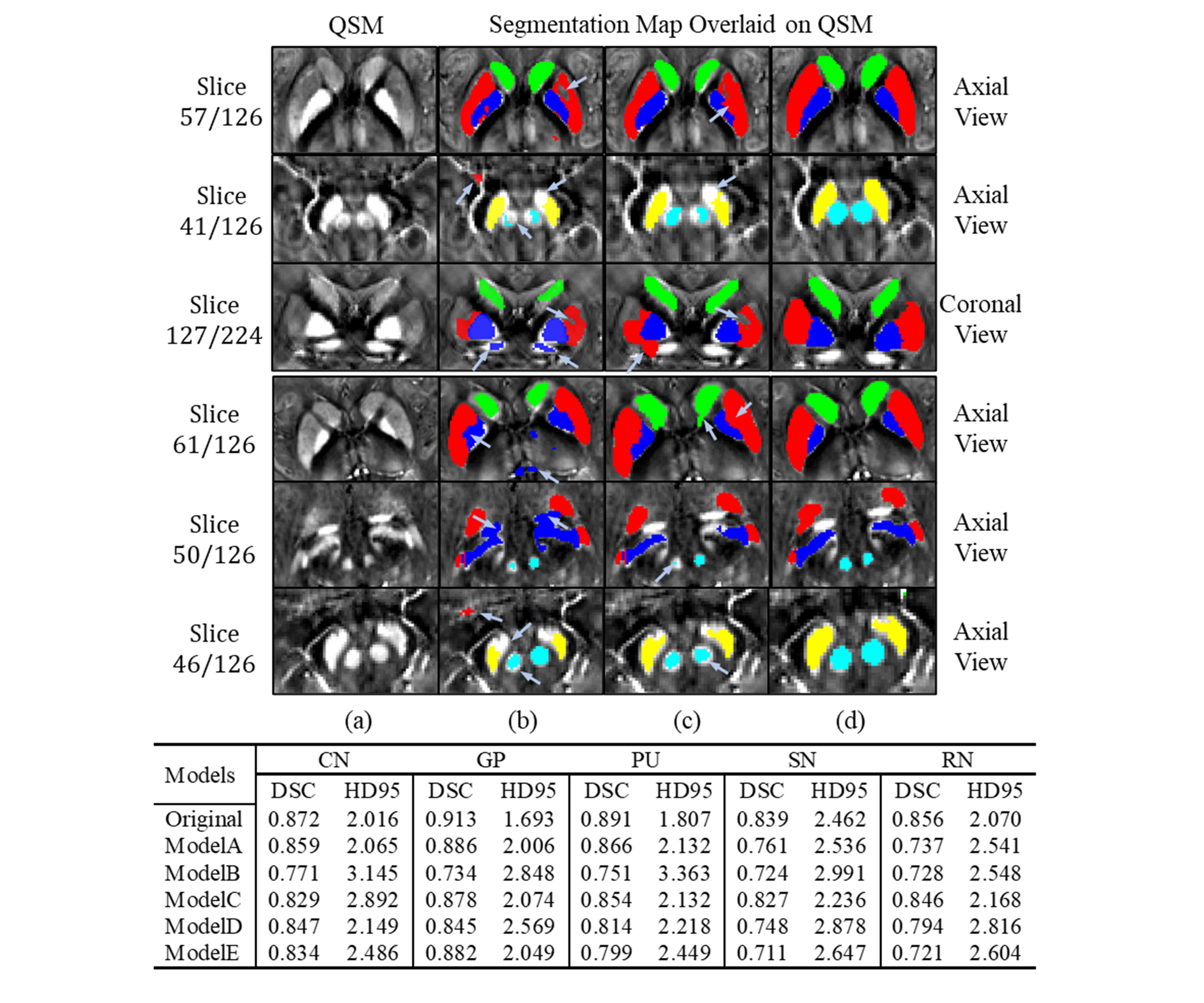
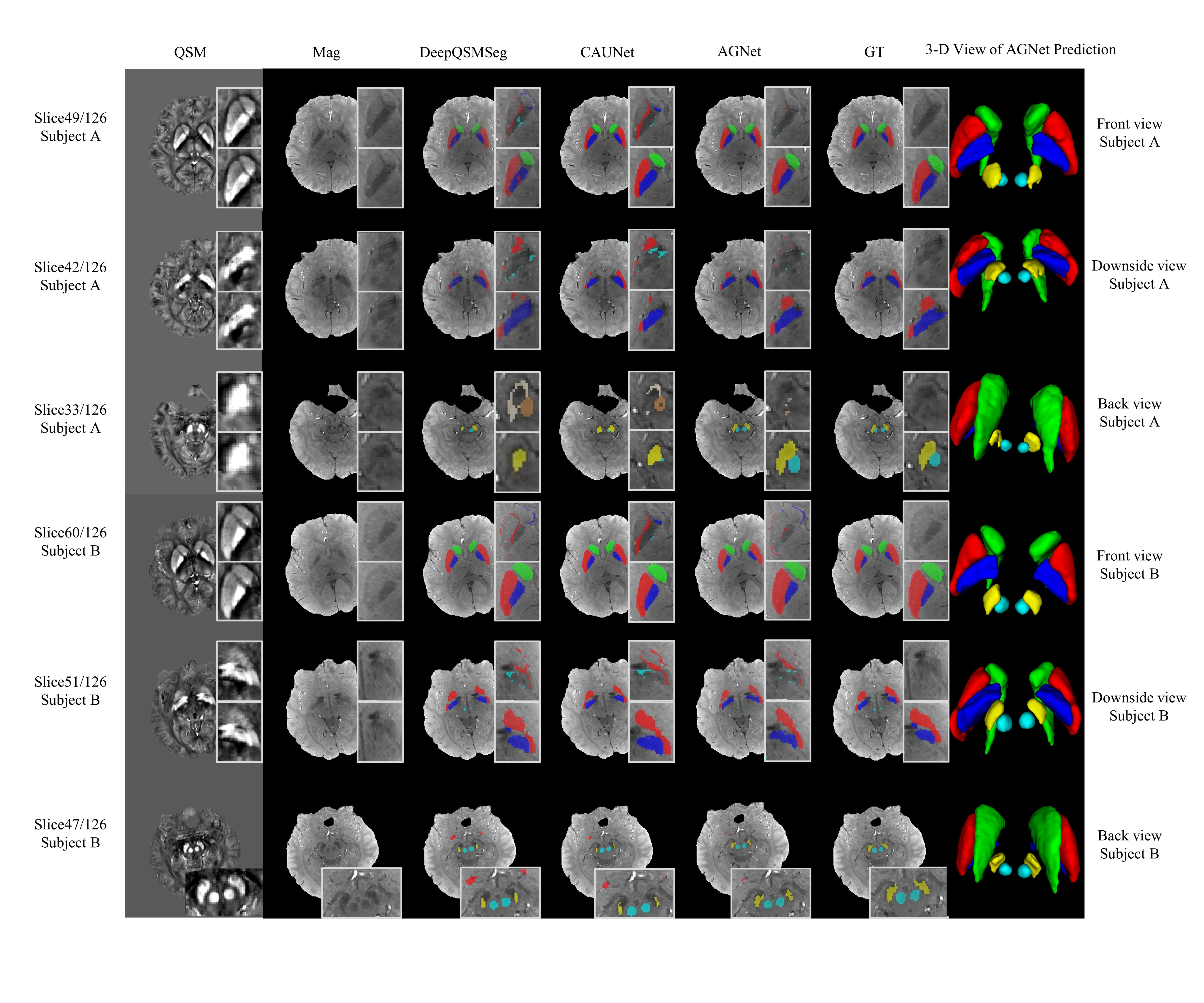
A healthy human dataset composed of 144 3T measurements and 44 7T measurements was employed to train and test the proposed AGNet3,4. An ablation study was performed to find out if the proposed modules function properly. The MIC module was replaced by convolution blocks with an approximate number of parameters, and a significant decrement ofsegmentation performance was observed. As shown in Table.1, the average Dice coefficient on five nuclei was dropped by 6.33% and the Harsdorff distance was increased by 10.93%. In the modified AGNet, in which the gradient branch was omitted, we replaced the GGM of the segmentation branch with trilinear interpolation to achieve the up-sampling function. The modification results in a 10.76% decrement in DC and a 24.28% increment in HD. After ablating the Grad-branch, the model tends to generate more false positive predictions and becomes severely inferior at distinguishing GP and Ventral Pallidum. Despite the reduction in overall parameter capacity, these significant performance differences still highlight the importance of the grad-branch. To further demonstrate the effectiveness of active sampling compared with fixed interval sampling, the interval parameter inside AGM was set to constant during inference. As stated in table inside Fig.3, comparing with all tested constant sample intervals, the model with active sampling achieves the best segmentation performance. AGNet was compared with four BG segmentation methods including DeepQSMSeg5, CAUNet6, VNet7, and Multi-atlas registration based algorithm8 on the test dataset composed of both 3T and 7T data. As results presented in Fig.5, AGNet has superior performance and generalization ability over existing methods.Discussion
The experiment results showcased commendable segmentation performance across both 3T and 7T datasets, aligning closely with annotations provided by seasoned medical practitioners. An experiment on clinical dataset with 19 epilepsy measurements has demonstrated good robustness and high sensitivity toward transfer learning.The core idea of employing gradient guidance to capture inter-slice information can be adopted in other medical image segmentation models to boost their performance.Acknowledgements
This work was supported in part by the National Natural Science Foundation of China under Grant 62071405.References
[1] Pierce J. E, Péron J. The basal ganglia and the cerebellum in human emotion. Social Cognitive and Affective Neuroscience, 2020; 15(5), 599-613.
[2] Jung W, Bollmann S, Lee, J. Overview of quantitative susceptibility mapping using deep learning: Current status, challenges and opportunities. NMR in Biomedicine,2022; 35(4), e4292.
[3] Shi Y, Feng R, Li Z, et al. Towards in vivo ground truth susceptibility for single-orientation deep learning QSM: A multi-orientation gradient-echo MRI dataset. NeuroImage, 2022; 261, 119522.
[4] Lai K W, Aggarwal M, van Zijl, et al. Learned proximal networks for quantitative susceptibility mapping. In Medical Image Computing and Computer Assisted Intervention–MICCAI 2020: 23rd International Conference, Lima, Peru, October 4–8, 2020, Proceedings, Part II 23 (pp. 125-135). Springer International Publishing.
[5] Guan Y, Guan X, Xu J, et al. . DeepQSMSeg: A Deep Learning-based Sub-cortical Nucleus Segmentation Tool for Quantitative Susceptibility Mapping. 2021; Paper presented at the 2021 43rd Annual International Conference of the IEEE Engineering in Medicine & Biology Society (EMBC).
[6] Chai C, Wu M, Wang H,et al. CAU-Net: A Deep Learning Method for Deep Gray Matter Nuclei Segmentation. Front Neurosci. 2022; 16, 918623.
[7] Milletari F, Navab N, Ahmadi S A. V-net: Fully convolutional neural networks for volumetric medical image segmentation. In 2016 fourth international conference on 3D vision (3DV). 2016; (pp. 565-571). Ieee.
[8] Li X, Chen L, Kutten K, et al. Multi-atlas tool for automated segmentation of brain gray matter nuclei and quantification of their magnetic susceptibility. NeuroImage, 2019; 191, 337-349.
Figures
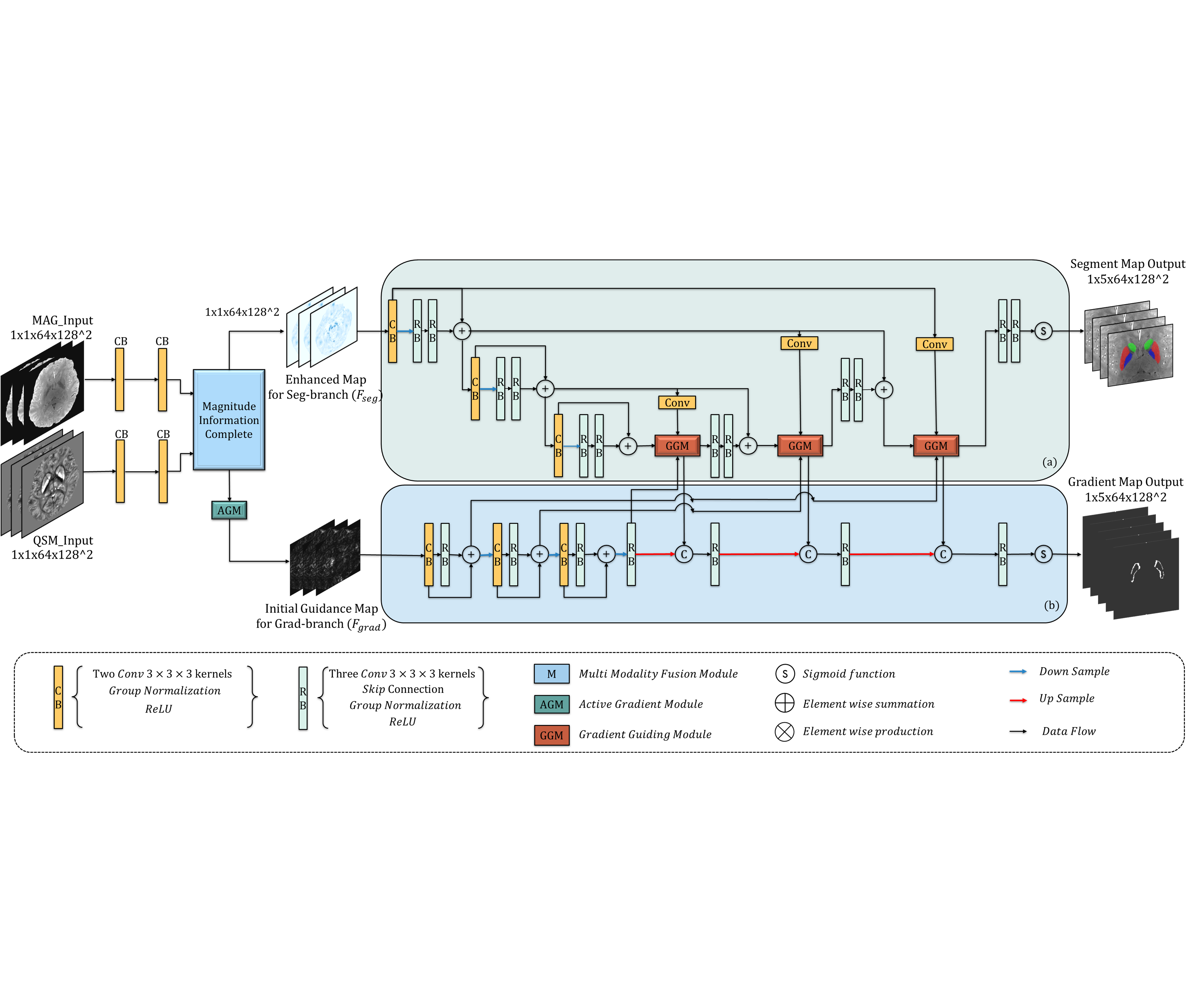
Figure1. The network architecture of AGNet with two inputs (Magnitude map and Quantitative Susceptibility map) and two outputs (Segment Map and Gradient Map of target nuclei): (a) Segment branch generates the final segmentation result under the guidance of multi-level gradient map. (b) Gradient branch reconstructs precise gradient maps from the initial gradient map of generated enhanced image.
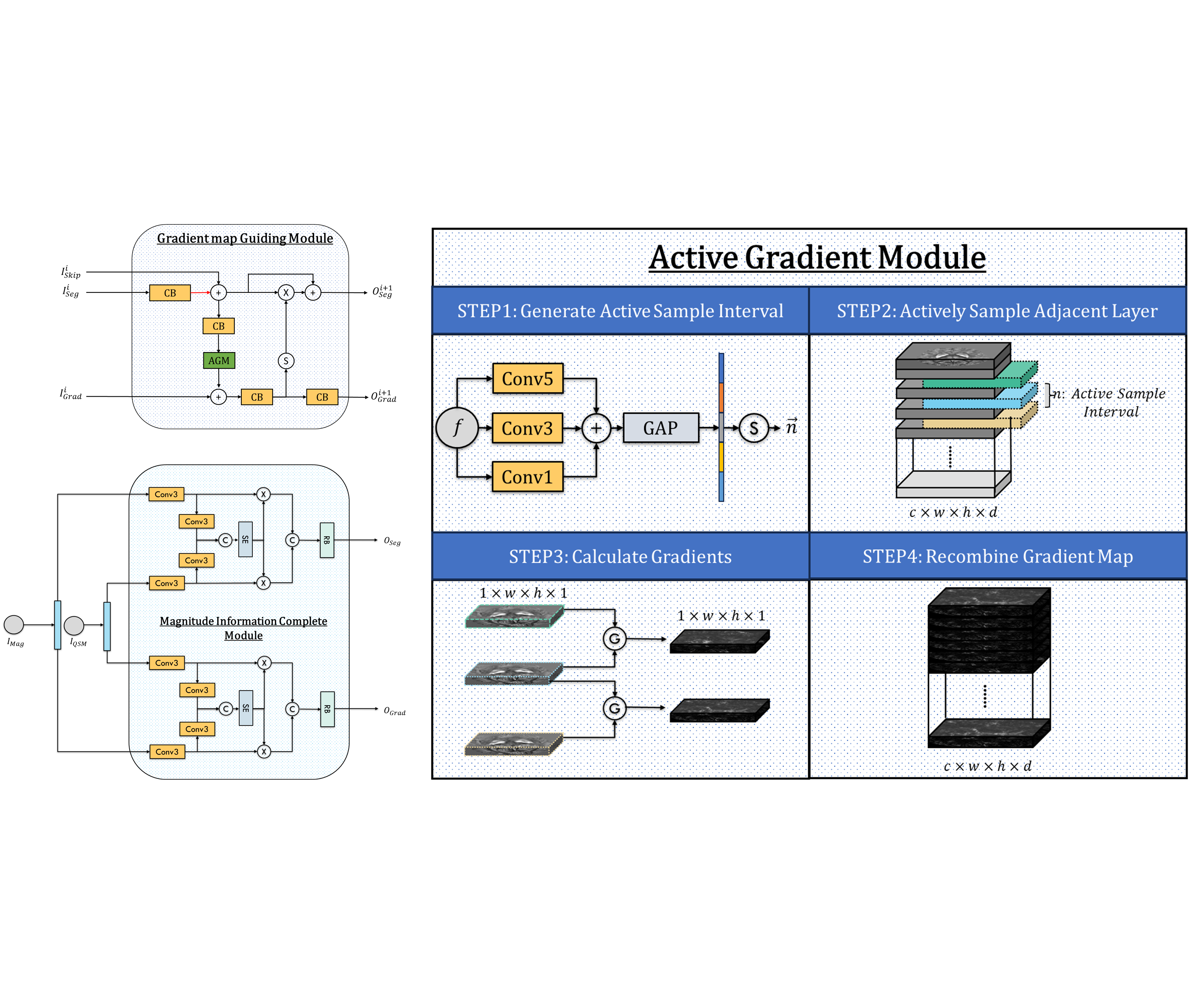
Figure2. Detail architectures of Proposed modules: (a)Active Gradient Module takes four steps to generate gradient maps of given feature maps by sampling them actively. (b)The Gradient Guiding Module serves as the information exchange path between Seg-branch and Grad-branch, with three inputs and two outputs. (c)The magnitude Information Complete module takes the Magnitude map and QSM as individual input and selectively fuses them to generate enhanced maps for downstream tasks.