1022
Segmentation of Brain Lesions Using Posterior Distributions Learned by Subspace-assisted Generative Model1School of Biomedical Engineering, Shanghai Jiao Tong University, Shanghai, China, 2Beckman Institute for Advanced Science and Technology, University of Illinois at Urbana-Champaign, Urbana, IL, United States, 3Department of Electrical and Computer Engineering, University of Illinois at Urbana-Champaign, Urbana, IL, United States
Synopsis
Keywords: Segmentation, Segmentation, Lesion segmentation; Generative model
Motivation: Deep learning shows great potential for brain lesion segmentation but poor generalization (due to limited training data) could lead to false positives.
Goal(s): Our goal was to improve the segmentation accuracy by learning target-specific posterior distributions.
Approach: We proposed a new Bayesian brain lesion segmentation method, leveraging posterior distributions learning, including both posterior normal and lesion distributions, through a subspace-assisted deep generative model.
Results: The proposed method achieved significantly improved segmentation performance across multiple public datasets with stroke, tumor, and multiple sclerosis lesions, in comparison with the state-of-the-art methods.
Impact: The proposed method significantly improved accuracy and robustness of lesions segmentation in brain MR images, which may provide a useful tool for brain lesion delineation in image processing and clinical applications.
Introduction
Accurate segmentation of brain lesions are essential for treatment planning of brain disorders1. Unsupervised learning methods, focusing on learning normative distributions from images of healthy subjects, are less dependent on lesion-labeled data, thus exhibiting better generalization capabilities2,3. A fundamental challenge in learning normative distributions of images lies in the high dimensionality if each pixel is treated as a random variable. Recent deep-learning based methods, such as Variational Auto-Encoders (VAEs) and Generative Adversarial Nets (GANs), have generated very encouraging results2,4. In this study, we further extended those methods using a subspace-assisted deep generative model to capture both posterior normal and lesion distributions. The proposed method achieved significantly improved segmentation performance across multiple public datasets with stroke, tumor, and multiple sclerosis lesions, in comparison with the state-of-the-art methods.Methods
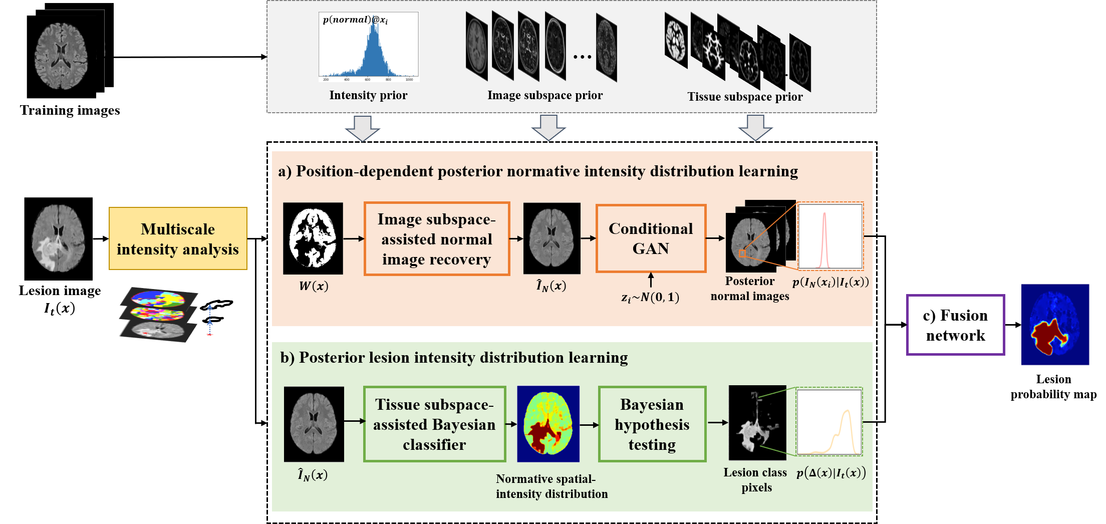
The overall framework of the proposed method is shown in Fig. 1, which contains three integral components: 1) posterior normative distribution learned by an image subspace-assisted deep generative model; 2) posterior lesion distribution learned by a tissue subspace-assisted Bayesian classifier; 3) a fusion network to generate the final segmentation.Problem formulation $$$\\$$$
Given an image with lesion $$$I_t(x)$$$, we represent it as $$$I_t(x)=I_N(x)+\Delta(x),$$$ where $$$I_N(x)$$$ represents an “hypothetical” lesion-free image and $$$\Delta(x)$$$ contains the lesion features. The following posterior distributions are estimated in our proposed method to capture the statistics of $$$I_N(x)$$$ and $$$\Delta(x)$$$ for improved segmentation: a) the position-dependent posterior normal intensity distribution, $$$p(I_N(x_i)|I_t(x))$$$ , at each pixel $$$i$$$; and b) the posterior lesion intensity distribution, $$$p(\Delta(x)|I_t(x))$$$.
Learning $$$\,p(I_N(x_i)|I_t(x))\\$$$
We proposed an image subspace-assisted conditional generative model to estimate $$$p(I_N(x_i)|I_t(x))$$$. After training, $$$p(I_N(x_i)|I_t(x))$$$ can be estimated based on the conditional samples from the learned generative model.
$$$\quad$$$In particular, we first obtained one posterior normal image $$$\hat{I}_N(x)$$$ as the condition of the generative model, leveraging a probabilistic image subspace model constrained by the pre-estimated neighboring normal pixels in $$$I_t(x)$$$ (denoted as $$$W(x)$$$). Specifically, $$$\hat{I}_N(x)$$$ was obtained by: $$\qquad\qquad\qquad\qquad\qquad\qquad\qquad\qquad\hat{I}_N(x)=(1-W(x))\odot\sum_{r=1}^Ra_r\phi_r(x)+W(x)\odot I_t(x), \qquad\qquad\qquad\qquad\qquad\qquad(1)$$where $$$W(x)$$$ is estimated leveraging prior intensity distributions of the normal training images $$$\{\rho_m(x)\}$$$ in a multiscale sense5, $$$\{\phi_r(x)\}$$$ is the pre-learned basis functions obtained by applying principal component analysis to $$$\{\rho_m(x)\}$$$, $$$a_r\sim p(\{a_r\})$$$ is the corresponding coefficients obtained by maximum-a-posteriori estimation: $$\qquad\qquad\qquad\qquad\qquad\qquad{\hat{a}_r}=\arg\min\limits_{\{a_r\}}\left\|W(x)\odot\left[I_t(x)-\sum_{r=1}^R a_r\phi_r(x)\right]\right\|_2-\lambda_{\text{coeff}} \log p(\{a_r\}). \qquad\qquad\qquad\qquad\qquad(2)$$$$$\quad$$$After $$$\hat{I}_N(x)$$$ was determined, we learned a conditional GAN to capture the nonlinear mapping from noise samples $$$z$$$ to a real image $$$I_N(x)$$$, conditioned on $$$\hat{I}_N(x)$$$. We adapted the Wasserstein GAN6 with improved loss function: $$L_{\text{cWGAN}}(G,D)=\mathbb{E}_{\hat{I}_N,z,I_N}\left[D(I_N)-D(G(z,\hat{I}_N))\right]+\lambda_{\text{GP}}L_{\text{GP}}(D)\\ \qquad\qquad\qquad\qquad\qquad\qquad\qquad\qquad\qquad\quad+\lambda_1L_{\text{L1}}(G,\hat{I}_N)+\lambda_2L_{\text{CE}}(G,\hat{I}_N)+\lambda_3L_{\text{VGG}}(G,\hat{I}_N), \qquad\qquad\qquad\qquad\;(3)$$where $$$L_{\text{GP}}$$$, $$$L_{\text{L1}}$$$, $$$L_{\text{CE}}$$$, $$$L_{\text{VGG}}$$$ is the gradient penalty, L1-loss, pixel-wise cross entropy loss and VGG loss, respectively. The patient-specific posterior normal images with high-fidelity details could be obtained by Monte Carlo sampling conditioned on $$$\hat{I}_N(x)$$$. $$$p(I_N(x_i)|I_t(x))$$$ could then be estimated from the sampled posterior images using a Mixture of Gaussian (MOG) model.
Estimation of $$$\,p(\Delta(x)|I_t(x))\\$$$
To estimate $$$p(\Delta(x)|I_t(x))$$$, we first obtained a global normative spatial-intensity probability distribution: $$\qquad\qquad\qquad\qquad\qquad\qquad\qquad\qquad p(\hat{I}_N(x_i),c(x_i);\pmb{\theta})=\sum_{k=1}^{3}p(\hat{I}_N(x_i)|c(x_i);\pmb{\theta}_k)\cdot p(c(x_i)), \qquad\qquad\qquad\qquad\qquad\quad\,(4)$$where $$$c$$$ represents the tissue class in $$$\hat{I}_N(x_i)$$$ and $$$p(\hat{I}_N(x_i)|c(x_i);\pmb{\theta}_k)$$$ is the likelihood of the observed intensity $$$p(\hat{I}_N(x_i))$$$ to be tissue class $$$k$$$. To obtain the posterior $$$p(c(x_i))$$$ of $$$p(\hat{I}_N(x_i))$$$, we incorporated a tissue subspace model into the Bayesian classifier for robust segmentation: $$\qquad\qquad\qquad\qquad\qquad\qquad\qquad\qquad\qquad\qquad\pi_k(c(x_i))=\sum_{r=1}^{R'}\beta_{r,k}\psi_{r,k}(c(x_i)), \qquad\qquad\qquad\qquad\qquad\qquad\qquad\qquad\quad\;(5)$$where $$$\{\psi_{r,k}(c)\}$$$ are the basis functions of tissue $$$k$$$ obtained on the tissue segmentation labels $$$\{c_1,c_2,\ldots,c_M\}$$$ of the training images. After $$$p(c(x_i))$$$ was obtained, $$$p(\hat{I}_N(x_i),c(x_i);\pmb{\theta})$$$ was fitted using the collection of pixels based on the predicted $$$c(x_i)$$$. With $$$p(\hat{I}_N(x_i),c(x_i);\pmb{\theta})$$$ estimated, the lesion class in $$$I_t(x)$$$ was obtained leveraging voxel-wise Bayesian hypothesis testing, from which $$$p(\Delta(x)|I_t(x))$$$ was estimated using an MOG model.
Fusion network$$$\\$$$
The final classification was determined by fusing the features derived from the learned posterior distributions using a patch-based convolutional neural network, including: 1) position-dependent posterior distributions; 2) multiscale region-level normal/lesion likelihood ratio; 3) multiscale posterior lesion distribution.
Results
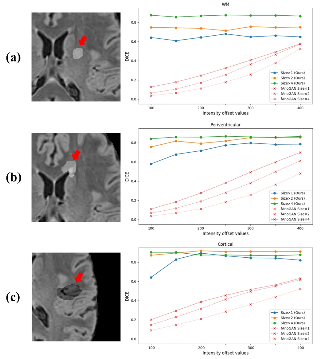
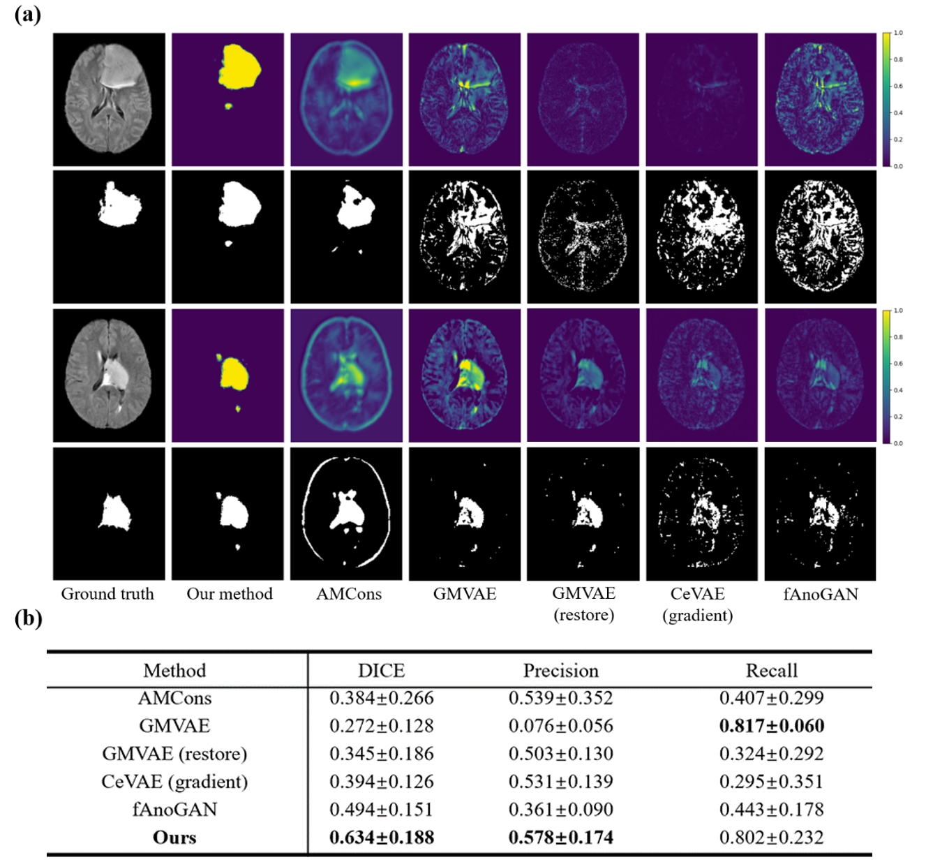
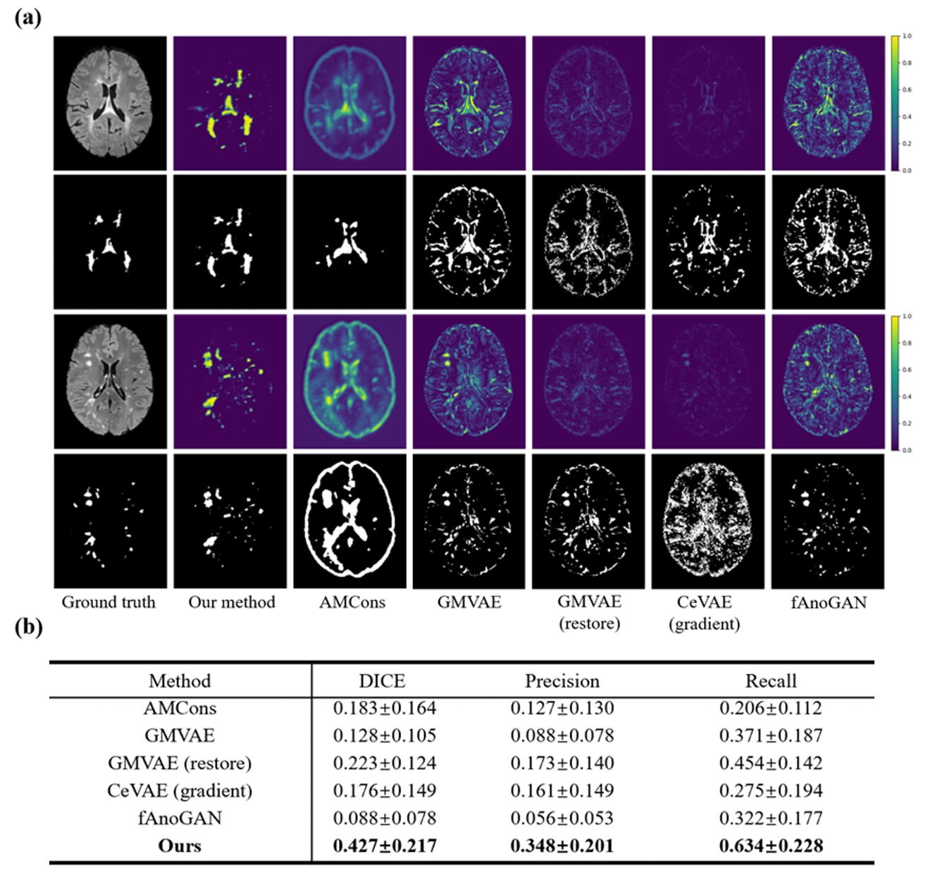
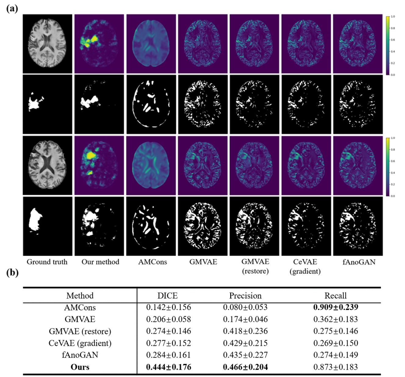
To obtain the normal image prior, we used 20000 T1w and 20000 FLAIR brain images from Biobank dataset7. The performance was evaluated on simulation data, BraTS2019 tumor (N=288)8, MSSEG2016 MS (N=53)9 and ATLAS stroke (N=236)10 datasets, respectively. Fig. 2 demonstrates our proposed method consistently and accurately segmented lesions across various sizes, locations and intensities. As shown in Figs 3-5, our proposed method outperformed the state-of-the-art deep learning-based abnormality detection methods2,11–15 for challenging lesion/normal interface (tumor), small and isolated lesions (MS) and atrophy brains (stroke). Quantitative analysis also indicated that the proposed method had improved performance compared with the state-of-the-art methods.Conclusions
This paper presents a new method for lesion segmentation leveraging posterior distributions learned using a subspace-assisted generative model. The proposed method showed significantly improved accuracy and robustness across multiple public brain MR image datasets with various lesion types.Acknowledgements
This work was supported by Shanghai Pilot Program for Basic Research—Shanghai Jiao Tong University (21TQ1400203), the Program for Professor of Special Appointment (Eastern Scholar) at Shanghai Institutions of Higher Learning, Key Program of Multidisciplinary Cross Research Foundation of Shanghai Jiao Tong University (YG2021ZD28, YG2023ZD22), National Natural Science Foundation of China (62001293).References
1. Prastawa M, Bullitt E, Ho S, Gerig G. A brain tumor segmentation framework based on outlier detection. Med Image Anal. 2004;8(3):275-283.
2. Chen X, You S, Tezcan KC, Konukoglu E. Unsupervised lesion detection via image restoration with a normative prior. Med Image Anal. 2020;64.
3. Van Leemput K, Maes F, Vandermeulen D, Colchester A, Suetens P. Automated segmentation of multiple sclerosis lesions by model outlier detection. IEEE Trans Med Imaging. 2001;20(8):677-688.
4. Baur C, Denner S, Wiestler B, Navab N, Albarqouni S. Autoencoders for unsupervised anomaly segmentation in brain MR images: a comparative study. Med Image Anal. 2021;69:101952.
5. Arbeláez P, Maire M, Fowlkes C, Malik J. Contour detection and hierarchical image segmentation. IEEE Trans Pattern Anal Mach Intell. 2011;33(5).
6. Isola P, Zhu JY, Zhou T, Efros AA. Image-to-image translation with conditional adversarial networks. In: Proceedings - 30th IEEE Conference on Computer Vision and Pattern Recognition, CVPR 2017. Vol 2017-January; 2017.
7. Alfaro-Almagro F, Jenkinson M, Bangerter NK, et al. Image processing and Quality Control for the first 10,000 brain imaging datasets from UK Biobank. Neuroimage. 2018;166:400-424.
8. Menze BH, Jakab A, Bauer S, et al. The multimodal brain tumor image segmentation benchmark (BRATS). IEEE Trans Med Imaging. 2014;34(10):1993-2024.
9. Liew SL, Anglin JM, Banks NW, et al. A large, open source dataset of stroke anatomical brain images and manual lesion segmentations. Sci Data. 2018;5(1):1-11.
10. Commowick O, Kain M, Casey R, et al. Multiple sclerosis lesions segmentation from multiple experts: The MICCAI 2016 challenge dataset. Neuroimage. 2021;244.
11. You S, Tezcan KC, Chen X, Konukoglu E. Unsupervised lesion detection via image restoration with a normative prior. In: International Conference on Medical Imaging with Deep Learning. PMLR; 2019:540-556.
12. Silva-Rodríguez J, Naranjo V, Dolz J. Constrained unsupervised anomaly segmentation. Med Image Anal. 2022;80.
13. Schlegl T, Seeböck P, Waldstein SM, Langs G, Schmidt-Erfurth U. f-AnoGAN: Fast unsupervised anomaly detection with generative adversarial networks. Med Image Anal. 2019;54:30-44.
14. Wyatt J, Leach A, Schmon SM, Willcocks CG. AnoDDPM: Anomaly Detection with Denoising Diffusion Probabilistic Models using Simplex Noise. In: IEEE Computer Society Conference on Computer Vision and Pattern Recognition Workshops. Vol 2022-June. IEEE Computer Society; 2022:649-655.
15. Zimmerer D, Kohl SAA, Petersen J, Isensee F, Maier-Hein KH. Context-encoding variational autoencoder for unsupervised anomaly detection. arXiv preprint arXiv:181205941. 2018.
Figures