1021
0.9mm isotropic 1min MPRAGE using highly-accelerated Deep learning Reconstruction for Brain Structural Analysis1Radiology, Kyoto prefectural university of medicine, Kyoto, Japan, 2radiology, Hirosaki university, Hirosaki, Japan, 3GE Healthcare, Tokyo, Japan, 4GE Healthcare, Menlo Park, CA, Japan
Synopsis
Keywords: Gray Matter, Machine Learning/Artificial Intelligence
Motivation: The project was driven by the need to reduce 3D T1-weighted MRI acquisition times, which are often prolonged, leading to motion artifacts and compromised image quality in structural nuroimaging analysis.
Goal(s): To evaluate whether deep learning reconstruction can shorten MRI scan times without significantly compromising image quality, facilitating efficient clinical and research neuroimaging.
Approach: We employed a deep learning technique, DL-speed, to reconstruct undersampled data from accelerated MRI scans, assessing image quality against conventional methods using a standardized rating system.
Results: Images with DL-speed maintained image quality, despite a slight quality trade-off, suggesting its viability for rapid, motion-artifact-reduced neuroimaging in various patient populations.
Impact: Our results impact clinicians and patients by enabling faster, high-quality MRIs, reducing patient discomfort and motion-related artifacts. This advance opens avenues for more efficient neuroimaging protocols, enhancing patient care and research productivity.
Introduction
High-resolution three-dimensional T1-weighted imaging (3D T1WI) plays a critical role in diagnosing various neurological disorders and structural neuroimaging research. However, its relatively long acquisition time leads to motion artifacts, compromising structural volume measurements. Traditional methods such as parallel imaging and compressed sensing have only partially mitigated these issues. Recently, deep learning techniques have been proposed to enhance MRI speed and quality. This study aims to investigate the potential of deep learning reconstruction in reducing 3D T1WI acquisition time while maintaining image quality for structural neuroimaging analysis.Method
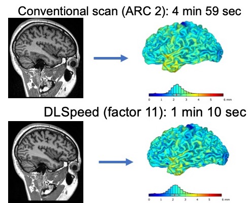
MRI scans were performed on six healthy volunteers and forty clinical cases using a 3.0-Tesla SIGNA Premier scanner (SIGNA Premier GE HealthCare, Waukesha, WI). The imaging protocol employed a Magnetization-Prepared Rapid Acquisition Gradient Echo (MPRAGE) pulse sequence with a resolution of 0.9mm isotropic. For the healthy volunteers, acceleration factors were varied from 2 to 16 to evaluate the potential of deep learning reconstruction in reducing scan time while maintaining image quality. In addition to these varied acceleration factors, both the volunteers and clinical cases were scanned using the ARC2 setting as a gold standard for comparison (Fig.1). The clinical cases were consistently scanned at an acceleration factor of 11. Additionally, in the clinical group, in-scanner motion was quantitatively measured to assess the impact of shorter scan times on motion artifacts. Image quality for all scans was assessed using the Computational Anatomy Toolbox 12 (CAT12), which provided a quantitative measure of image integrity.Result
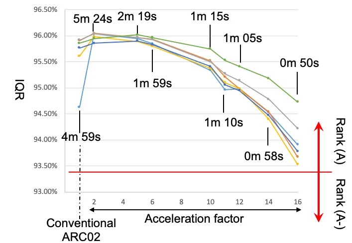
We evaluated the trade-off between scan duration and image quality in high-resolution 3D T1-weighted MRI scans across various acceleration factors. For healthy volunteers, the results demonstrated that image quality was preserved across a range of acceleration factors, maintaining a CAT12 quality rating of Rank A even at higher speeds (Fig.2). For the disease group, the DL-speed technique was applied with an acceleration factor of 11, resulting in a mean image quality score of 93.50% and a standard deviation of 0.93%. On the other hand, the mean head total vector changes during acquisition with DL-speed was significantly reduced to 50.8±11.9, compared to 137.7±34.7 in conventional imaging (p < 0.05).Discussion
The significance of 3D T1-weighted imaging (3D T1WI) in clinical and research neuroimaging is well-established, particularly for detailed brain structure analysis such as hippocampal volume measurements. Its clinical adoption for brain volume assessments is on the rise, aiding in the diagnosis and management of neurological conditions. However, the long acquisition time associated with 3D T1WI often results in motion artifacts, which compromise image quality and precision in voxel-based morphometry (VBM) analyses. The introduction of deep learning reconstruction techniques like DL-speed, which utilize neural networks for data reconstruction, presents a promising avenue to overcome these limitations by improving MRI acquisition speed and quality. In healthy volunteers, our analysis validates the effectiveness of deep learning reconstruction in maintaining image quality ranked as A rank in CAT 12 image quality assessment with increased acceleration factors, up to an acquisition time of 0m50s. In clinical scenarios, despite a statistical reduction in image quality for a 1m10s DL-speed scan compared to conventional imaging, the practical utility remains comparable due to the significant reduction in motion artifacts. This is especially pertinent for patients with movement disorders or cognitive impairments, where shorter scan times can mitigate the patient motion.Conclusion
Deep learning techniques like DL-speed can significantly reduce MRI acquisition times without substantially compromising image quality, offering a valuable tool in clinical settings.Acknowledgements
No acknowledgement found.References
No reference found.Figures