1020
MRF-SEG: Accelerated Brain MRI Acquisition and Segmentation1Radiology, Stanford University, Stanford, CA, United States, 2Massachusetts Institute of Technology, Cambridge, MA, United States
Synopsis
Keywords: Segmentation, Segmentation
Motivation: Despite the availability of rapid high-resolution MRF sequences that can be used to synthesize MPRAGE, acquiring MPRAGE scans remains necessary for accurate downstream segmentation.
Goal(s): The aim is to perform accurate-segmentation directly on MRF time-resolved data, eliminating the need for a lengthy MPRAGE scan, resulting in significant time savings, and providing quantitative tissue parameter maps.
Approach: We used deep learning to directly segment MRF time-resolved data and generate multi-tissue brain segmentation maps.
Results: Our findings indicate that deep learning segmentation methods trained directly on MRF data, both quantitatively and qualitatively perform better than segmentation on synthesized MPRAGE.
Impact: Applying deep learning directly on MRF data improves MRF segmentation compared to synthesizing MPRAGE and performing segmentation on it. This strengthens the validation of MRF and enhances its clinical potential by rapidly acquiring and segmenting brain images.
Introduction
The 3D spiral-projection-MRF acquisition method has been developed and used with subspace reconstruction to enable rapid high-resolution whole-brain mapping. The resulting reconstructed subspace coefficient maps provide a compact representation of the MRF time-series images, which can be used with dictionary matching to generate quantitative multi-parameter maps1-3.For traditional neuroimaging brain segmentation analysis, Magnetization-Prepared Rapid Acquisition Gradient Echo (MPRAGE) scans are acquired, which can then be segmented into multiple tissues using FreeSurfer4.
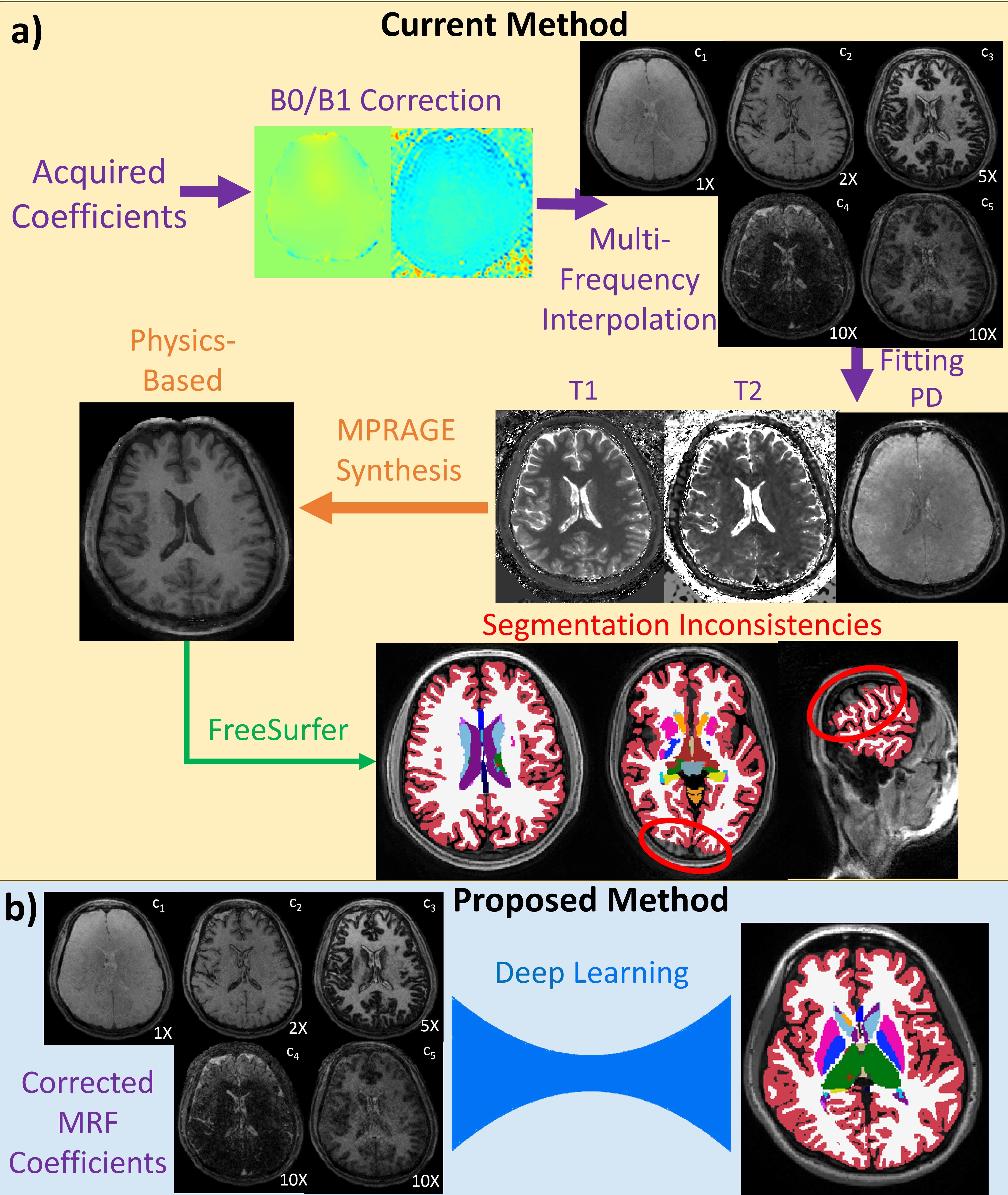
Currently, it is possible to synthesize MPRAGE from MRF subspace coefficients through dictionary-based fitting and physics-based modeling using the resulting quantitative maps (Figure 1a). This requires compressing higher-dimensional MRF coefficient information by calculating T1, T2, and PD maps followed by fitting to compute the MPRAGE. Such compression leads to loss of partial volume information and additional time-series contrast information that could be useful for segmentation, which can result in segmentation inconsistencies (Figure 1a).
Consequently, we here propose to generate MPRAGE segmentation maps directly from MRF coefficients using deep learning (Figure 1b). MRF coefficients inherently contain multi-tissue-compartment information, providing a richer source of data for the model to interpret and lead to more robust segmentation.
Methods
Dataset and Pre-ProcessingWhole-brain 1-mm MRF data was acquired in two minutes using 3D spiral-projection-MRF on a 3T-MRI2. Subspace reconstruction was performed using MFI5 on DL-generated B0 maps6 and interpolated PhysiCal7 B1 maps. This technique provides five, high-quality MRF coefficients and mitigates B0 blurring artifacts caused by spiral acquisition. Data from 14 healthy volunteers was acquired for this study and divided into training (9), validation (2), and testing datasets (3). Synthesized MPRAGE was fitted from MRF coefficients using a physics-based synthesis.
These multi-frequency B0 corrected MRF coefficients were used as training input for the model. The ground truth for the model was multi-tissue FreeSurfer segmentation maps that were computed from acquired MPRAGE.
CNN Models and Approach Overview
Previous experiments from non-MFI MRF coefficients indicated that 2.5D and 3D CNN segmentation approaches are viable methods for MRF segmentation. We therefore experimented training an axial 2.5D network using MONAI’s U-Net8 and 3D full-resolution cascading U-Net using nnUNet9. The 2.5D U-Net model was set up using the following parameters: 63 input channels (7 slice sliding window; 9 MRF coefficients), 40 output channels (one-hot), channels sequence of (32, 64, 128, 256, 512, 1024), 5 residual units, and 0.2 dropout probability. The nnUNet architecture closely resembles the original 3D U-Net with smaller patch sizes for initial low-resolution training9. These models were then compared to ground truth MPRAGE segmentation and evaluated on various overlap and distance segmentation metrics including Dice similarity coefficient (DSC) and average symmetric surface distance (ASSD)10,11. Additionally, we evaluated the models on a subject who underwent both MRF and MPRAGE acquisitions in two different head positions, aiming to assess model repeatability.
Results
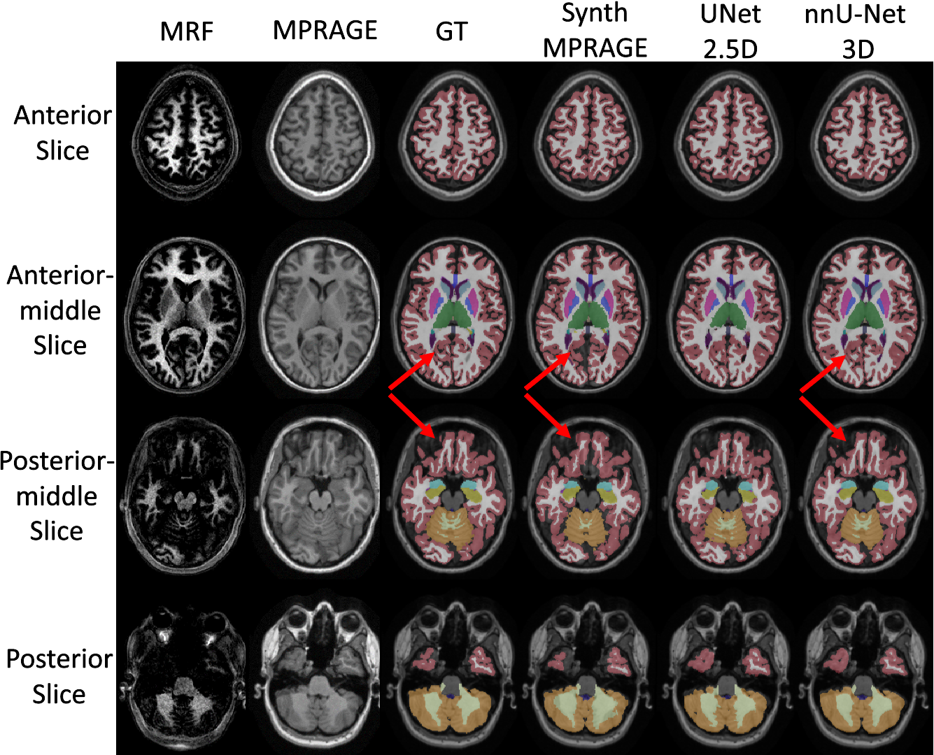
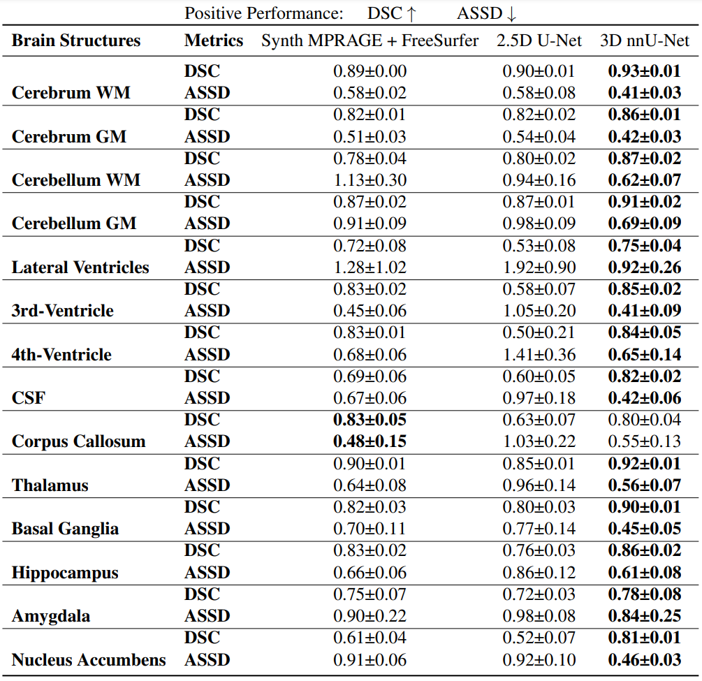
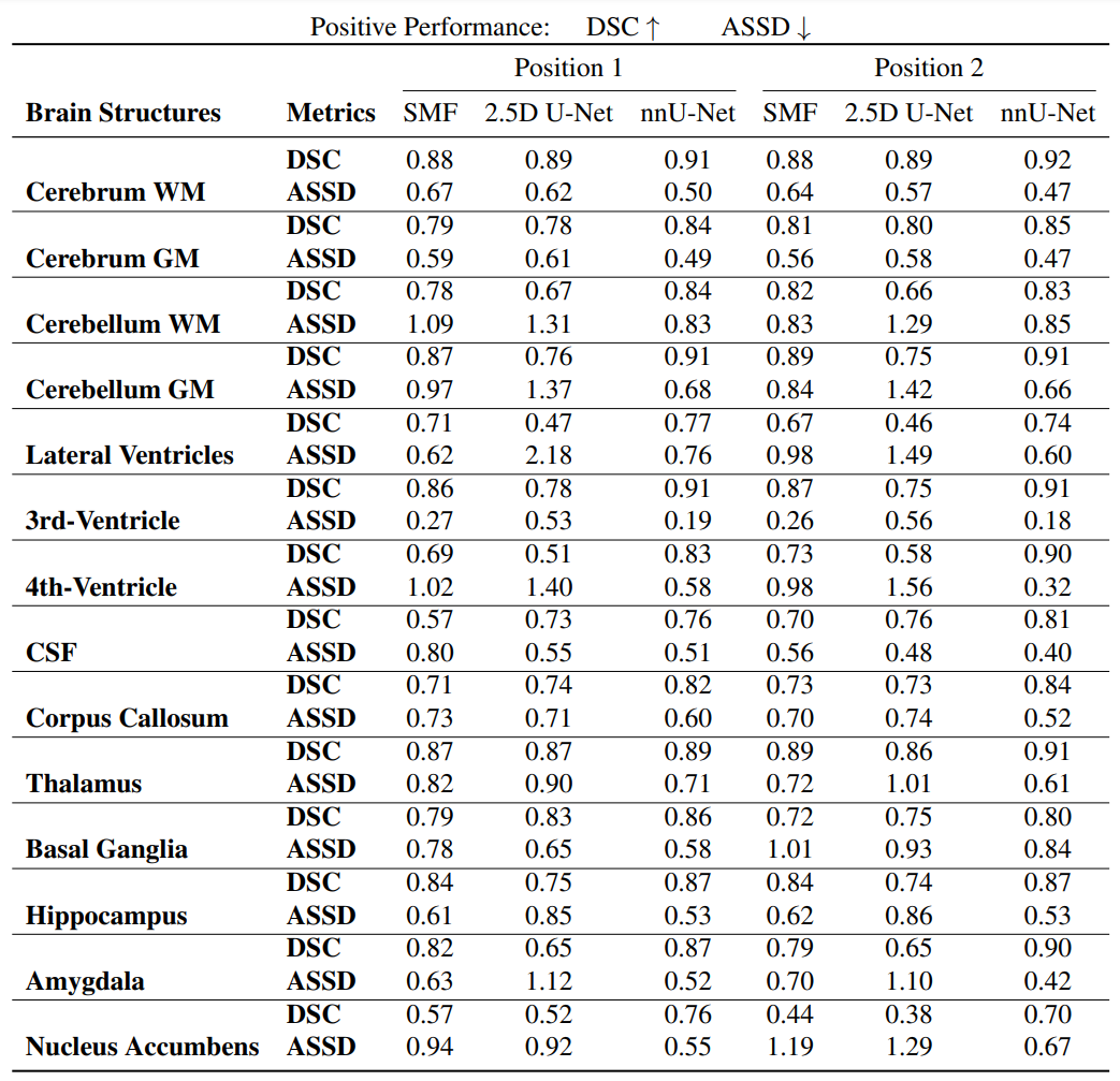
nnUNet maintains segmentation robustness across the parenchymannUNet trained directly on MRF coefficients exhibits superior performance with higher DSC and reduced ASSD in comparison to the segmentation results obtained from 2.5D U-Net and synthetic MPRAGE (Figure 4). Notably, nnUNet achieves improved segmentation of the sulci and gyri (Figure 2).
DL corrects segmentation inconsistencies observed from fitted segmentation
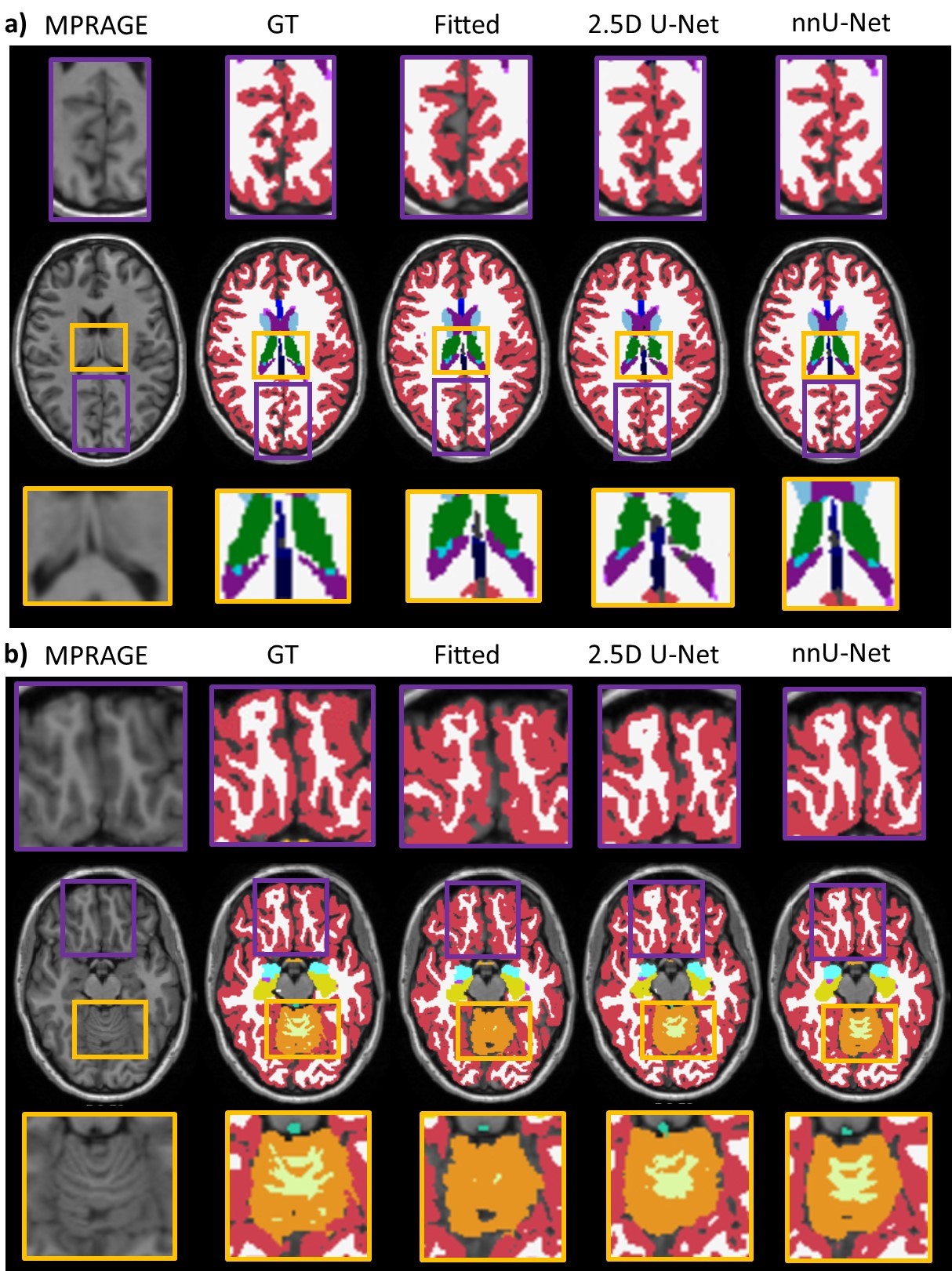
The DL approaches demonstrate improved qualitative segmentation compared to the synthesized MPRAGE images, e.g, more precise thalamus segmentation (Figure 3a) and enhanced cerebellar WM segmentation (Figure 3b).
Segmentation approaches remain consistent even in different head positions
We found that the DL-based methods maintain consistent segmentation performance across two different head positions though the 2.5D U-Net had some inconsistencies, particularly in the nucleus accumbens (Figure 5).
Discussion
nnUNet showed improved segmentation throughout the parenchyma compared to segmentation on synthetic MPRAGE. This underscores the substantial quantitative and qualitative advantages of volumetric segmentation combined with a tailored data augmentation pipeline, particularly in brain structures with limited representation.We opted for multi-atlas FreeSurfer segmentation as our chosen GT in the absence of better GT and it being the typical method of choice in the community for MPRAGE-segmentation. Furthermore, it is worth noting that multi-atlas FreeSurfer has undergone extensive validation12 and continues to be a widely adopted brain segmentation method.
Additionally, there has been promising work focused on generating multi-parametric scans directly from MRF coefficients using DL13. However, this research does not include a demonstration of segmentation performance on FreeSurfer labels.
Conclusion and Future Work
We here observed that DL segmentation methods, specifically nnUNet, trained directly on MRF coefficients can surpass quantitative and qualitative segmentation performance compared to segmentation on synthesized MPRAGE. In future research, we plan to develop a multi-task network, which could potentially facilitate the generation of MPRAGE segmentation maps and the synthesis of MPRAGE scans.Acknowledgements
This research was supported, in part, by R01MH116173, R01EB019437, U01EB025162, P41EB030006, R01EB033206, U24NS129893. A.K. was supported by the Tau Beta Pi and Stanford Knight-Hennessy fellowship.References
- Dan Ma, Vikas Gulani, Nicole Seiberlich, Kecheng Liu, Jeffrey L Sunshine, Jeffrey L Duerk, and Mark A Griswold. Magnetic resonance fingerprinting. Nature, 495(7440):187–192, 2013.
- Xiaozhi Cao, Huihui Ye, Congyu Liao, Qing Li, Hongjian He, and Jianhui Zhong. Fast 3d brain mr fingerprinting based on multi-axis spiral projection trajectory. Magnetic resonance in medicine, 82(1):289–301, 2019.
- Xiaozhi Cao, Congyu Liao, Siddharth Srinivasan Iyer, Zhixing Wang, Zihan Zhou, Erpeng Dai, Gilad Liberman, Zijing Dong, Ting Gong, Hongjian He, et al. Optimized multi-axis spiral projection MR fingerprinting with subspace reconstruction for rapid whole-brain high-isotropic-resolution quantitative imaging. Magnetic Resonance in Medicine, 88(1):133–150, 2022.
- Bruce Fischl. Freesurfer. Neuroimage, 62(2):774–781, 2012.
- Jason Ostenson, Ryan K Robison, Nicholas R Zwart, and E Brian Welch. Multi-frequency interpolation in spiral magnetic resonance fingerprinting for correction of off-resonance blurring. Magnetic resonance imaging, 41:63–72, 2017.
- Mengze Gao, Xiaozhi Cao, Daniel Abraham, Zihan Zhou, and Kawin Setsompop. Sequence adaptive deep learning framework to improve accuracy and robustness of MRF quantification via retrospective estimation and correction of B1+ and B0 inhomogeneities. Submitted 2023.
- Siddharth Srinivasan Iyer, Congyu Liao, Qing Li, Mary Katherine Manhard, Avery Berman, Berkin Bilgic, and Kawin Setsompop. Physical: A rapid calibration scan for B0, B1+, coil sensitivity and eddy current mapping. In Proceedings of the 28th Annual Meeting of ISMRM, Sydney/Virtual, page 0661, 2020.
- M Jorge Cardoso, Wenqi Li, Richard Brown, Nic Ma, Eric Kerfoot, Yiheng Wang, Benjamin Murrey, Andriy Myronenko, Can Zhao, Dong Yang, et al. Monai: An open-source framework for deep learning in healthcare. arXiv preprint arXiv:2211.02701, 2022.
- Fabian Isensee, Paul F Jaeger, Simon AA Kohl, Jens Petersen, and Klaus H Maier-Hein. nnu-net: a selfconfiguring method for deep learning-based biomedical image segmentation. Nature methods, 18(2):203– 211, 2021.
- Ferran Prados, John Ashburner, Claudia Blaiotta, Tom Brosch, Julio Carballido-Gamio, Manuel Jorge Cardoso, Benjamin N Conrad, Esha Datta, Gergely David, Benjamin De Leener, et al. Spinal cord grey matter ´ segmentation challenge. Neuroimage, 152:312–329, 2017.
- Minho Lee, JeeYoung Kim, Regina EY Kim, Hyun Gi Kim, Se Won Oh, Min Kyoung Lee, Sheng-Min Wang, Nak-Young Kim, Dong Woo Kang, ZunHyan Rieu, et al. Split-attention u-net: a fully convolutional network for robust multi-label segmentation from brain MRI. Brain Sciences, 10(12):974, 2020.
- Dhivya Srinivasan, Guray Erus, Jimit Doshi, David A Wolk, Haochang Shou, Mohamad Habes, Christos Davatzikos, Alzheimer’s Disease Neuroimaging Initiative, et al. A comparison of freesurfer and multi-atlas muse for brain anatomy segmentation: Findings about size and age bias, and inter-scanner stability in multisite aging studies. Neuroimage, 223:117248, 2020.
- Shihan Qiu, Sen Ma, Lixia Wang, Yuhua Chen, Zhaoyang Fan, Franklin G Moser, Marcel Maya, Pascal Sati, Nancy L Sicotte, Anthony G Christodoulou, et al. Direct synthesis of multi-contrast brain MR images from MR multitasking spatial factors using deep learning. Magnetic Resonance in Medicine, 2023.
Figures