1017
Accelerated multi-shell diffusion MRI with Gaussian processes estimated reconstruction of multi-band imaging1Wellcome Centre for Integrative Neuroimaging, FMRIB, Nuffield Department of Clinical Neurosciences, Univeristy of Oxford, Oxford, United Kingdom
Synopsis
Keywords: Diffusion Reconstruction, Diffusion/other diffusion imaging techniques
Motivation: Advanced diffusion MRI models that utilize multi-shell data provide higher specificity about tissue microstructure but require longer scan time, hindering wider application.
Goal(s): To increase the acquisition speed of multi-shell diffusion MRI for rapid tissue microstructure mapping.
Approach: We integrated multi-band imaging with the extended multi-shell Diffusion Acceleration with Gaussian process Estimated Reconstruction (ems-DAGER), including eddy-current corrected joint k-q reconstruction. The method was evaluated with in vivo data.
Results: Simulated and in-vivo results demonstrate that ems-DAGER method can significantly improve the image quality of reconstructed dMRI data with both in-plane and slice-wise acceleration to enable advanced multi-shell diffusion analysis.
Impact: Highly accelerated dMRI with the proposed method can shorten the scan time of multi-shell dMRI without sacrificing quality compared to conventional practice. This may facilitate a wider application of advanced dMRI models in basic and clinical neuroscience.
Introduction
Advanced diffusion MRI models provide higher specificity about tissue microstructure1-3 than conventional DTI4. However, to fit those advanced models requires acquisitions of many diffusion directions across several b shells that incurs higher scan time burden, which hinders its wider application in human scans. Various methods have been proposed to accelerate dMRI5-10. Diffusion Acceleration with Gaussian process Estimated Reconstruction (DAGER)11 is a joint k-q reconstruction method that achieves high acceleration by identifying and exploiting smoothness between different directions. Recently, a simulation work extended DAGER for multi-shell acquisition (ems-DAGER) for improving reconstruction of data with in-plane under-sampling12. Here, we further extend ems-DAGER to incorporate simultaneous-multi-slice (SMS) imaging. We demonstrate ems-DAGER with in-vivo scanning using both SMS and in-plane acceleration.Method
ems-DAGERDAGER uses Gaussian Processes (GP) to model smoothness of dMRI signals in q-space11and only considers covariance between signals within a single shell. For multi-shell dMRI data, the covariance between signals at different q space locations is extended to multi-shell as:
$$c(x,x') = C_\theta(\theta;a)exp(-(\frac{(logb-logb')^2}{2l^2})) (1)$$
$$C_{\theta}(\theta;a)=\begin{cases}1-\frac{3\theta}{2a}+\frac{\theta^3}{2a^3} & \theta \leq a\\0 & \theta > a\end{cases} (2)$$
Where $$$x$$$ and $$$x'$$$ refer to dMRI signals at two b shells ($$$b$$$ and $$$b'$$$ ) with an angular distance of $$$\theta$$$ . $$$a$$$ and $$$l$$$ are hyperparameters controlling signal smoothness within and between shells, respectively.
ems-DAGER incorporates GP estimated multi-shell dMRI signal as a prior and solves the following reconstruction problem:
$$u=\underset{u}{\operatorname{argmin}} (\frac{1}{2\sigma_k^2}\parallel Au-d\parallel^2_2+\frac{1}{2}(u-\mu)^H(\sum \otimes I_N)^{-1}(u-\mu))(3)$$
where $$$u$$$ is the unknown image, $$$\sigma$$$ is the noise standard deviation, $$$A$$$ maps $$$u$$$ to the acquired k-space data $$$d$$$ (including sensitivity encoding, Fourier transform and k-space under-sampling). Eddy currents are also incorporated in $$$A$$$. $$$\mu$$$ is the mean of GP prediction,$$$\sum$$$ is a covariance matrix generated from (1), and $$$I_N$$$ is an identity matrix . $$$H$$$ is the conjugate operation and $$$\otimes$$$ is Kronecker product. The problem is solved using gradient descent method. An initial reconstruction with shell-by-shell DAGER11 without using antipodal symmetric property is used to estimate eddy current induced field with FSL’s Eddy13-14.
Simulation
Simulations were performed based on HCP15 dMRI data. The data were fit with a ball-and-stick model16, which was then used to generate simulation data as forward model. A 100-direction (staggered across shells) dataset was generated with 50 b=1000s/mm2 and 50 b=2000s/mm2 images. A 64-direction dataset and a 36-direction dataset were generated by uniformly downsampling the 100-direction dataset on the unit q-space sphere. Eddy-current-induced $$$\triangle B_0$$$ was measured from a phantom using the same diffusion protocols on a 3T Siemens Prisma scanner and incorporated to simulate eddy-current distortion. k-q undersampling was optimized with a graph model based approach11 where a cross-shell q-space neighborhood has different k-space undersampling patterns. Multi-channel datasets with in-plane/SMS=3/4 (total acceleration12) were simulated with sensitivity maps from an 8-channel head coil.
In vivo
In vivo data from seven subjects were acquired on a Siemens 7T scanner with a 32-channel receive coil. A 2D DW-SE sequence was modified to include SMS acquisition with blipped-CAIPI encoding. dMRI data were acquired with 12X acceleration (in-plane/SMS=3/4) at 1.25mm and 1.5mm isotropic resolutions for different subjects. TR=3500ms, TE=70/67ms and 6/8 partial Fourier was used. Varying number of q-space points (100,66,36) were acquired. Single-band data with 2 repetitions were acquired as reference.
Results
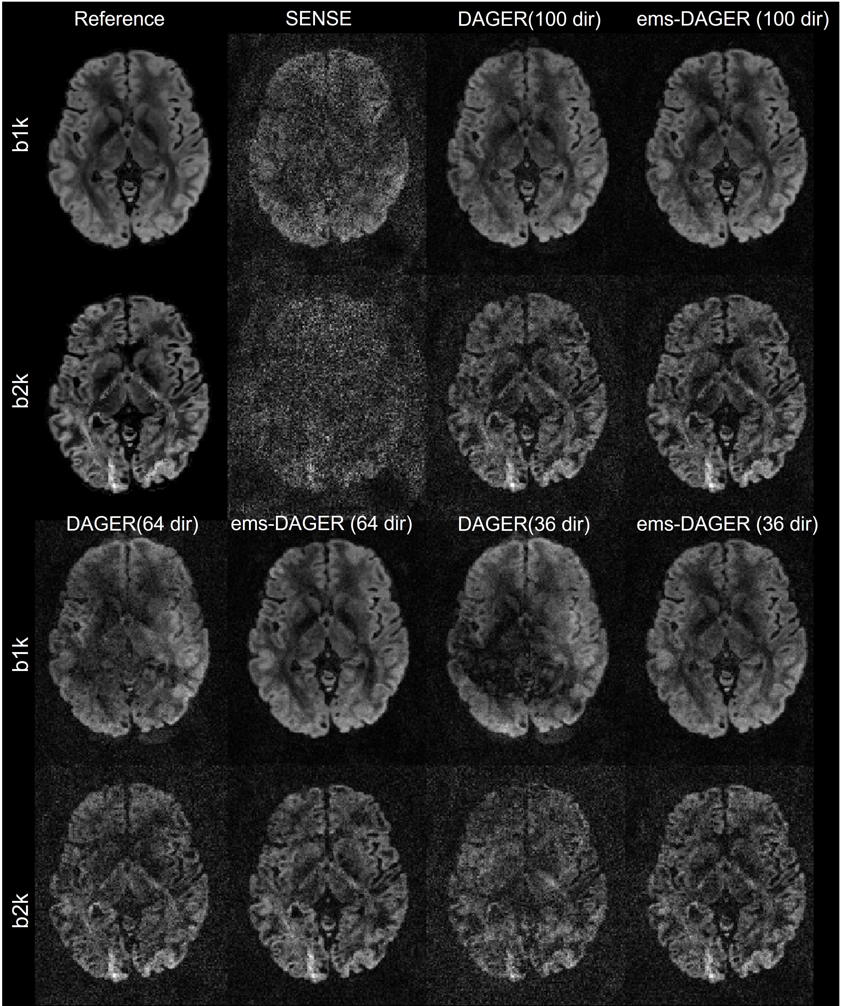
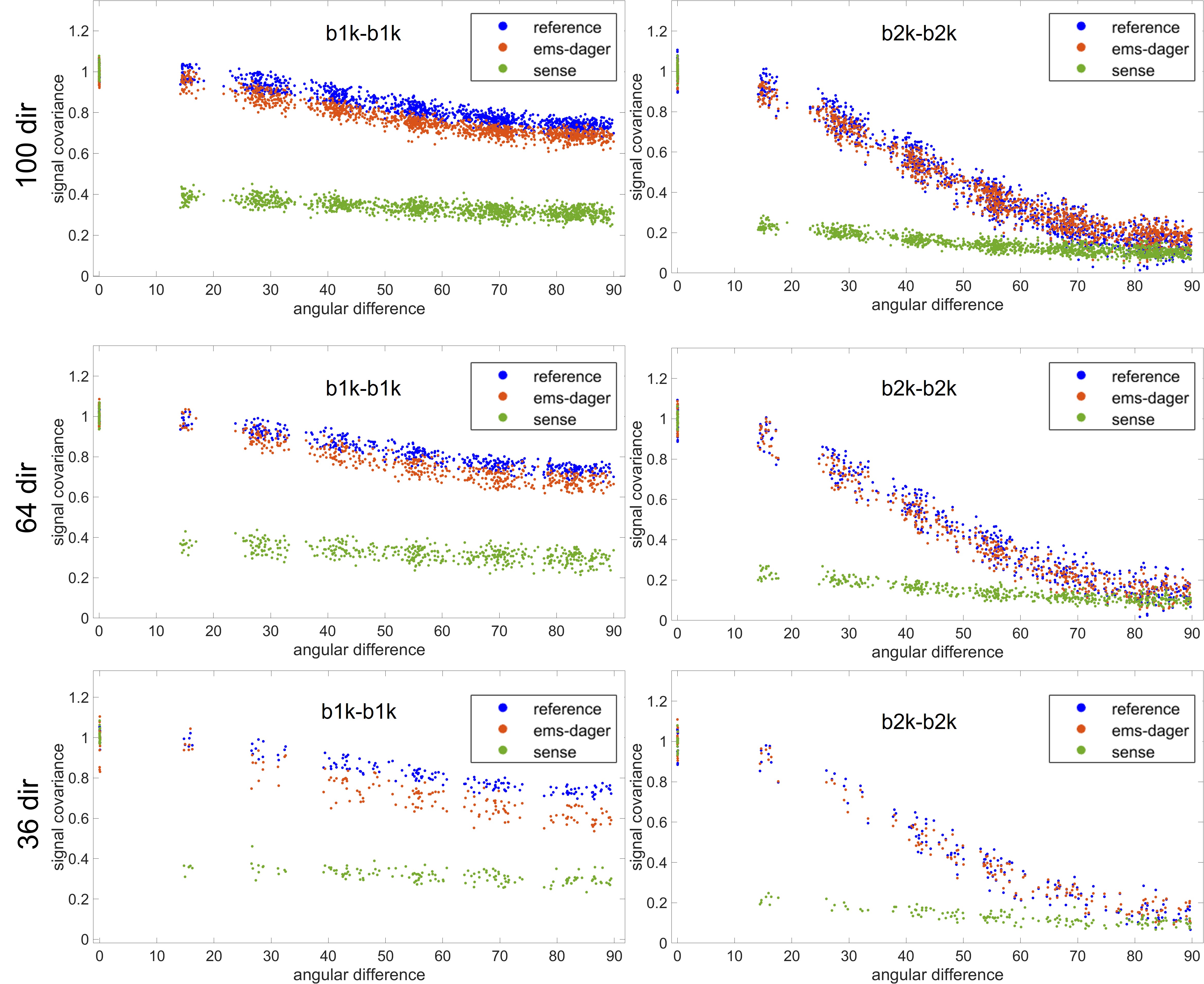
Fig.1 shows the 100,64 and 36 direction simulation results. ems-DAGER enables improved image quality compared to SENSE and single-shell DAGER by exploiting information across shells, particularly with a small number of directions.Fig.2 shows the covariance of the simulated dMRI signal as a function of the angle between them in q‐space, and smoothing would lead to increased covariance. ems-DAGER demonstrates highly consistent covariance compared to the reference, indicating preservation of angular resolution.
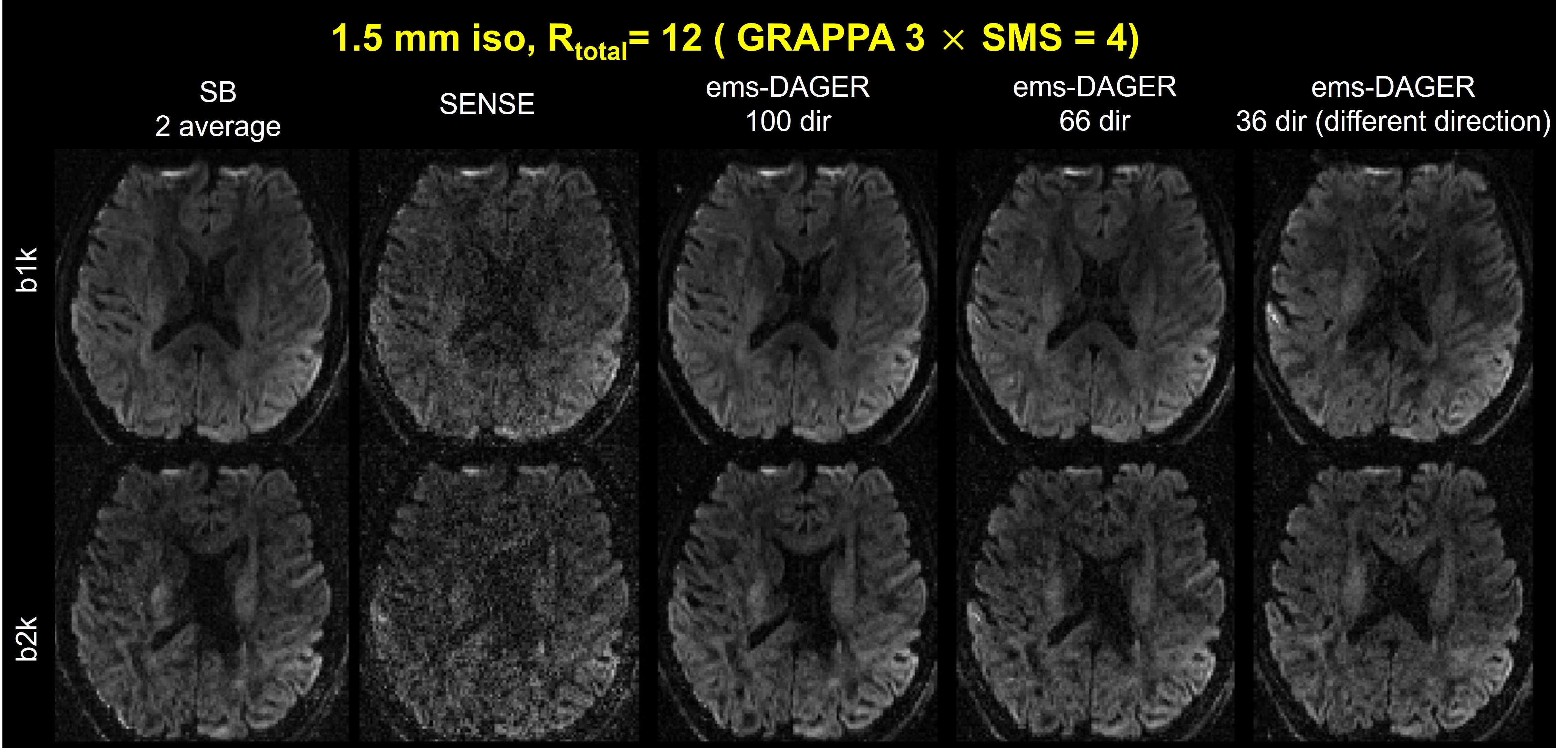
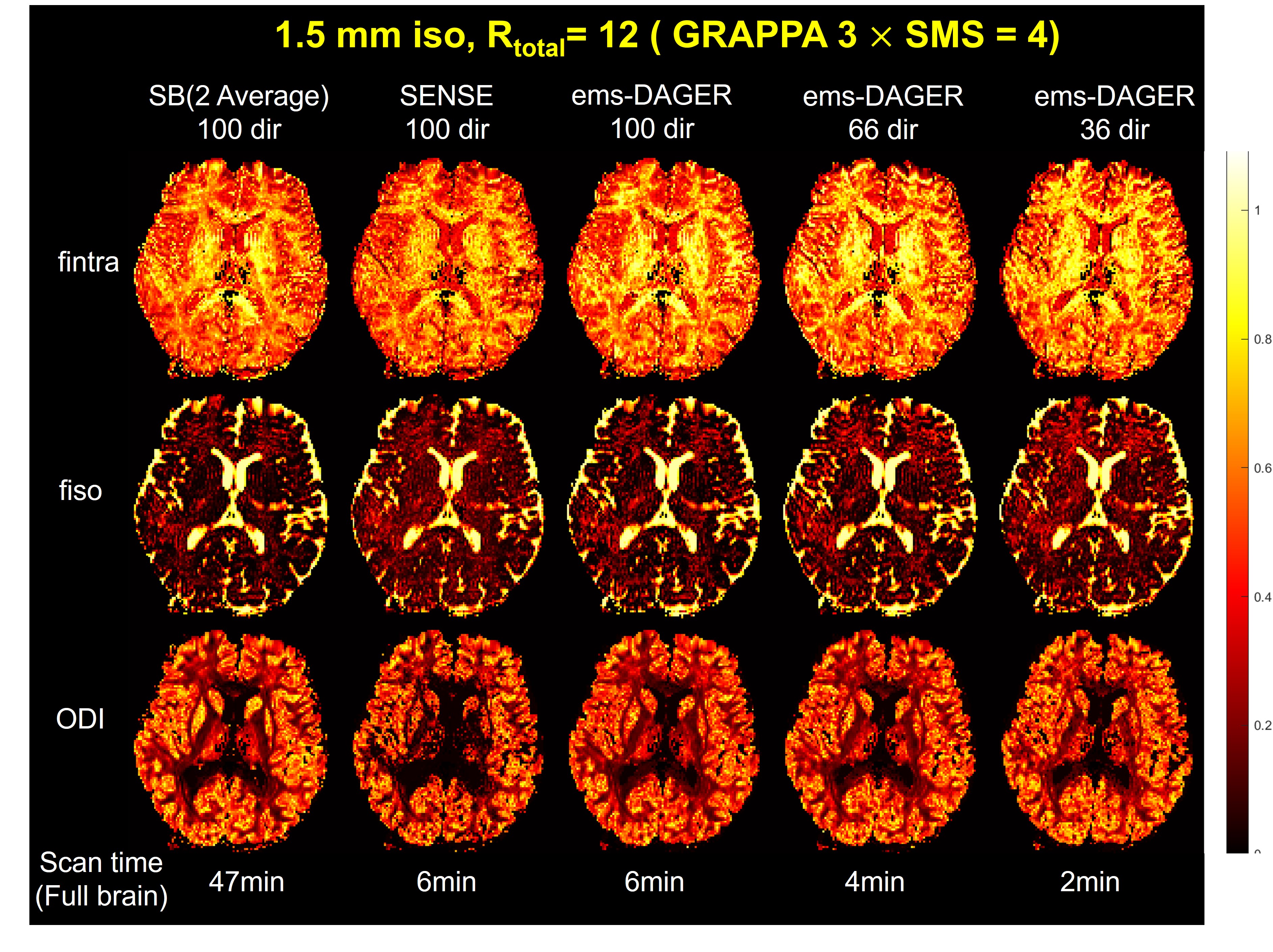
The reconstruction results for the 1.5 mm in vivo data with 100-dir, 66-dir and 36-dir are shown in Fig.3. Compared to the SENSE results, ems-DAGER provides significantly improved SNR and reduced artifacts, with comparable quality to the single-band reference. The improved image quality translates to the NODDI results as shown in Fig.4, where ems-DAGER produces consistent parameter maps compared to the single-band reference.
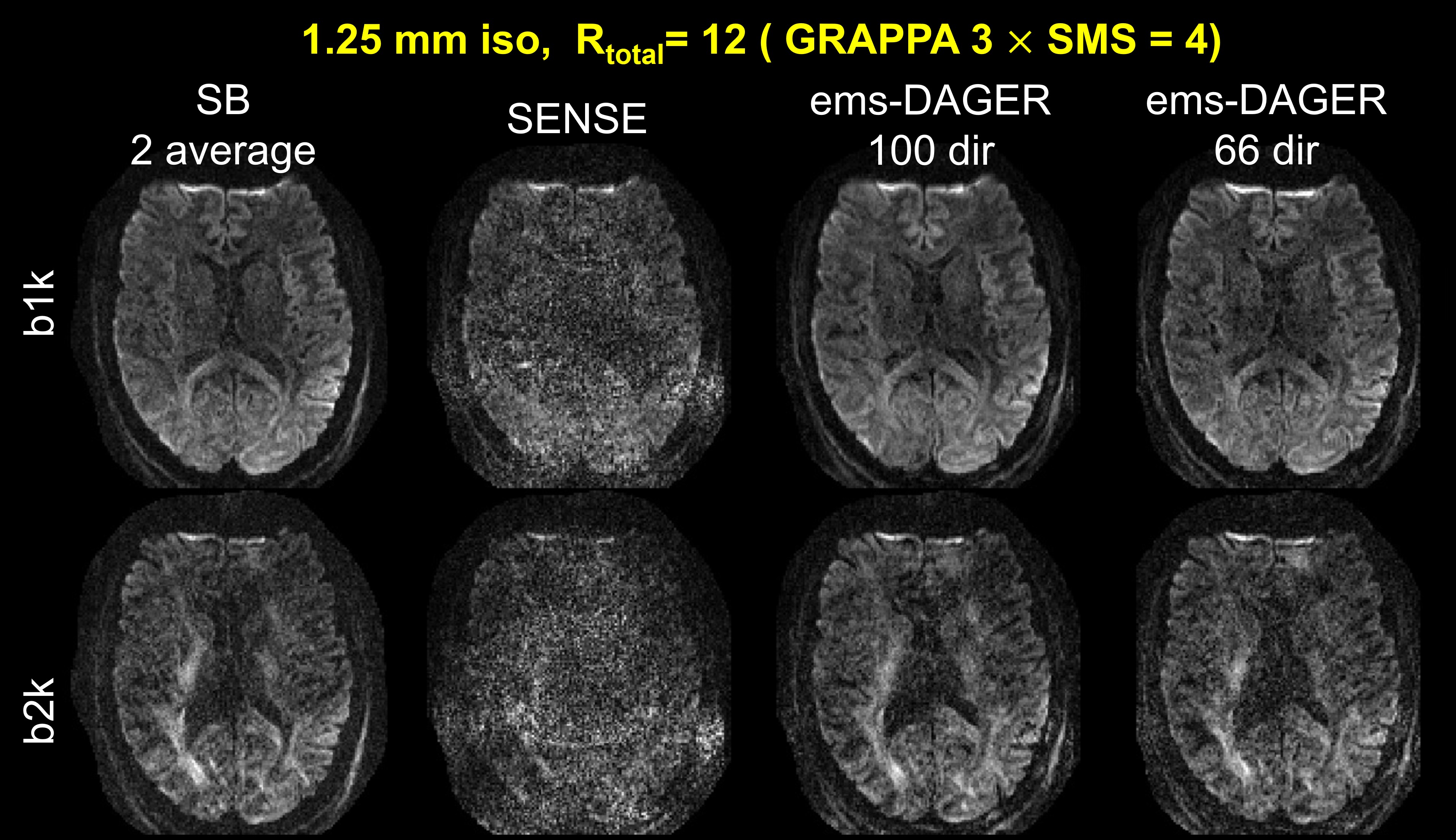
Fig.5 shows the reconstruction results for the 1.25mm in vivo data. Although the image quality is extremely poor in SENSE reconstruction, ems-DAGER significantly improved the image quality by incorporating GP prior information both for 100-dir data and 66-dir data.
Discussion
We extended the DAGER method to jointly reconstruct multi-band, multi-shell dMRI images with integrated eddy-current correction. Simulation and in vivo results demonstrated the proposed method can increase SNR and suppress artifacts with high acceleration factor. Eddy current effects were not obvious with our in-plane acceleration factor 3; we will further investigate this with lower in-plane accelerations.Acknowledgements
W.W. is supported by the Royal Academy of Engineering (RF\201819\18\92). K.L.M. is supported by the Wellcome Trust (WT202788/Z/16/A). This study is supported by the NIHR Oxford Health Biomedical Research Centre (NIHR203316). The views expressed are those of the author(s) and not necessarily those of the NIHR or the Department of Health and Social Care. The Wellcome Centre for Integrative Neuroimaging is supported by core funding from the Wellcome Trust (203139/Z/16/Z and 203139/A/16/Z).References
1. Jensen, J.H., Helpern, J.A., Ramani, A., Lu, H. and Kaczynski, K., 2005. Diffusional kurtosis imaging: the quantification of non‐gaussian water diffusion by means of magnetic resonance imaging. Magnetic Resonance in Medicine: An Official Journal of the International Society for Magnetic Resonance in Medicine, 53(6), pp.1432-1440.
2. Kaden E, Kelm ND, Carson RP, Does MD, Alexander DC. Multi-compartment microscopic diffusion imaging. Neuroimage 2016;139:346-359.
3. Zhang, H., Schneider, T., Wheeler-Kingshott, C.A. and Alexander, D.C., 2012. NODDI: practical in vivo neurite orientation dispersion and density imaging of the human brain. Neuroimage, 61(4), pp.1000-1016.
4. Basser, P.J., Mattiello, J. and LeBihan, D., 1994. MR diffusion tensor spectroscopy and imaging. Biophysical journal, 66(1), pp.259-267.
5. Griswold MA, Jakob PM, Heidemann RM, et al. Generalized autocalibrating partially parallel acquisitions (GRAPPA). Magn Reson Med. 2002;47:1202–1210.
6. Pruessmann KP, Weiger M, Scheidegger MB, Boesiger P. SENSE: sensitivity encoding for fast MRI. Magn Reson Med. 1999;42:952–962.
7.Setsompop K, Gagoski BA, Polimeni JR, Witzel T, Wedeen VJ, Wald LL. Blipped-controlled aliasing in parallel imaging for simultaneous multislice echo planar imaging with reduced g-factor penalty. Magn Reson Med 2012;67(5):1210-1224.
8. Menzel, M.I., Tan, E.T., Khare, K., Sperl, J.I., King, K.F., Tao, X., Hardy, C.J. and Marinelli, L., 2011. Accelerated diffusion spectrum imaging in the human brain using compressed sensing. Magnetic Resonance in Medicine, 66(5), pp.1226-1233.
9. Mani M, Jacob M, Guidon A, Magnotta V, Zhong J. Acceleration of high angular and spatial resolution diffusion imaging using compressed sensing with multichannel spiral data. Magn Reson Med 2015;73(1):126-138.
10.Shi X, Ma X, Wu W, Huang F, Yuan C, Guo H. Parallel imaging and compressed sensing combined framework for accelerating high-resolution diffusion tensor imaging using inter-image correlation. Magn Reson Med 2015;73(5):1775-1785.
11.Wu W, Koopmans PJ, Andersson JLR, Miller KL. Diffusion Acceleration with Gaussian process Estimated Reconstruction (DAGER). Magn Reson Med 2019;82(1):107-125.
12. Ye X, Miller KL, Wu W. Extended multi-shell diffusion acceleration with Gaussian processes estimated reconstruction (ems-DAGER). In Proceedings of ISMRM 2023, 2023. P3960.
13. Andersson JL, Sotiropoulos SN. Non-parametric representation and prediction of single- and multi-shell diffusion-weighted MRI data using Gaussian processes. Neuroimage 2015;122:166-176.
14. Jenkinson M, Beckmann CF, Behrens TE, Woolrich MW, Smith SM. Fsl. Neuroimage. 2012;62(2):782-790.
15. Sotiropoulos SN, Jbabdi S, Xu JQ, et al. Advances in diffusion MRI acquisition and processing in the Human Connectome Project. Neuroimage 2013;80:125-143.
16. Behrens TEJ, Berg HJ, Jbabdi S, Rushworth MFS, Woolrich MW. Probabilistic diffusion tractography with multiple fibre orientations: what can we gain? NeuroImage. 2007;34:144–155
Figures