1016
Improved SLIPEN (iSLIPEN) for Three-Dimensional Multi-slab Diffusion-Weighted Imaging by Partial Fourier and Prior Information1Department of Diagnostic Radiology, The University of Hong Kong, Hong Kong, Hong Kong, 2Department of Biomedical Engineering, The Chinese University of Hong Kong, Hong Kong, Hong Kong, 3Multi-Scale Medical Robotics Center, Hong Kong, Hong Kong
Synopsis
Keywords: DWI/DTI/DKI, Diffusion Tensor Imaging
Motivation: SLIPEN is a promising technique to obtain 3D multi-slab DWI without suffering slab boundary artifacts. However, its performance is degraded when encountering limited signal SNR.
Goal(s): An improved SLIPEN is desired to achieve robust perfomance regardless of limited signal SNR.
Approach: Partial Fourier was applied to design an optimized sampling pattern and prior information was also incorporated into the model to improve the performance.
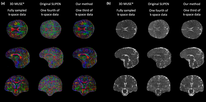
Results: The improved SLIPEN could achieve comparable results to gold standard for in-vivo DWI images and DTI maps, with the need of only one third of gold standard data.
Impact: 3D isotropic high-resolution DWI without suffering from slab boundary artifacts can be robustly achieved by our method with the use of 2D navigator, therefore benefiting the neuroscience study in evaluating crossing and kissing fibers.
Introduction
Three-dimensional multi-slab diffusion-weighted imaging (3D-ms-DWI)[1-3] is a promising technique to acquire diffusion data with high-resolution and high-SNR, and therefore can be used to resolve crossing fibers or kissing fibers in neuroscience study[4-6]. However, it suffers from slab boundary artifacts (SBA) due to slab aliasing and crosstalk effect[7]. The originally proposed sliding-slab profile encoding (SLIPEN), conducting on a 3T scanner, dramatically alleviated SBA by reconstructing the 3D-ms-DWI data acquired with multiple separate acquisitions and enlarged gap between adjacent slabs[8]. Nevertheless, the limited and insufficient SNR of each thin slab at 1.5T may degrade the SLIPEN reconstruction performance. To optimize the SLIPEN for its application at 1.5T MRI, an improved SLIPEN (iSLIPEN) is proposed to incorporate partial Fourier and prior information into the original SLIPEN model.Material and Method
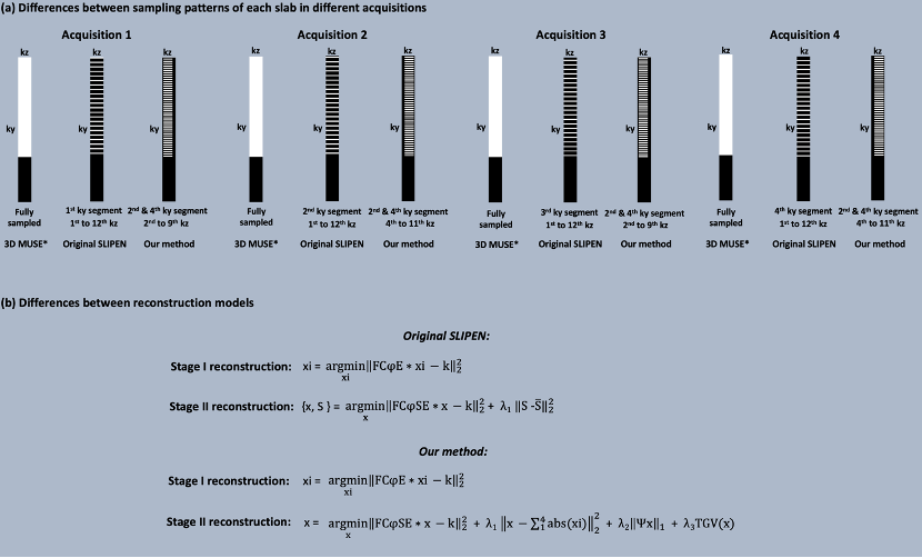
Differences between improved and original SLIPEN frameworksIn data acquisition aspect, both methods share the same sliding-slab strategy to acquire the 3D-ms-DWI data from four separate acquisitions with data under-sampling (Figure 2a), but different under-sampling patterns of each acquisition are applied among them (Figure 1a). Original SLIPEN acquires four different ky-segments respectively for four separate sliding-slab acquisitions, with an under-sampling factor equals to 4 for each acquisition. In contrast, the proposed iSLIPEN acquires two fixed ky-segments (e.g., 2nd and 4th ky-segment) with kz partial Fourier for four separate sliding-slab acquisitions, and the corresponding under-sampling factor is equal to 3 for each acquisition.
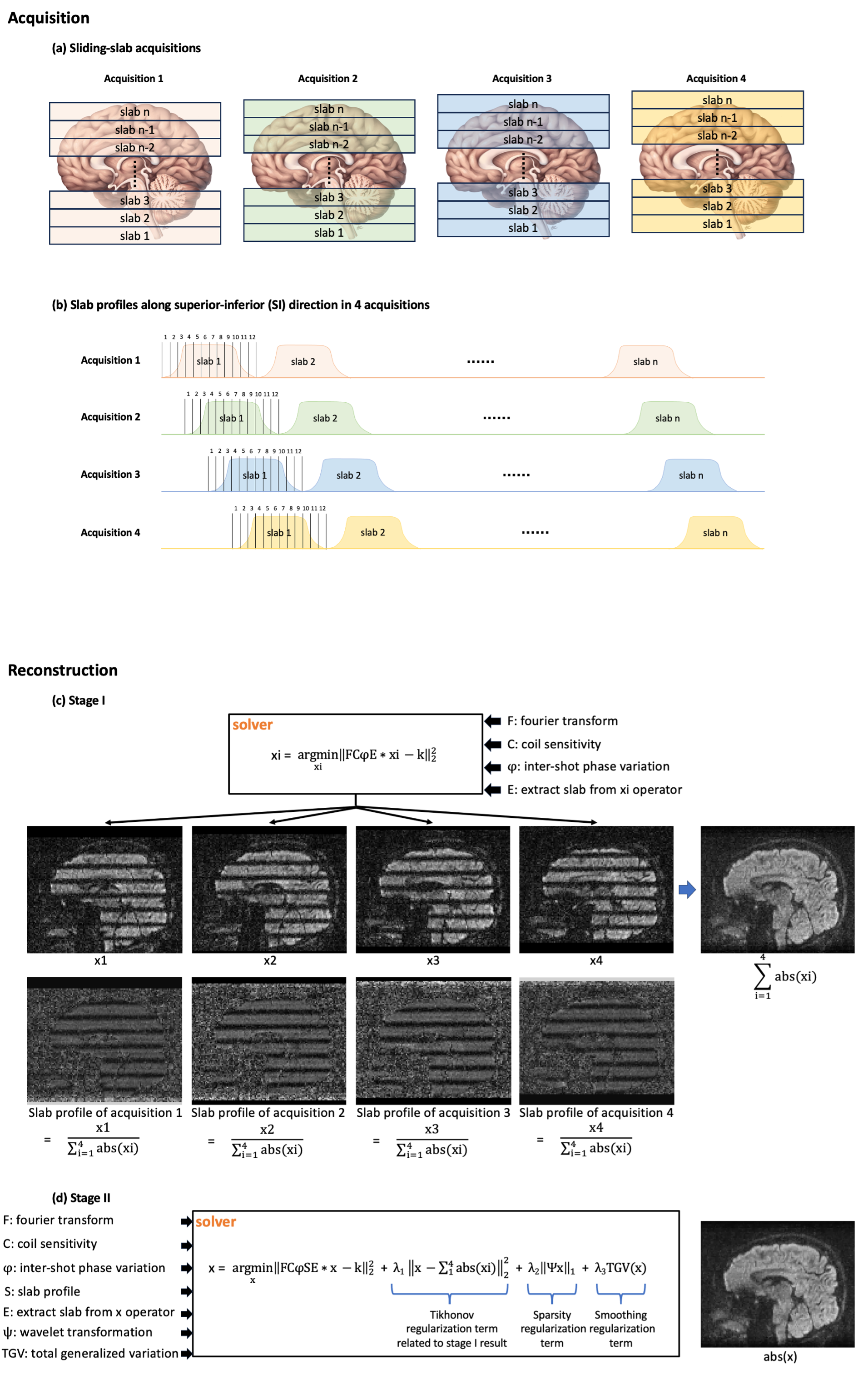
Regarding the reconstruction model, instead of updating slab profiles with an averaging constraint, iSLIPEN applies three regularization terms to stabilize and optimize the result in stage II (Figure 1b and Figure 2d) based on prior information. Because of the fact that the results generated in stage II and stage I should be close, the Tikhonov regularization term[9] is incorporated into stage I to make the solver in stage II converge faster and more stable. The second sparsity regularization term that relies on the data sparsity in wavelet domain is used as the prior information to reduce the undesired noise in image domain[10]. Total generalized variation (TGV) is the third regularization term, which takes first order and higher order differentiation information into consideration, can perform further denoising to the result without staircasing effect arising from smoothing[11].
Pipeline of iSLIPEN
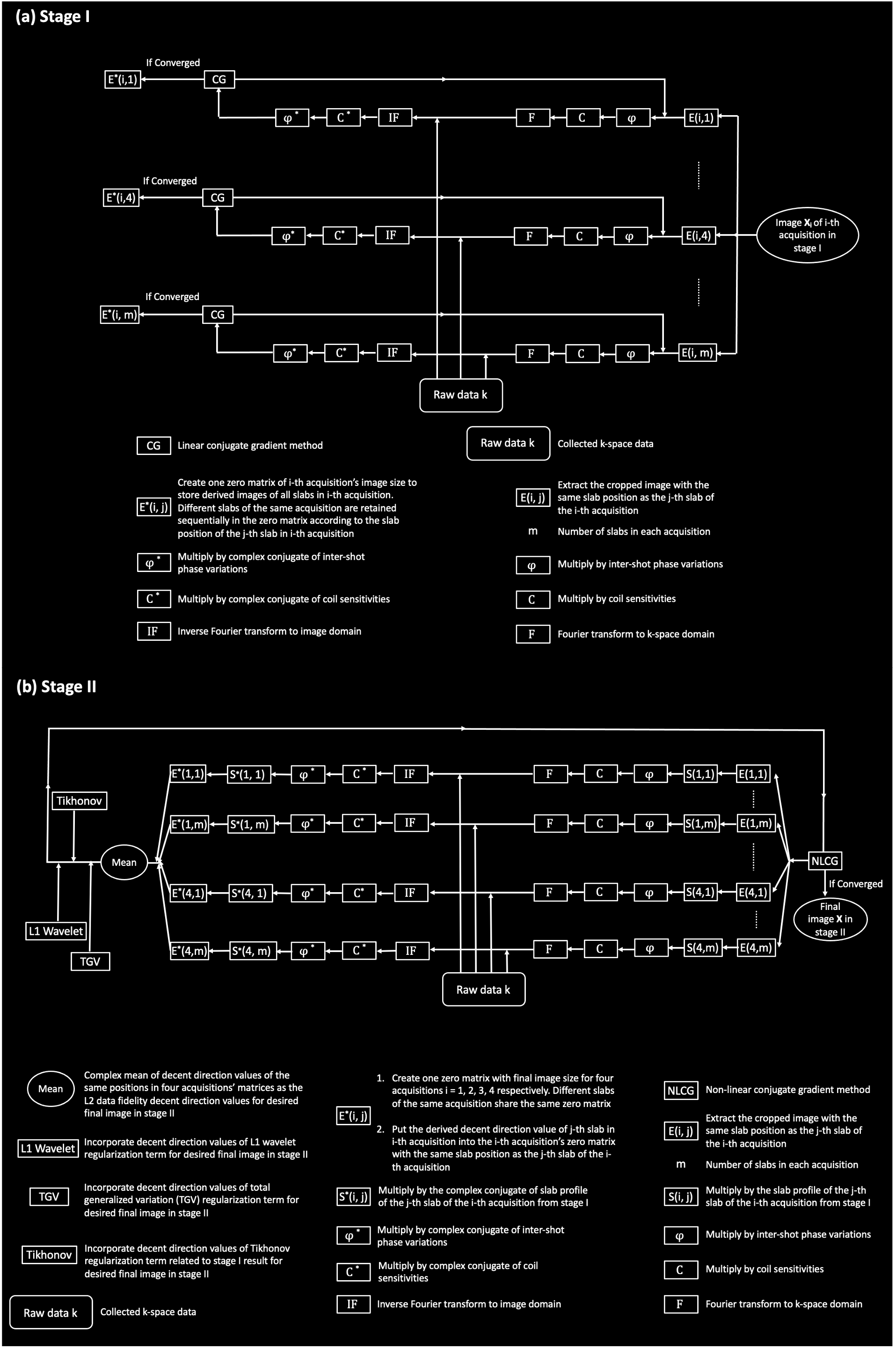
Figure 2 illustrates the entire pipeline of iSLIPEN for 3D-ms-DWI from data acquisition to reconstruction. During data acquisition (Figure 2a), total 4 separate sliding-slab acquisitions are used to cover the whole brain and the sliding-slab shift between adjacent acquisitions is one fourth of slab FOV along z direction (Figure 2b). The images of each acquisition are then produced by the solver in the stage I of reconstruction pipeline, and the slab profiles of all sliding-slab acquisitions can be estimated (Figure 2c). In stage II, the final optimized images can be derived by taking into account the Fourier transform F, coil sensitivity C, inter-shot phase variation φ, slab profile S, extract slab operator E, and all three regularization terms. Figure 3a and 3b respectively illustrate the flow chart of the use of either linear conjugate gradient or non-linear conjugate gradient algorithm to solve the images in stage I and stage II.
In-vivo experiment
Three sets of human brain DTI data with 1.6mm isotropic resolution and 6 diffusion directions were collected from a healthy subject at a 1.5T GE MRI scanner using 3D-ms-DWI acquisition with sliding-slab encoding. The identical sliding-slab parameters and slab locations were used to acquire these three datasets, but with different design of sampling pattern for the data acquisition of each slab (Figure 1a). The first dataset acquired fully sampled k-space data of each slab for 4 sliding-slab acquisitions. Afterwards, slabs in each sliding-slab acquisition were individually reconstructed by 3D MUSE[12], and the final volume 3D DWI data was obtained by averaging all sliding-slab data together (denoted as 3D MUSE* as the gold standard without SAB). The other two datasets were acquired with the sampling patterns proposed in original SLIPEN and iSLIPEN, respectively, and then reconstructed with corresponding pipelines.
Results
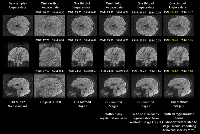
Figure 4 compares the representative DWI images reconstructed by 3D MUSE*, original SLIPEN and at different stages of iSLIPEN. Figure 5 compares the DTI results of 3D MUSE*, original SLIPEN and iSLIPEN in terms of color-FA maps and mean diffusivity (MD) maps.Discussion and Conclusion
This study demonstrates an improved sliding-slab profile encoding technique (i.e., iSLIPEN), incorporating partial Fourier and prior information, for enabling three-dimensional multi-slab diffusion-weighted imaging with robust suppression of SAB. It can dramatically optimize the results compared to original SLIPEN when encountering limited signal SNR (Figure 4).Acknowledgements
The work was in part supported by grants from Hong Kong Research Grant Council (GRF17106820, GRF17125321, GRF14206723, and ECS24213522).References
- Engstrom, M. and S. Skare, Diffusion-weighted 3D multislab echo planar imaging for high signal-to-noise ratio efficiency and isotropic image resolution. Magn Reson Med, 2013. 70(6): p. 1507-14.
- Frost, R., et al., 3D multi-slab diffusion-weighted readout-segmented EPI with real-time cardiac-reordered K-space acquisition. Magn Reson Med, 2014. 72(6): p. 1565-79.
- Wu, W.C., et al., High-resolution diffusion MRI at 7T using a three-dimensional multi-slab acquisition. Neuroimage, 2016. 143: p. 1-14.
- Jeurissen, B., et al., Diffusion MRI fiber tractography of the brain. NMR Biomed, 2019. 32(4): p. e3785.
- Basser, P.J., et al., In vivo fiber tractography using DT-MRI data. Magn Reson Med, 2000. 44(4): p. 625-32.
- Auriat, A.M., et al., Comparing a diffusion tensor and non-tensor approach to white matter fiber tractography in chronic stroke. Neuroimage Clin, 2015. 7: p. 771-81.
- Wu, W., et al., Reducing slab boundary artifacts in three-dimensional multislab diffusion MRI using nonlinear inversion for slab profile encoding (NPEN). Magn Reson Med, 2016. 76(4): p. 1183-95.
- Xiaoxi Liu , D.C., Xucheng Zhu , Edward S. Hui , Queenie Chan , Peder E.Z. Larson , and Hing-Chiu Chang, Sliding-Slab Profile Encoding (SLIPEN) for Eliminating Slab Boundary Artifact in Three-Dimensional Multi-slab Diffusion-Tensor Imaging, in ISMRM & SMRT Virtual Conference & Exhibition. 2020.
- Lin, F.H., et al., Parallel imaging reconstruction using automatic regularization. Magn Reson Med, 2004. 51(3): p. 559-67.
- Lustig, M., D. Donoho, and J.M. Pauly, Sparse MRI: The application of compressed sensing for rapid MR imaging. Magn Reson Med, 2007. 58(6): p. 1182-95.
- Knoll, F., et al., Second order total generalized variation (TGV) for MRI. Magn Reson Med, 2011. 65(2): p. 480-91.
- Chang, H.C., et al., Human brain diffusion tensor imaging at submillimeter isotropic resolution on a 3Tesla clinical MRI scanner. Neuroimage, 2015. 118: p. 667-75.
Figures