1014
Towards Genuine Three-dimensional Diffusion Imaging with Second Order Gradient Moment Nulling1MR Research Collaboration Team, Siemens Healthineers Ltd., Beijing, China, 2Siemens Shenzhen Magnetic Resonance Ltd., Shenzhen, China, 3Department of Biomedical Engineering, School of Medicine, Tsinghua University, Beijing, China
Synopsis
Keywords: Diffusion Acquisition, Pulse Sequence Design
Motivation: While 3D acquisition has the advantages of achieving high resolution and signal-to-noise ratio and has been established for most sequences, diffusion imaging, has predominantly adhered to 2D acquisition or partial 3D such as multi-slab acquisition for over 50 years.
Goal(s): We aim to bring diffusion imaging to the next era by implementing a genuine 3D diffusion imaging sequence.
Approach: The sequence was implemented using gradient moment nulling and 3D EPI acquisition and basic reconstruction methods.
Results: Whole brain 3D diffusion imaging was achieved at isotropic sub-millimeter resolution and a practical scan time.
Impact: This work liberates diffusion imaging from 2D or partial 3D acquisition to true 3D acquisition.
Introduction
While 3D acquisition has the advantages of achieving high resolution and signal-to-noise ratio and has been established for most sequences, diffusion imaging1, has predominantly adhered to 2D acquisition or partial 3D such as multi-slab acquisition2 for over 50 years. Multi-slab method remains a hybrid approach that is prone to the occurrence of slab boundary artifacts3-5. Fundamentally, addressing the critical aspect of diffusion imaging with multiple shots invariably involves tackling motion-induced random phase variations. An emerging technique is gradient moment nulling6, which substantially reduces signal loss for myocardium diffusion imaging. In this study, we propose to use gradient moment nulling to achieve true 3D diffusion imaging.Methods
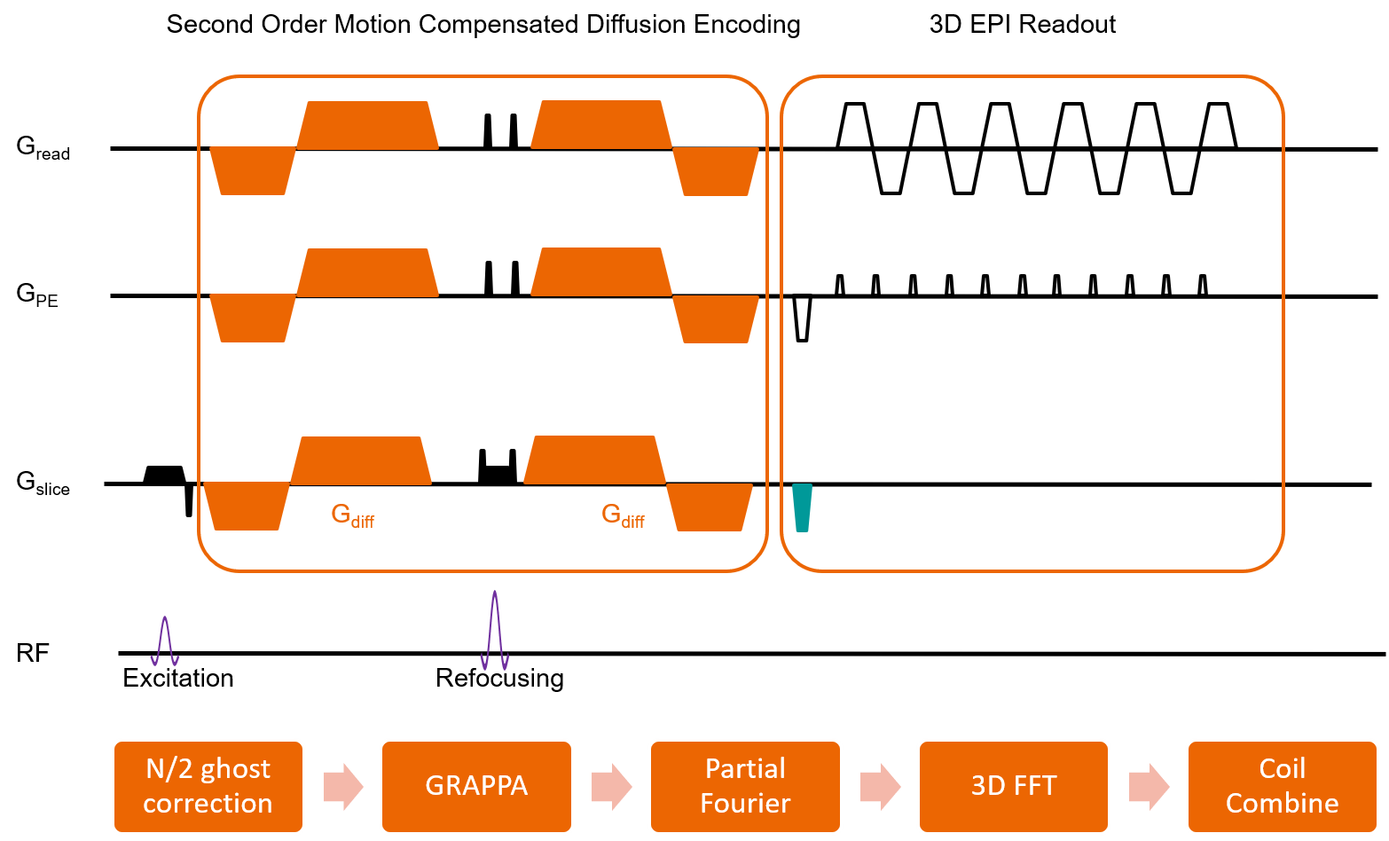
Sequence Design:As shown in Figure 1, the design of the diffusion encoding gradients were adopted from Welsh’s work6 to null the gradient moment up to the second order. The 3D k-space was acquired with a 3D echo planar imaging (EPI) trajectory. After each excitation of the whole 3D volume, one kz plane is acquired.
Data Acquisition
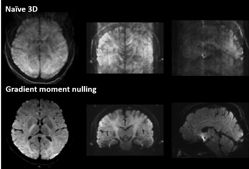
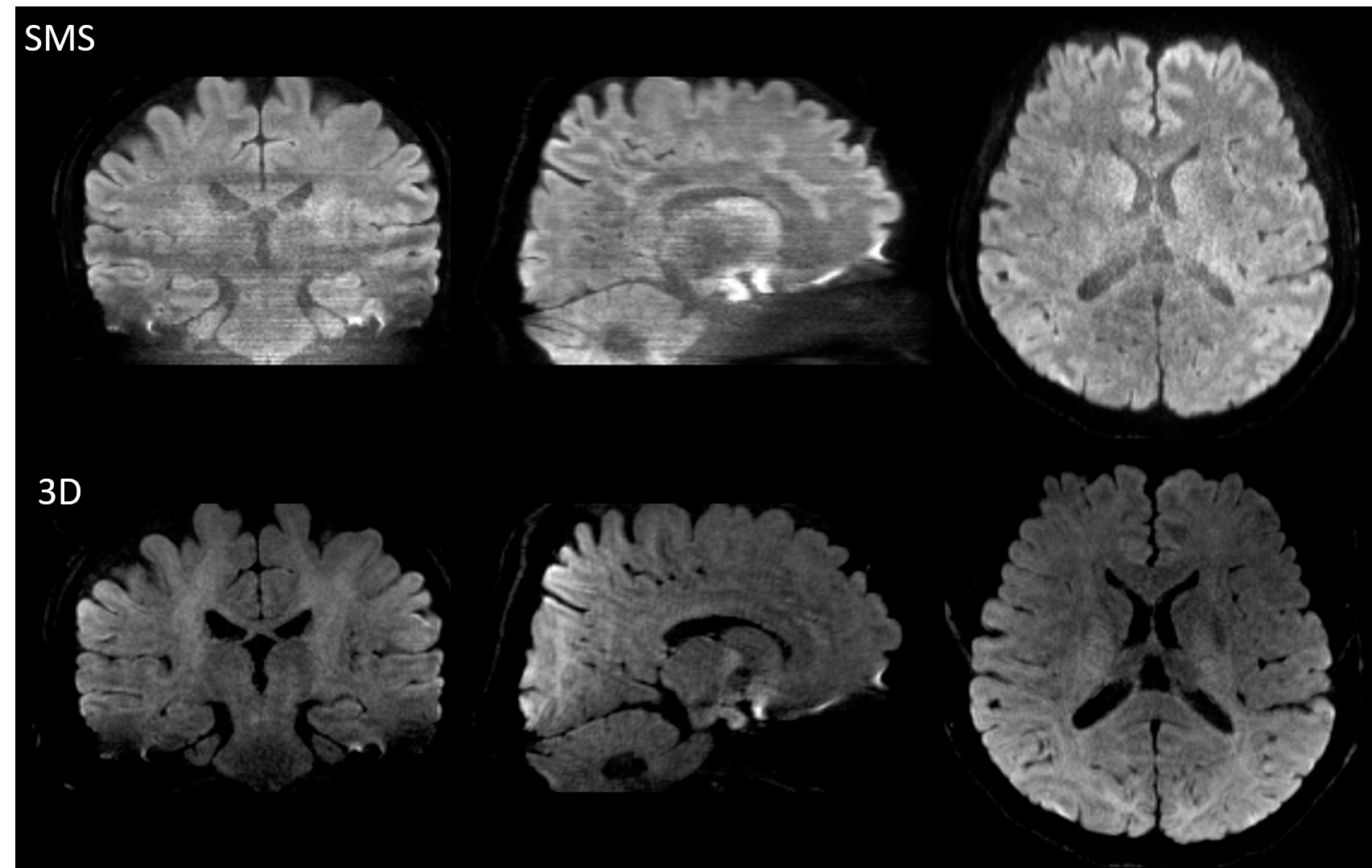
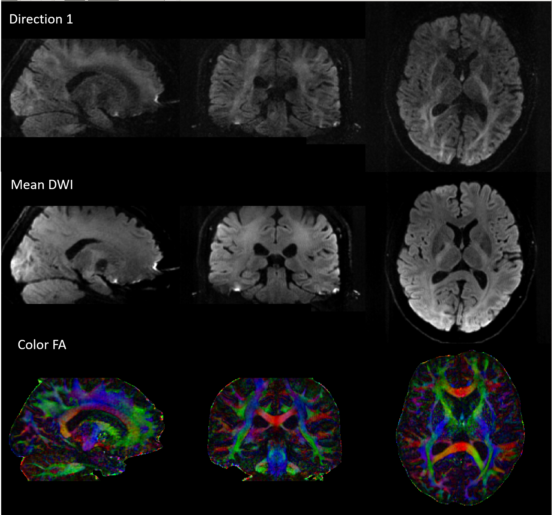
All data were acquired on a 3T MAGNETOM Prisma (Siemens Healthcare, Erlangen, Germany) using a 64 channel head and neck coil. Three healthy volunteers were recruited for the MRI scan. To illustrate the efficacy of the proposed method compared to a naïve 3D DWI acquisition with monopolar diffusion gradients, one subject underwent two 3D DWI sequences at an isotropic spatial resolution of 2 mm3: 1) monopolar diffusion encoding with 0th moment nulled (M0), 2) second order gradient moment nulled (M2). Other sequence parameters were identical: FOV = 240*240 mm2, 60 slices, one b=0 s/mm2 and 3 b=1000 s/mm2 using 3-scan trace, TR = 1500 ms, TE = 68 ms, slice oversampling = 20%, ipat =2 in the phase encoding direction. The scan time for each sequence was 7 min. The second subject underwent two high resolution DWI scans, a 2D simultaneous multi-slice (SMS) sequence which is the current state-of-the-art 2D DWI sequence and the proposed 3D DWI sequence. Both sequences used identical spatial resolution of 0.9 mm3 and almost identical scan time of 4.5 minutes. For the 3D DWI sequence, TR = 750 ms, TE = 88 ms, ipat = 3, phase partial Fourier = 5/8 and slice partial Fourier = 5/8. For the SMS sequences, ipat = 3 and phase partial Fourier = 5/8 were used the same as 3D DWI, and SMS = 3 was used for slice acceleration, TE = 61ms, TR=5300 ms. The third subject underwent a 3D DTI scan with 12 diffusion encoding directions at isotropic 1.2 mm3 spatial resolution, ipat = 3, phase partial Fourier = 6/8, slice partial Fourier = 5/8, TR = 750 ms, TE = 100 ms, 90 slices and slice oversampling = 20%. The scan time was 10 mins.
Image Reconstruction and Processing
All 3D DWI images were reconstructed with a simple pipeline shown in Figure 1: after correction of Nyquist ghost, GRAPPA7 was performed to restore the missing data and partial Fourier was reconstructed using a POCS algorithm. The image reconstruction was carried out using MATLAB (Mathworks, Natick, MA, USA). The 2D SMS images were reconstructed online using the product algorithm. The DTI data was fitted using FSL.
Results
Figure 2 showed the reconstructed axial mean diffusion weighted images as well as reformatted coronal and sagittal views. It was obvious that, without any motion control, a plain 3D acquisition resulted in severe ghost artifacts. M2 substantially reduced the ghost artifacts and generated image with high fidelity. Figure 3 showed the 0.9 mm3 isotropic diffusion images using SMS and the proposed method. With similar scan time, the 3D sequence generated diffusion image with high fidelity in all three views. The 2D SMS images were hampered by low SNR and parallel imaging artifacts. Figure 4 showed the original diffusion images as well as color coded FA maps using the proposed sequence, showing fine structural details in three views.Discussion and conclusion
In this study, a novel yet a simple method was proposed to achieve genuine 3D diffusion imaging with motion control. The capability of acquiring sub-millimeter isotropic diffusion imaging using the proposed method was also demonstrated. There was no need for estimating phase variation using either navigator acquisition or self-navigator, which has been widely used for both 2D multi-shot diffusion imaging and 3D multi-slab diffusion imaging. The k-space from different acquisition were directly assembled without any extra phase correction. Since the acquisition is in a true 3D format, no slab boundary artifacts exist. This method may pave the way for a new generation of diffusion imaging technical development and applications.Acknowledgements
No acknowledgement found.References
1 Stejskal, E. O. & Tanner, J. E. Spin Diffusion Measurements: Spin Echoes in the Presence of a Time‐Dependent Field Gradient. Journal of Chemical Physics 42, 288-292, doi:10.1063/1.1695690 (1965).
2 Engstrom, M. & Skare, S. Diffusion-weighted 3D multislab echo planar imaging for high signal-to-noise ratio efficiency and isotropic image resolution. Magnetic resonance in medicine 70, 1507-1514, doi:10.1002/mrm.24594 (2013).
3 Parker, D. L., Yuan, C. & Blatter, D. D. MR angiography by multiple thin slab 3D acquisition. Magnetic resonance in medicine 17, 434-451, doi:https://doi.org/10.1002/mrm.1910170215 (1991).
4 Van, A. T. et al. Slab profile encoding (PEN) for minimizing slab boundary artifact in three-dimensional diffusion-weighted multislab acquisition. Magnetic resonance in medicine 73, 605-613, doi:10.1002/mrm.25169 (2015).
5 Wu, W., Koopmans, P. J., Frost, R. & Miller, K. L. Reducing slab boundary artifacts in three-dimensional multislab diffusion MRI using nonlinear inversion for slab profile encoding (NPEN). Magnetic resonance in medicine 76, 1183-1195, doi:10.1002/mrm.26027 (2016).
6 Welsh, C. L., DiBella, E. V. & Hsu, E. W. Higher-Order Motion-Compensation for In Vivo Cardiac Diffusion Tensor Imaging in Rats. IEEE Trans Med Imaging 34, 1843-1853, doi:10.1109/TMI.2015.2411571 (2015).
7 Griswold, M. A. et al. Generalized autocalibrating partially parallel acquisitions (GRAPPA). Magnetic resonance in medicine 47, 1202-1210, doi:10.1002/mrm.10171 (2002).
Figures