1012
Low-distortion Spine DWI with Ultra-high Shot and Navigator-free Reconstruction1Xiamen University, Xiamen, China, 2Philips Healthcare, Beijing, China, 3United Imaging Healthcare, Shanghai, China, 4Department of Radiology, Zhongshan Hospital (Xiamen), Fudan University, Xiamen, China, 5Xiamen University of Technology, Xiamen, China
Synopsis
Keywords: Diffusion Reconstruction, Diffusion/other diffusion imaging techniques
Motivation: State-of-the-art low-rank methods recover multi-shot DWI with 2D structured matrix completion, but are hindered by long computation time (tens of minutes per image) and unsatisfactory ultra-high shot reconstruction (no more than 8-shot).
Goal(s): Fast and reliable ultra-high (above 8-shot) DWI reconstruction.
Approach: ODLRS: A 1D low-rank Hankel reconstruction method with self-adaptive subspace.
Results: ODLRS is a novel 1D low-rank framework for multi-shot DWI reconstruction. Compared to conventional low-rank methods, ODLRS achieves 109 times accelerated reconstruction, and low-distortion spine DWI with 12 shots.S
Impact: This work achieves fast (109 times acceleration) and reliable ultra-high (10 and 12 shots) DWI reconstruction, reducing the deformation of conventional spinal cord DWI significantly.
Introduction
Multi-shot interleaved echo planer imaging (ms-iEPI) can obtain DWI with reduced distortions [1, 2] than single-shot EPI. The higher the number of excitations, the lower the image distortion [3]. State-of-the-art low-rank methods with 2D structured matrix completion, such as MUSSELS [4] and PAIR [5], have shown nice performance on high-shot number data reconstructions (6~8 shots). However, extremely long computational time (tens of minutes per image) are huge limitations for the practical application of these methods. Moreover, when the shot number increased further (10 and 12 shots), these methods may be unsatisfactory. In this work, we propose a 1D low-rank model [6, 7] with self-adaptive subspace (ODLRS) for low-distortion DWI reconstruction, obtaining 109 times computation acceleration and stable performance at ultra-high shot reconstruction (12-shot).Method
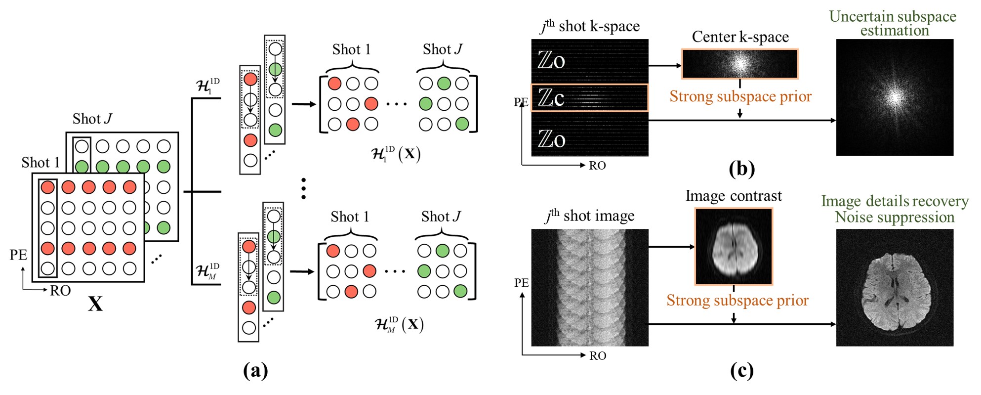
In this work, we attempt to establish a framework to enable fast and reliable 1D low-rank ms-iEPI DWI reconstructions. Inspired by separable low-rank methods in fast MRI, we first propose ODLR, which replaces the large 2D block Hankel matrix with multiple small 1D Hankel matrices (Fig. 1(a)). Since the SVD time of multiple 1D Hankel matrices is largely reduced than the original 2D block Hankel matrix, ODLR accelerates the reconstruction by about 4 times. However, ODLR has two weaknesses that can be further optimized. The former is the slow convergence speed, and the latter is the unstable performance under ultra-high shot reconstruction. We further propose the ODLR with self-adaptive subspace (ODLRS) reconstruction, which obtain about 109 times acceleration and better reconstruction performance. In the first step of ODLRS, strong subspace is calculated from the pre-reconstructed k-space center (Fig. 1(b)), and incorporated into the reconstruction model. In the second step, uncertain subspace is estimated with 1D low-rank constraint. This adaptive subspace strategy greatly reduces the degree of freedom and reconstruction difficulty of the problem to be solved. The proposed subspace reconstruction in DWI can be further employed in other MRI reconstruction problems, such as fast MRI.Results
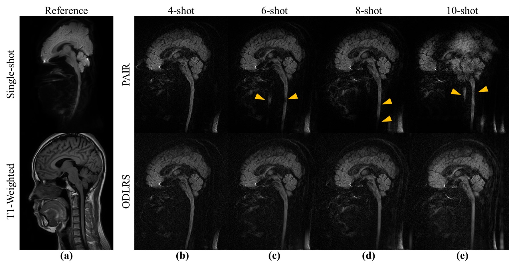
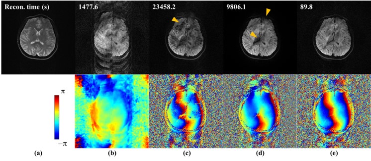
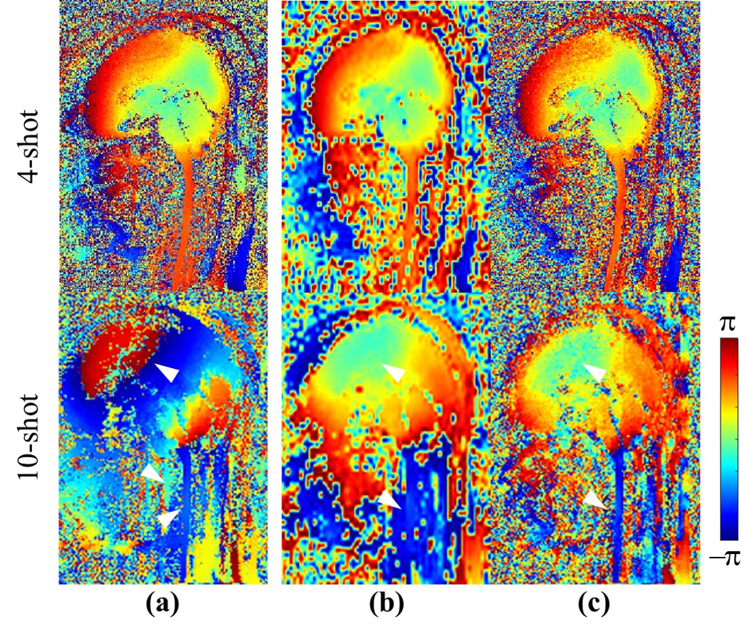
We conducted extensive experiments on two datasets of healthy volunteers to validate the performance of ODLRS. The DATASET I is obtained on the United Imaging 3.0T uMR 790 scanner, channel = 24, shot = 12, matrix size = 224×224, diffusion direction = 3, b-value = 0, and 1000 s/mm2, slice thickness = 4 mm. The DATASET II is obtained on the Philips 3.0T Ingenia CX scanner, channel = 16, shot = 4, 6, 8, 10, matrix size = 168×262, diffusion direction = 3, b-value = 0, and 1000 s/mm2, slice thickness = 4 mm. In Fig. 2, the widely employed MUSE [9] failed to provide reasonable results on 12-shot reconstructions (Fig. 2(b)). MUSSELS [4] and PAIR [5] are reported to achieve better performance than MUSE under high shot number scenarios (4-8 shots). However, for 12-shot DWI reconstruction, they are also hard to provide satisfactory results and produce structural mistakes (yellow arrows in Fig. 2(c) and (d)). Moreover, the computational time of MUSSELS and PAIR reach 23458.2 and 9806.1 seconds per image, respectively. This is unacceptable in practical applications. ODLRS removes the motion artifacts better than all comparison methods and provides the best reconstructed 12-shot DWI image (Fig. 2(e)). Moreover, it improves the reconstruction speed by about a factor of 100 (89.8 seconds). The extensive study on the cervical spine DWI (Fig. 3) demonstrates that ODLRS performs well on multi-shot iEPI DWI from 4-shot to 10-shot, which brings great EPI distortion suppressions. However, PAIR cannot reconstruct spine DWI images well when the shot number is high (Fig. 3(c)-(e)). We further explore why ODLRS performs better than PAIR at ultra-high shot reconstructions through shot phase analysis. The shot phase provided by PAIR is good at 4-shot but poor at 10-shot (Fig. 4(a)). While in the ODLRS (Fig. 4(c)), shot phases are significantly better than PAIR at 10-shot data (Fig. 4(b)), which may be attributed to reliable pre-reconstruction (strong subspace extracted from pre-reconstruction visualized in Fig. 4(b)).Conclusion
In conclusion, we propose ODLRS, a novel 1D low-rank DWI reconstruction model with a self-adaptive subspace. Extensive experiments show that ODLRS achieves fast (109 times acceleration) and reliable ultra-high (10 and 12 shots) DWI reconstruction, reducing the deformation of conventional spinal cord DWI significantly.Acknowledgements
This work was supported in part by the National Natural Science Foundation of China under grants 61971361, 62122064, and 62331021, Natural Science Foundation of Fujian Province of China under grants 2023J02005, President Fund of Xiamen University under grant 20720220063, and Xiamen University Nanqiang Outstanding Talents Program.References
[1] H. An, X. Ma, Z. Pan, H. Guo, and E. Y. P. Lee, “Qualitative and quantitative comparison of image quality between single-shot echo-planar and interleaved multi-shot echo-planar diffusion-weighted imaging in female pelvis,” European Radiology, vol. 30, no. 4, pp. 1876-1884, 2020.
[2] G. C. Baxter, A. J. Patterson, R. Woitek, I. Allajbeu, M. J. Graves, and F. Gilbert, “Improving the image quality of DWI in breast cancer: Comparison of multi-shot DWI using multiplexed sensitivity encoding to conventional single-shot echo-planar imaging DWI,” The British Journal of Radiology, vol. 94, no. 1119, p. 20200427, 2021.
[3] X. Ma, Z. Zhang, E. Dai, and H. Guo, “Improved multi-shot diffusion imaging using GRAPPA with a compact kernel,” NeuroImage, vol. 138, pp. 88-99, 2016.
[4] M. Mani, H. K. Aggarwal, V. Magnotta, and M. Jacob, “Improved MUSSELS reconstruction for high-resolution multi-shot diffusion weighted imaging,” Magnetic Resonance in Medicine, vol. 83, no. 6, pp. 2253-2263, 2020.
[5] C. Qian et al., “A paired phase and magnitude reconstruction for advanced diffusion-weighted imaging,” IEEE Transactions on Biomedical Engineering, DOI: 10.1109/TBME.2023.3288031, 2023.
[6] X. Zhang et al., “Accelerated MRI reconstruction with separable and enhanced Low-Rank Hankel regularization,” IEEE Transactions on Medical Imaging, vol. 41, no. 9, pp. 2486-2498, 2022.
[7] Z. Wang et al., “One-dimensional deep Low-rank and sparse network for accelerated MRI,” IEEE Transactions on Medical Imaging, vol. 42, no. 1, pp. 79-90, 2023.
[8] D. Guo et al. “XCloud-VIP: Virtual peak enables highly accelerated NMR spectroscopy and faithful quantitative measures,” IEEE Transactions on Computational Imaging, DOI: 10.1109/TCI.2023.3330298, 2023.
[9] N. k. Chen, A. Guidon, H. C. Chang, and A. W. Song, “A robust multi-shot scan strategy for high-resolution diffusion weighted MRI enabled by multiplexed sensitivity-encoding (MUSE),” NeuroImage, vol. 72, pp. 41-47, 2013.
Figures