1010
PRIME: Phase Reversed Interleaved Multi-Echo acquisition enables highly accelerated distortion-free diffusion MRI1Athinoula A. Martinos Center for Biomedical Imaging, Massachusetts General Hospital, Boston, MA, United States, 2Department of Radiology, Harvard Medical School, Boston, MA, United States, 3Department of Psychiatry, Brigham and Women’s Hospital, Boston, MA, United States, 4Pediatric Imaging Research Center, Massachusetts General Hospital, Boston, MA, United States, 5Zhejiang University, Hangzhou, China, 6Harvard/MIT Health Sciences and Technology, Cambridge, MA, United States, 7Department of Radiology, Brigham and Women’s Hospital, Boston, MA, United States
Synopsis
Keywords: Diffusion Acquisition, Diffusion/other diffusion imaging techniques
Motivation: Current distortion-free multishot diffusion MRI (dMRI) techniques rely on interim reconstructions to estimate a fieldmap, whose quality deteriorates at high accelerations, thus precluding high-resolution imaging.
Goal(s): To develop a distortion-free acquisition that reaches high accelerations with high fidelity.
Approach: We propose PRIME, which incorporates a second echo acquired at lower resolution and acceleration, but with matching echo spacing as the first echo. This yields high-fidelity fieldmaps to be used in 10-fold accelerated scans.
Results: PRIME enables high-quality distortion-free dMRI at Rinplane×SMS=5×2 and 1mm3 resolution without prolonging the scan thanks to utilizing the dead time in gSlider RF-encoded acquisitions.
Impact: We propose a distortion-free dMRI sequence, PRIME, that reaches Rinplane×SMS=5×2 at 1mm3 resolution with high fidelity owing to its ability to estimate a high-quality fieldmap from a second echo inserted without prolonging the TR in gSlider RF-encoded acquisitions.
Introduction
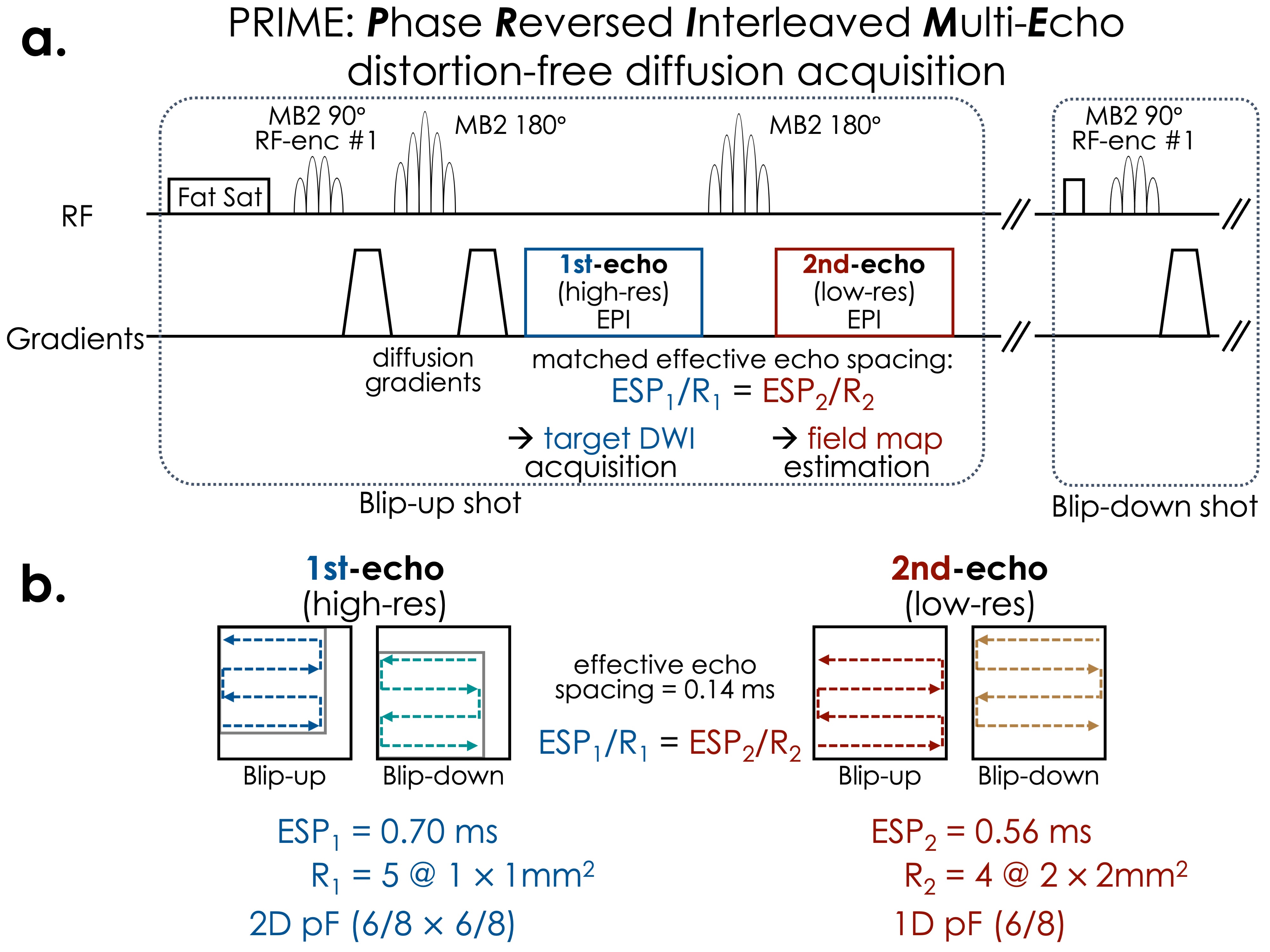
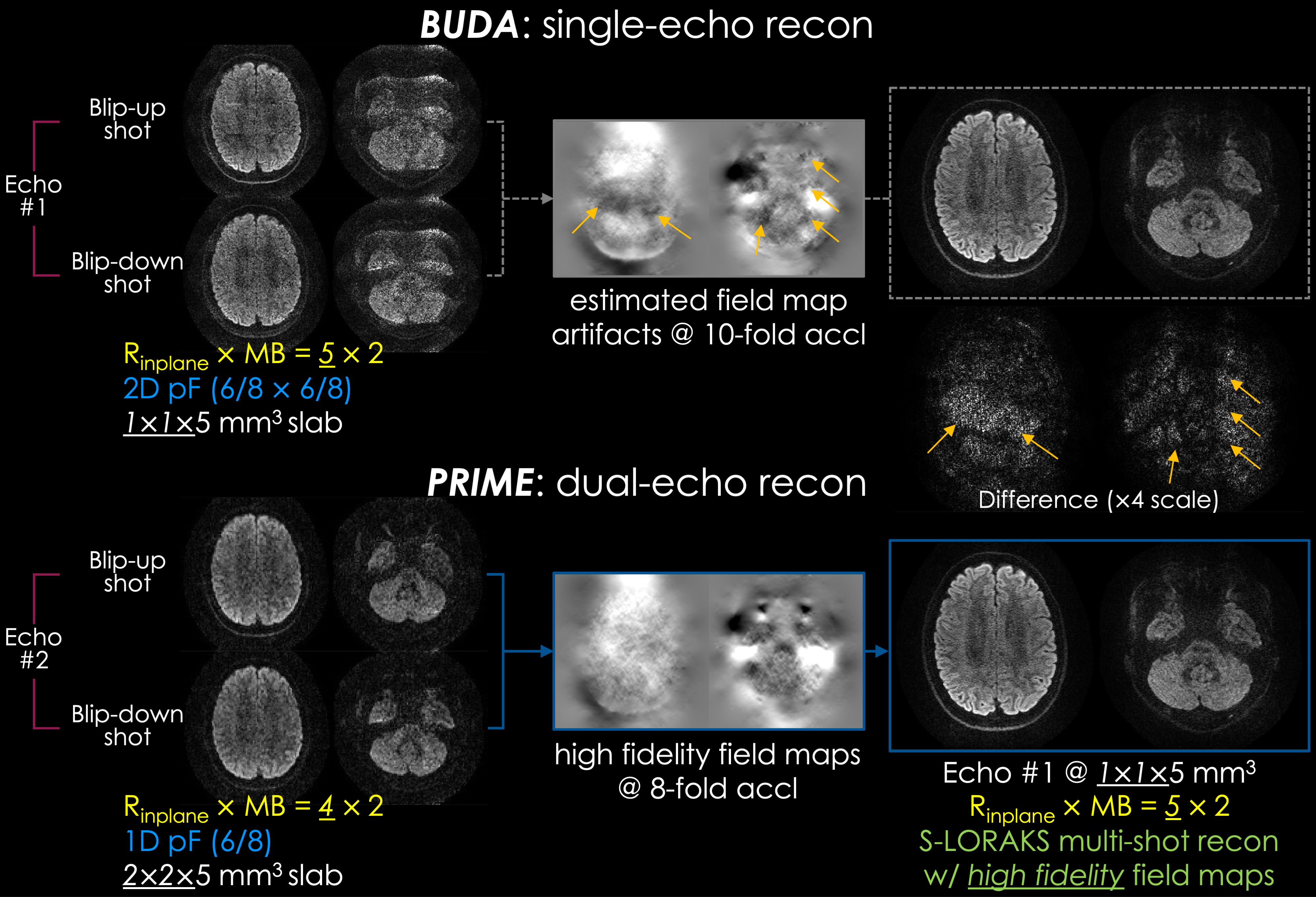
Blip-up & -down acquisition (BUDA)1 is a distortion-free dMRI technique where two shots of EPI with alternating phase encoding polarities are acquired. A limitation of BUDA is the achievable acceleration per shot, since artifacts in the interim SENSE2 images propagate to the estimated fieldmaps using FSL topup3,4 (Fig. 2 upper row). This constrains BUDA to use Rinplane of 4-fold or lower. To address this drawback, we propose a new pulse sequence, PRIME (Phase Reversed Interleaved Multi-Echo).PRIME inserts an additional echo (Fig. 1) to estimate a high-fidelity fieldmap. The second echo is made at a lower resolution, thus boosting its SNR, while matching the effective echo spacing of the first readout. This ensures that both readouts have identical distortion, but also allows the second readout to use lower Rinplane so that the fieldmaps are estimated from higher quality interim reconstructions. The first echo data from blip-up and -down shots are then jointly reconstructed using S-LORAKS5,6 with the estimated fieldmaps.
The addition of the second echo does not incur a scan time penalty when gSlider RF-encoding is used7. Due to spin history-related slab boundary artifacts, gSlider benefits from long repetition times, which leaves dead time, especially when simultaneous multi-slice (SMS)8 is employed. We use this dead time in PRIME and demonstrate distortion-free, high-quality dMRI while using 2-dimensional partial Fourier (pF) to minimize echo time (TE) and relaxation-related blurring. The sequence is implemented using Pulseq9.
Sequence and reconstruction codes: https://anonymous.4open.science/r/PRIME/
Methods
Pulse SequenceFig. 1 illustrates the pulse sequence diagram of PRIME. gSlider RF encoding is used for resolving thick slabs into isotropic resolution. 2-dimensional pF used to reduce the echo spacing (ESP) and TE. The blip-up and -down shots cover complementary positions, and aided by the virtual coil constraints in S-LORAKS6, all quadrants of k-space are covered. The effective echo spacing is matched between the readouts using R1=5 and R2=4 for the two readouts, thereby obtaining ESPeffective=0.14ms on a clinical system. VERSE10 was used to mitigate SAR constraints.
Image Reconstruction
Fig. 2. top demonstrates the standard BUDA pipeline at Rinplane×SMS=5×2 where interim SENSE reconstructions are input to topup. Due to artifacts and g-factor loss at 10-fold acceleration, the estimated fieldmaps suffer from artifacts, as indicated by arrows. Fig. 2. bottom shows that PRIME uses the second echo acquired at lower resolution and in-plane acceleration (Rinplane×SMS=4×2) to boost SNR and mitigate artifacts, enabling higher fidelity topup estimation. The two high-resolution shots from the first echo are then reconstructed with S-LORAKS.
Data Acquisition
3T Siemens Prisma: a volunteer was scanned using 32ch array with the parameters FOV=220x220x130mm3, TR/TE1/TE2=4000/54/108ms, 1mm3 resolution using gSlider, b=1000s/mm2, and 6 directions.
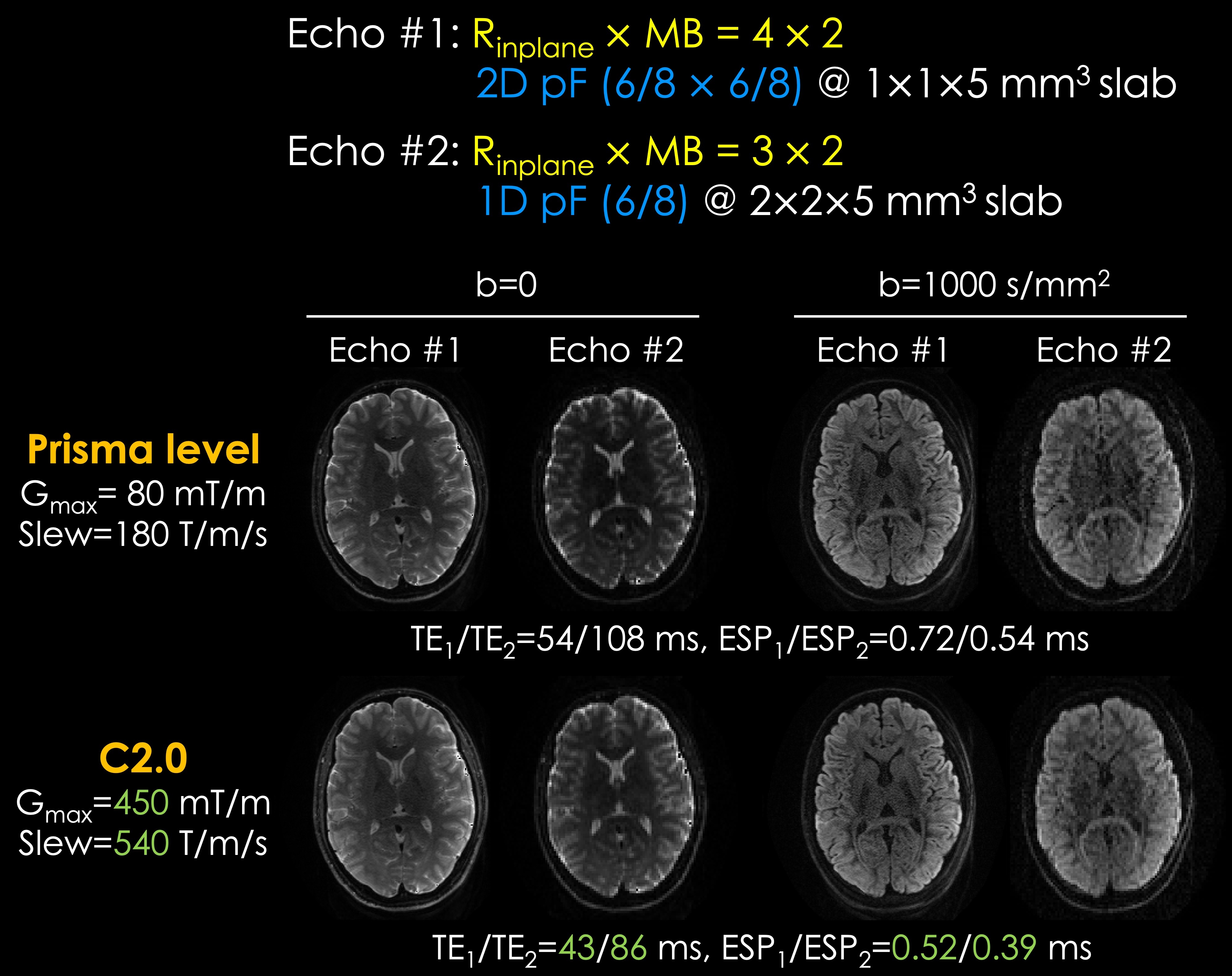
3T Siemens Connectome 2.0: a second volunteer was scanned where Gmax=450mT/m and Slew=540T/m/s were used to reduce the TEs and echo spacing to obtain TR/TE1/TE2=5500/43/86ms and ESP1/ESP2=0.52/0.39ms. Other parameters were kept the same.
Results
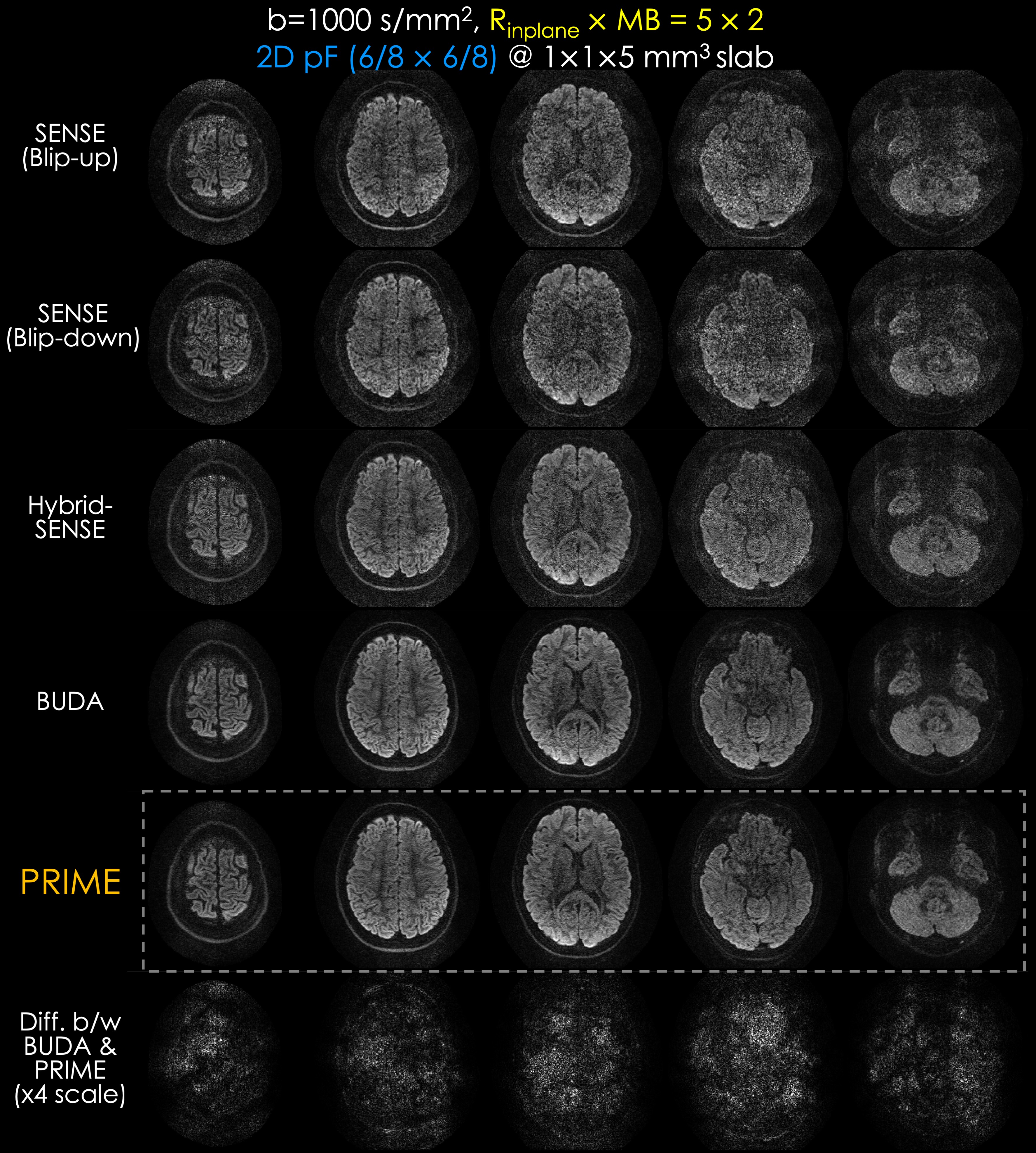
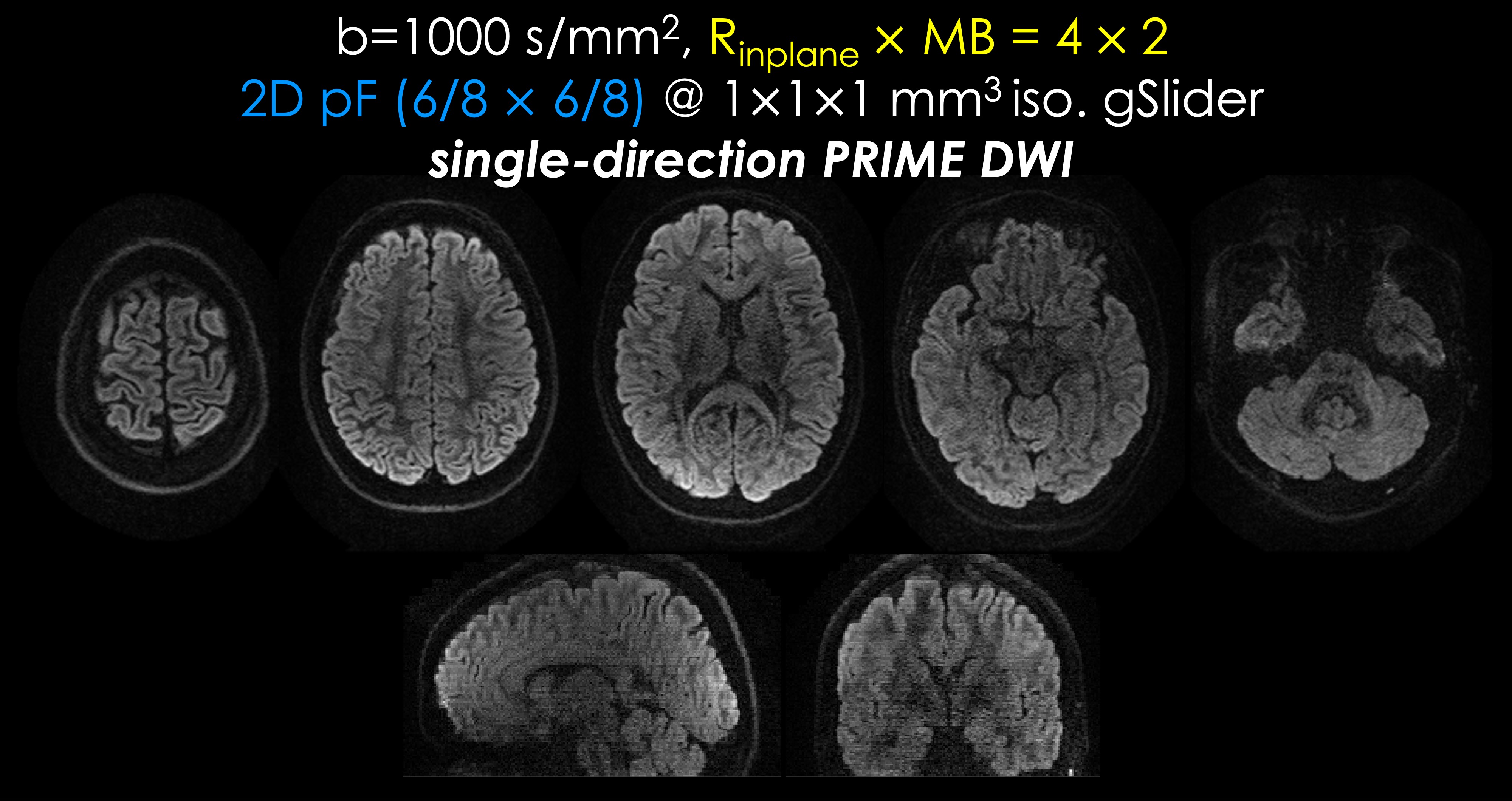
Fig. 3 shows results from the Prisma system. SENSE reconstructions on the individual shots suffer from noise amplification and artifacts due to the 10-fold acceleration. Hybrid-SENSE11, which uses the fieldmap and shot-to-shot phase difference estimated from interim SENSE reconstructions, has residual artifacts at this acceleration. While BUDA and PRIME showed improved quality, the imperfect estimation of the fieldmap in BUDA leads to differences between PRIME as shown in the difference images.Fig. 4 depicts a single-direction volume (b=1000s/mm2) at 1mm3 resolution obtained using gSlider reconstruction from PRIME data. High geometric fidelity and SNR are observed.
Fig. 5 demonstrates results from Connectome 2.0 at the same resolution with PRIME and gSlider encoding with comparisons between Prisma level of Gmax and slew rate and those of Connectome 2.0. Due to reduced TEs and echo spacing using increased Gmax and slew rate, b0 and diffusion-weighted images from Connectome 2.0 have less signal decayed, especially for the second echo images.
Discussion and Conclusion
We proposed a new distortion-free sequence, PRIME, which bypasses the initial, poorly-conditioned reconstruction and reconstructs highly accelerated, high-resolution data with fieldmap estimated from the calibration echoes. We were able to insert an additional echo and a refocusing pulse without prolonging the TR, and without triggering the SAR monitor at 2-fold SMS using VERSE.By capitalizing on the Connectome 2.0 gradients, we further reduced the echo spacing of both readouts to 0.52/0.39 ms. This offers the flexibility to use both readouts at the same high resolution for diffusion relaxometry with adequate TEs. Deployment of PRIME in diffusion relaxometry will enable the utilization of advanced models, including multi-echo NODDI12 and SANDI13,14 at high resolution.
Acknowledgements
This work was supported by research grants NIH R01 EB028797, P41 EB030006, U01 EB026996, R03 EB031175, R01 EB032378, UG3 EB034875, and NVidia Corporation for computing support.References
1. Liao C, Bilgic B, Tian Q, et al. Distortion-free, high-isotropic-resolution diffusion MRI with gSlider BUDA-EPI and multicoil dynamic B0 shimming. Magn. Reson. Med. 2021;86:791–803.
2. Pruessmann KP, Weiger M, Scheidegger MB, Boesiger P. SENSE: sensitivity encoding for fast MRI. Magn. Reson. Med. 1999;42:952–962.
3. Andersson JLR, Skare S, Ashburner J. How to correct susceptibility distortions in spin-echo echo-planar images: application to diffusion tensor imaging. Neuroimage 2003;20:870–888.
4. Smith SM, Jenkinson M, Woolrich MW, et al. Advances in functional and structural MR image analysis and implementation as FSL. Neuroimage 2004;23 Suppl 1:S208–19.
5. Haldar JP. Low-Rank Modeling of Local k-Space Neighborhoods (LORAKS) for Constrained MRI. IEEE Trans. Med. Imaging 2014;33:668–681.
6. Liao C, Yarach U, Cao X, et al. High-fidelity mesoscale in-vivo diffusion MRI through gSlider-BUDA and circular EPI with S-LORAKS reconstruction. Neuroimage 2023;275:120168.
7. Setsompop K, Fan Q, Stockmann J, et al. High-resolution in vivo diffusion imaging of the human brain with generalized slice dithered enhanced resolution: Simultaneous multislice (gSlider-SMS). Magn. Reson. Med. 2018;79:141–151.
8. Setsompop K, Gagoski BA, Polimeni JR, Witzel T, Wedeen VJ, Wald LL. Blipped-controlled aliasing in parallel imaging for simultaneous multislice echo planar imaging with reduced g-factor penalty. Magn. Reson. Med. 2012;67:1210–1224.
9. Layton KJ, Kroboth S, Jia F, et al. Pulseq: A rapid and hardware-independent pulse sequence prototyping framework. Magn. Reson. Med. 2017;77:1544–1552.
10. Hargreaves BA, Cunningham CH, Nishimura DG, Conolly SM. Variable-rate selective excitation for rapid MRI sequences. Magn. Reson. Med. 2004;52:590–597.
11. Zahneisen B, Aksoy M, Maclaren J, Wuerslin C, Bammer R. Extended hybrid-space SENSE for EPI: Off-resonance and eddy current corrected joint interleaved blip-up/down reconstruction. Neuroimage 2017;153:97–108.
12. Gong T, Tong Q, He H, Sun Y, Zhong J, Zhang H. MTE-NODDI: Multi-TE NODDI for disentangling non-T2-weighted signal fractions from compartment-specific T2 relaxation times. Neuroimage 2020;217:116906.
13. Palombo M, Ianus A, Guerreri M, et al. SANDI: A compartment-based model for non-invasive apparent soma and neurite imaging by diffusion MRI. Neuroimage 2020;215:116835.
14. Gong T, Tax CMW, Mancini M, Jones DK, Zhang H, Palombo M. Multi-TE SANDI: Quantifying compartmental T2 relaxation times in the grey matter. In: ISMRM 2023. ; p. 0766.
Figures