1003
Thin slice positive source QSM improves deep learning based paramagnetic rim detection in multiple sclerosis lesions1Weill Cornell Medicine, New York, NY, United States
Synopsis
Keywords: Diagnosis/Prediction, Quantitative Susceptibility mapping
Motivation: Rim lesions are important subset of chronic active MS lesions that show strong correlation to patient disability. Rim identification by experts is time consuming.
Goal(s): Develop tool for supporting the expert in Rim identification using 1 mm QSM.
Approach: We developed an automated deep learning-based network for PRL detection on thin-slice 1mm QSMp. We evaluated the improvement in performance compared with networks trained using 1mm QSM and 3mm QSMp.
Results: Use of high-resolution positive susceptibility source maps improves detection of Rim in MS patients compared to 1mm QSM and 3mm QSMp. The network does not require a precise QSM lesion mask to operate.
Impact: Using the Deep learning for detecting rim on 1mm QSMp, enabling reducing workload for human in detecting rim.
Introduction
Innate immune activity in chronic active lesions is a key promotor of progressive cognitive and ambulatory decline in multiple sclerosis (MS)1. Paramagnetic rim lesions (PRLs) form a subset of chronic active lesions which are specific to MS and independently associated with myelin injury and clinical disability1,2. PRLs show a dense rim of iron-laden pro-inflammatory immune cells on histology and can be detected on QSM as having a hyperintense rim appearance3. However, while visual PRL identification by humans on all MS lesions is very time-consuming, PRLs are relatively rare, accounting for only about 10% of MS lesions4. Furthermore, simultaneous myelin loss and iron accumulation both increase hyperintensity on QSM5, which may reduce rim contrast and leads to suboptimal rim detection, especially when the imaging slices are thick.Purpose
) To develop an automated deep learning-based network for PRL detection on thin-slice 1mm positive source QSM images (1 mm QSMp); 2) to evaluate the improvement in performance compared with networks trained using 1mm QSM and thick-slice 3mm QSMp images.Materials and Methods
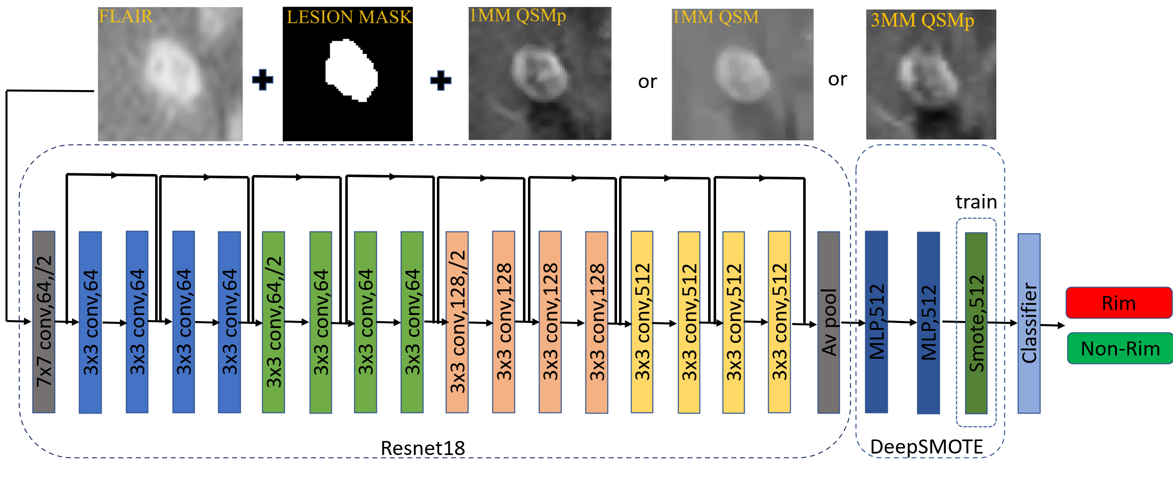
The patient cohort consisted of 78 patients. MRI was performed at 3T (Magnetom Skyra, Siemens, Erlangen, Germany) using a 20-channel head/neck coiland included 1mm T2FLAIR (FLAIR) and two 3D multi-echo GRE acquisitions for QSM with acquisition parameters: 1) 3 mm QSMp: voxels = 0.75x0.75x3 mm3; 2) 1 mm QSM: voxel = 0.375x0.375x1 mm3. 1mm QSM and 3mm QSM images were reconstructed using morphology-enabled dipole inversion method with global CSF referencing (MEDI+0)7. An R2*-based QSM source separation algorithm 7,8 was then applied to QSM to derive QSMp images. Lesion masks were automatically segmented on FLAIR images using AllNet9, which were check manually and corrected if necessary, by a trained reader. They were then coregistered to QSM. A neuroradiologist with over 25 years of experience classified each lesion as having rim (rim+) or not (rim-) on 1mm QSM and 1mm QSMp images in two reading sessions two weeks apart. Only lesions that were identified as rim+ on both QSM and QSMp were considered as rim+ and used as ground truth labels for network training. To reduce network training time, each lesion was cropped into an image patch of 64x64x24 for 1mm QSM and QSMp, and 32x32x16 for 3mm QSMp. The implemented network (Fig.1) consists of a Resnet backbone18 for deep feature extraction and deepSMOT10 for addressing imbalance data between rim+ and rim-. Image patches from FLAIR and QSMp/QSM images were concatenated and used as network input. The images from 49 and 29 subjects were selected as training and testing set, respectively, for evaluating the performance of the network (Table 1). Augmentation techniques such as rotation, transformation and blurring were applied to enrich the patches during training10, which used a batch size of 32. Ensemble-learning training of three random seeds was performed to obtain the models used for major voting prediction on the test set. Other training hyperparameters were reused from our previous study11.Results
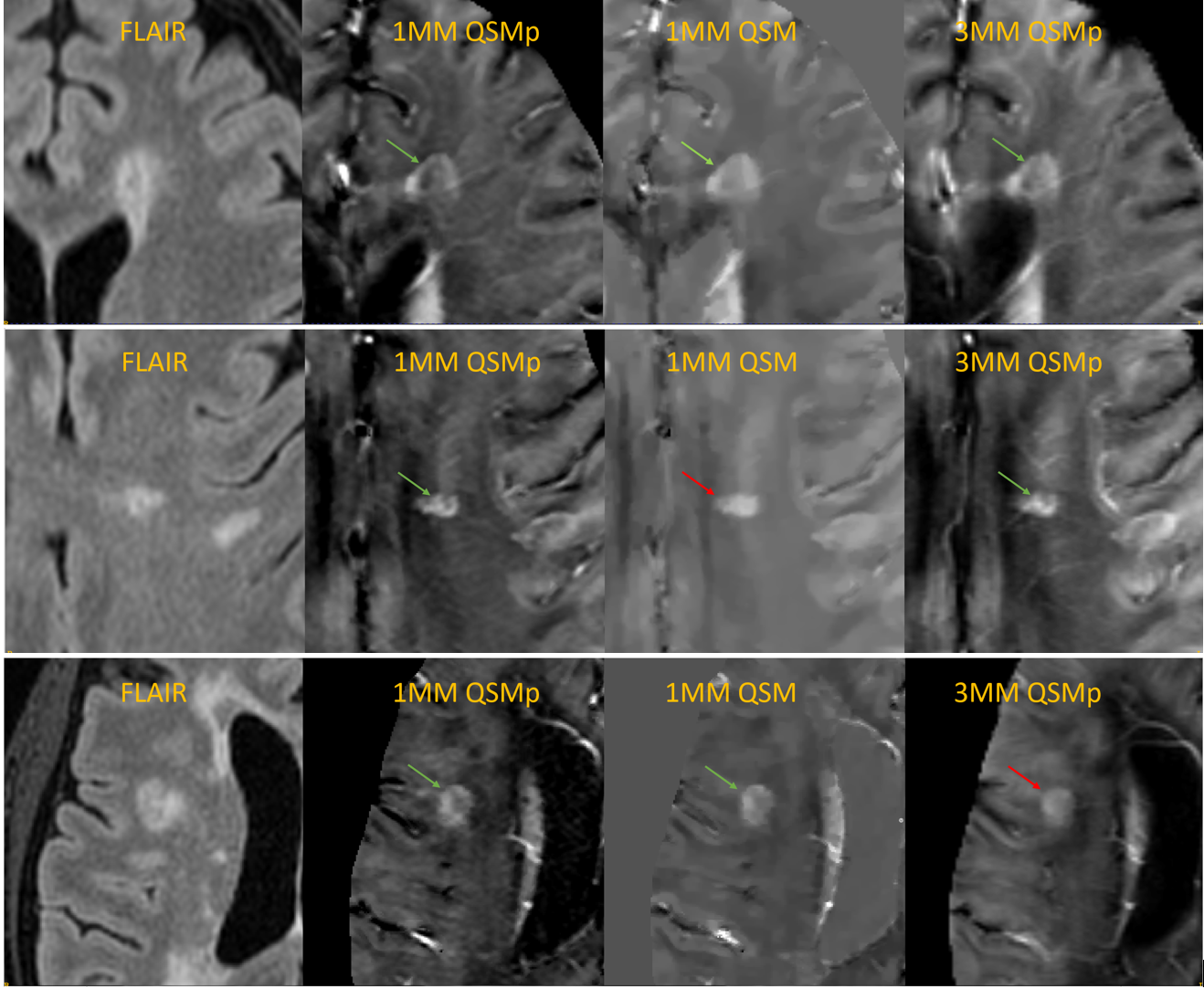
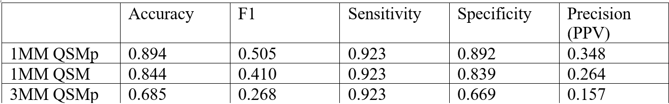
A total of 1792 lesions were identified, of which 64 were classified as rim+. The network trained on 1mm QSMp images achieved the best detection performance with AUC=0.965 compared to that obtained from 1mm QSM (0.926) and 3mm QSMp (0.896) images. Similarly, using 1mm QSMp images outperformed with regards to PR AUC (0.585 vs. 0.461 by 1mm QSM and 0.402 by 3mm QSMp). The improvement in precision (PPV)by 1mm QSMp network is more striking at high detection sensitivity level (>0.9), therefore making it more suitable as a lesion rim screening tool. For example, for a detection sensitivity of 0.923, using 1mm QSMp improves the precision (PPV) from 0.157 by 3mm QSMp and 0.264 by 1mm QSM to 0.348 (Table 2). Figures 3 shows examples of rim classification of the three networks, demonstrating improved rim contrast on 1mm QSMp images.Conclusion
We found that 1MM QSMp enables the deep learning network to achieve better performance in PRL identification. Our results support further investigation and use of QSMp to detect Rim in MS patients.Summary of main findings
Use of high-resolution positive susceptibility source maps improves detection of Rim in MS patients using deep learning compared to 1mm QSM and 3mm QSMp.Synopsis
Rim lesions are important subset of chronic active MS lesions that show strong correlation to patient disability. Rim identification by experts is time consuming and deep learning is a promising tool for supporting the expert rim identification. 3 mm slice thickness and contribution of demyelination may distract the performance of the network. Our goal in this project is to improve overall rim detection using neural networks through utilization of 1mm paramagnetic source maps.Acknowledgements
No acknowledgement found.References
1. Gillen KM, Mubarak M, Park C, et al. QSM is an imaging biomarker for chronic glial activation in multiple sclerosis lesions. Ann Clin Transl Neurol 2021;8:877-86.
2. Hametner S, Wimmer I, Haider L, Pfeifenbring S, Bruck W, Lassmann H. Iron and neurodegeneration in the multiple sclerosis brain. Ann Neurol 2013;74:848-61.
3. Yao Y, Nguyen TD, Pandya S, et al. Combining quantitative susceptibility mapping with automatic zero reference (QSM0) and myelin water fraction imaging to quantify iron-related myelin damage in chronic active MS lesions. AJNR Am J Neuroradiol 2018;39:303-10.
4. Absinta M, Sati P, Masuzzo F, et al. Association of chronic active multiple sclerosis lesions with disability in vivo. JAMA Neurol 2019;76:1474-83.
5. Huang W, Sweeney EM, Kaunzner UW, Wang Y, Gauthier SA, Nguyen TD. Quantitative susceptibility mapping versus phase imaging to identify multiple sclerosis iron rim lesions with demyelination. J Neuroimaging. 2022 Jul;32(4):667-675.
6. Shin HG, Lee J, Yun YH, Yoo SH, Jang J, Oh SH, Nam Y, Jung S, Kim S, Fukunaga M, Kim W, Choi HJ, Lee J. χ-separation: Magnetic susceptibility source separation toward iron and myelin mapping in the brain. Neuroimage. 2021 Oct 15;240:118371. doi: 10.1016/j.neuroimage.2021.118371. Epub 2021 Jul 6. PMID: 34242783.
7. Dimov, A. V., T. D. Nguyen, P. Spincemaille, E. M. Sweeney, N. Zinger, I. Kovanlikaya, B. H. Kopell, S. A. Gauthier, and Y. Wang. "Global Cerebrospinal Fluid as a Zero-Reference Regularization for Brain Quantitative Susceptibility Mapping." J Neuroimaging (2021).
8. Dimov, A. V., T. D. Nguyen, K. M. Gillen, M. Marcille, P. Spincemaille, D. Pitt, S. A. Gauthier, and Y. Wang. "Susceptibility Source Separation from Gradient Echo Data Using Magnitude Decay Modeling." J Neuroimaging (2022).
9 Zhang H, Zhang J, Li C, Sweeney EM, Spincemaille P, Nguyen TD, Gauthier SA, Wang Y, Marcille M. ALL-Net: Anatomical information lesion-wise loss function integrated into neural network for multiple sclerosis lesion segmentation. Neuroimage Clin. 2021;32:102854
10. Zhang H, Nguyen TD, Zhang J, Marcille M, Spincemaille P, Wang Y, Gauthier SA, Sweeney EM. QSMRim-Net: Imbalance-aware learning for identification of chronic active multiple sclerosis lesions on quantitative susceptibility maps. Neuroimage Clin. 2022;34:102979
11. Luu H, Gauthier S, Kovanlikaya I, Wang Y, Spincemaille P, Sisman M and Nguyen T. “Quantitative susceptibility source separation improves the performance in the identification of chronic active multiple sclerosis lesions using deep learning-based method”. Whatever (2023).
Figures