1002
Sleep and Cardiovascular Risk Variables Predict Perivascular Space Morphological Alterations in the Aging Brain1Neurology, Laboratory of NeuroImaging, Stevens Neuroimaging and Informatics Institute, Keck School of Medicine, University of Southern California, Los Angeles, CA, United States, 2Scripps College, Claremont, CA, United States, 3Health Promotion and Disease Prevention Studies, Keck School of Medicine, University of Southern California, Los Angeles, CA, United States, 4Neurology, Chonnam National University Medical School and Hospital, Gwangju, Korea, Republic of, 5Pusan National University School of Medicine, Busan, Korea, Republic of, 6Neurology, College of Medicine, Catholic University of Korea, Seoul, Korea, Republic of, 7Department of Biological Sciences,University of Southern California, Los Angeles, CA, United States, 8University of California, Los Angeles, Los Angeles, CA, United States, 9Neurology, Samsung Medical Center, Sungkyunkwan University School of Medicine, Seoul, Korea, Republic of, 10Neurology, Konkuk University, Seoul, Korea, Republic of
Synopsis
Keywords: Diagnosis/Prediction, Aging
Motivation: Enlarged perivascular space (PVS) has been brought into attention in aging populations. However, which cardiovascular risk factors contribute to enlarged PVS are not well understood.
Goal(s): This study aims to quantify PVS morphology and investigate which cardiovascular risk factors contribute the PVS deformity in aging populations.
Approach: We employed random forest to predict PVS morphological changes using 9 cardiovascular risk factors and computed the importance index for all predictive factors.
Results: Our findings highlighted the significant role of sleep quality, being the best predictor to PVS count, linearity, and diameter. Cardiovascular risk factors such as triglycerides best predicted PVS tortuosity.
Impact: Our study is the first to investigate which cardiovascular risk factors are predictive of atypical PVS morphology. Our discovery provides valuable insights into the mechanism underlying PVS deformity and their subsequent impact on glymphatic system and cerebral vascular diseases.
Introduction
Perivascular Spaces (PVS) are fluid-filled cavities that envelop blood vessels in the brain (Fig. 1a). They have emerged as a focal point of research in the context of age-related cognitive decline and neurodegenerative disorders, including Alzheimer’s Disease1. Increasing evidence suggests that morphological changes, such as enlargement PVS (ePVS), may serve as indicators of disruptions in glymphatic function2. Recent studies underscore that PVS deformity should be characterized not only by ePVS but also other various attributes, including tortuosity, diameter, linearity, and curvature, suggesting the potential of atypical PVS morphology as a biomarker for poor sleep, glymphatic-related pathologies, cerebrovascular diseases, and brain aging process3. The PVS evaluation has been mostly using qualitative scoring systems, and the precise connections between cardiovascular health and PVS deformity remain to be elucidated. This study aims to quantify PVS morphology and investigate which sleep and cardiovascular risk factors contribute the PVS deformity in preclinical aging populations.Methods
In this study, 394 subjects from The Human Connectome Project Aging (HCP-A) dataset were examined4. PVS were automatically segmented from multimodal T1w and T2w images (resolution=0.8x0.8x0.8mm3) using a semi-supervised U-Net deep learning model5.A depth-first search algorithm separated the segmented PVSs into individual PVS instances. For the 10 largest PVS instances, the following morphological features were calculated: tortuosity, linearity and cross-sectional diameter. A skeletonizing algorithm was used to create a skeleton for tortuosity, and mean-cross sectional diameter calculations3,6. PVS tortuosity was calculated as the skeleton length (red line in Fig1.c) to its end-to-end Euclidean distance ratio (black line in Fig1.c). Linearity was then defined as the mean Euclidian distance between the center of the voxel and the PVS axial direction (Fig1.d). To calculate cross-sectional diameter7, the vertices of the skeleton (vertices shown as circles, connected by red lines in Fig1.e) are assigned the voxels closest to them (color-coded). The diameter was computed as $$$D = 2\sqrt{\frac{N}{l *\pi}}$$$ where $$$N$$$ is the number of assigned voxels and $$$l$$$ is taken from the average distance to neighboring vertices. All the features were calculated using Python.
To explore the potential causative factors of PVS deformity, we employed Pittsburgh sleep quality index (PSQI) and cardiovascular factors including systolic, diastolic, triglycerides, total cholesterol, body mass index (BMI), hemoglobin A1c (HbA1c), glucose and high-density lipoprotein (HDL). We trained the Bootstrap random forest regression (RFR), with the following training parameters: number of trees = 100, number of features sampled for each split = 7, minimum number of split per tree = 10. We included age and sex as covariates. We performed a 5-fold nested cross validation and predicted all five PVS deformity measures using the 10 clinical features above. We computed the importance indices from the RFR to rank the 10 features in terms of their contributions to the prediction.
Results and Discussion
Table 1 presents the demographic and clinical characteristics of the studied cohort. The top row of Fig.2 plots the true PVS deformity metrics and predicted values using RFR. The R-values range from 0.70 to 0.75. The bottom row of Fig.2 shows the top three most contributing factors in predicting PVS deformity using the importance index from the RFR.We explored the factors contributing to PVS deformity in preclinical aging cohorts. Our findings highlighted the significant role of sleep quality (PSQI), which was the best predictor to PVS count, linearity, and diameter. Additionally, cardiovascular risk factors such as blood fat (triglycerides) and body fat (BMI) best predicted PVS tortuosity.
Our results align with prior visual evaluation studies emphasizing the significant relationship between enlarged PVS, sleep quality9, and cardiovascular risk factors10. Intriguingly, our top 10 largest PVS were identified mostly in the superficial white matter of parietal and occipital lobes. To clarify whether the PVS deformity led by sleep disturbance or cardiovascular risks are predominantly observed in these areas, investigating the PVS morphological features in different brain regions can be a focus of future work.
Hypertension, a hallmark of cardiovascular risk factors, has previously been linked to slower glymphatic flow11. It has been suggested that PVS enlargement and deformity can lead to diminished glymphatic flux12 . Our findings not only corroborate the established connection between hypertension and PVS deformation but also extend this relationship to encompass a range of cardiovascular risk factors that may contribute to arterial thickening, subsequent inflammation, and the deformity of arteries and veins and their related PVS13.
Conclusion
In summary, our study is the first to investigate the potential causative factors of PVS deformity. The discovery of cardiovascular risk factors contributing to PVS deformation provides valuable insights into our understanding of glymphatic function and neurodegeneration.Acknowledgements
The image computing resources provided by the Laboratory of Neuro Imaging Resource (LONIR) at USC are supported by National Institutes of Health (NIH) National Institute of Biomedical Imaging and Alzheimer’s Disease Research Center (ADRC) at USC.References
- Sepehrband, Farshid, et al. "Perivascular space fluid contributes to diffusion tensor imaging changes in white matter." Neuroimage 197 (2019): 243-254.
- Bouvy, Willem H., et al. "Visualization of perivascular spaces and perforating arteries with 7 T magnetic resonance imaging." Investigative radiology 49.5 (2014): 307-313.
- Lynch, Kirsten M., et al. "Brain perivascular space imaging across the human lifespan." Neuroimage 271 (2023): 120009.
- Bookheimer, Susan Y., et al. "The lifespan human connectome project in aging: an overview." Neuroimage 185 (2019): 335-348.
- Chai, Yaqiong, et al. "Semi-supervised learning in perivascular space segmentation using MRI images." Medical Imaging with Deep Learning, short paper track. 2023
- Zong, Xiaopeng, et al. "Morphology of perivascular spaces and enclosed blood vessels in young to middle-aged healthy adults at 7T: Dependences on age, brain region, and breathing gas." Neuroimage 218 (2020): 116978.
- Barisano, Giuseppe, et al. "Imaging perivascular space structure and function using brain MRI." NeuroImage. 257 (2022): 119329.
- Yang, Yuyuan, et al. "Enlarged Perivascular Spaces and Age-Related Clinical Diseases." Clinical Interventions in Aging (2023): 855-867.
- Del Brutto, Oscar H., et al. "Enlarged basal ganglia perivascular spaces and sleep parameters. A population-based study." Clinical neurology and neurosurgery 182 (2019): 53-57.
- Gutierrez, J., et al. "Perivascular spaces are associated with atherosclerosis: an insight from the Northern Manhattan Study." American Journal of Neuroradiology 34.9 (2013): 1711-1716.
- Kikuta, J., et al. "An investigation of water diffusivity changes along the perivascular space in elderly subjects with hypertension." American Journal of Neuroradiology 43.1 (2022): 48-55.
- Benveniste, Helene, and Maiken Nedergaard. "Cerebral small vessel disease: A glymphopathy?." Current opinion in neurobiology 72 (2022): 15-21.
- Li, Yue, et al. "The relationship between blood–brain barrier permeability and enlarged perivascular spaces: a cross-sectional study." Clinical interventions in aging (2019): 871-878.
Figures
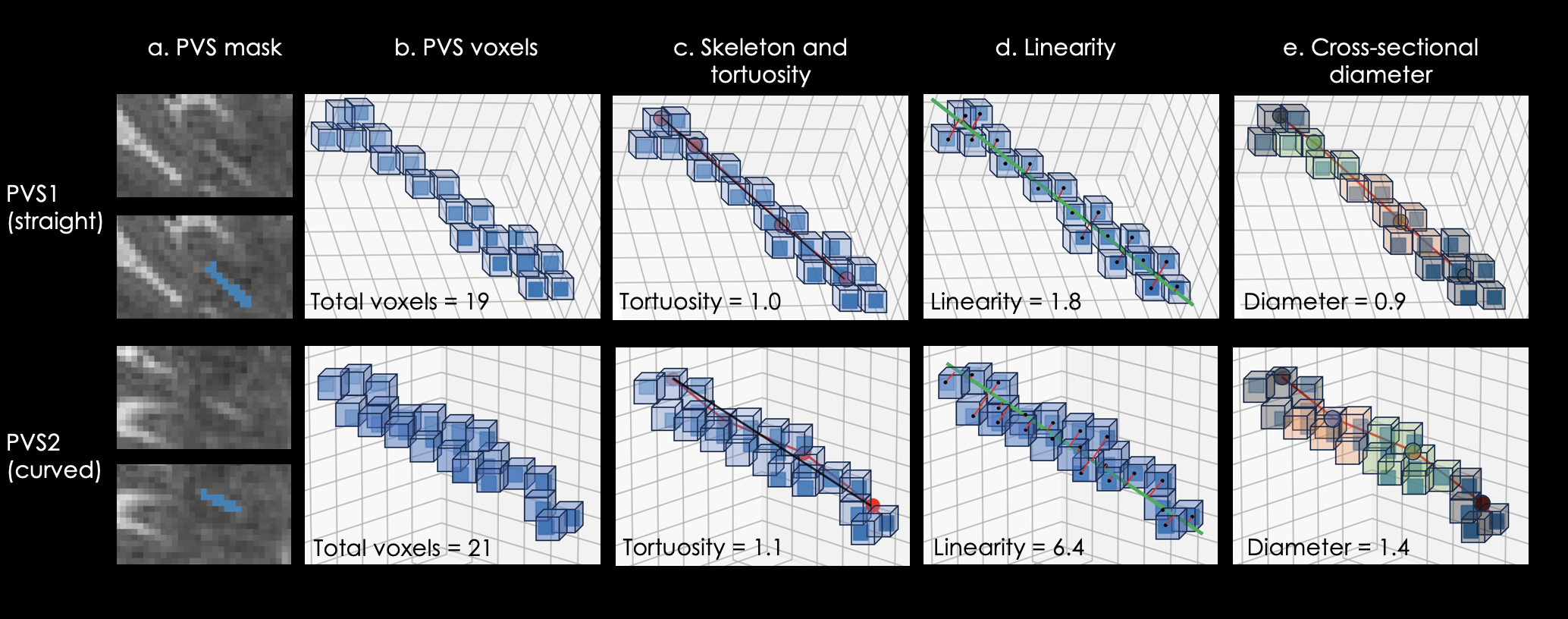
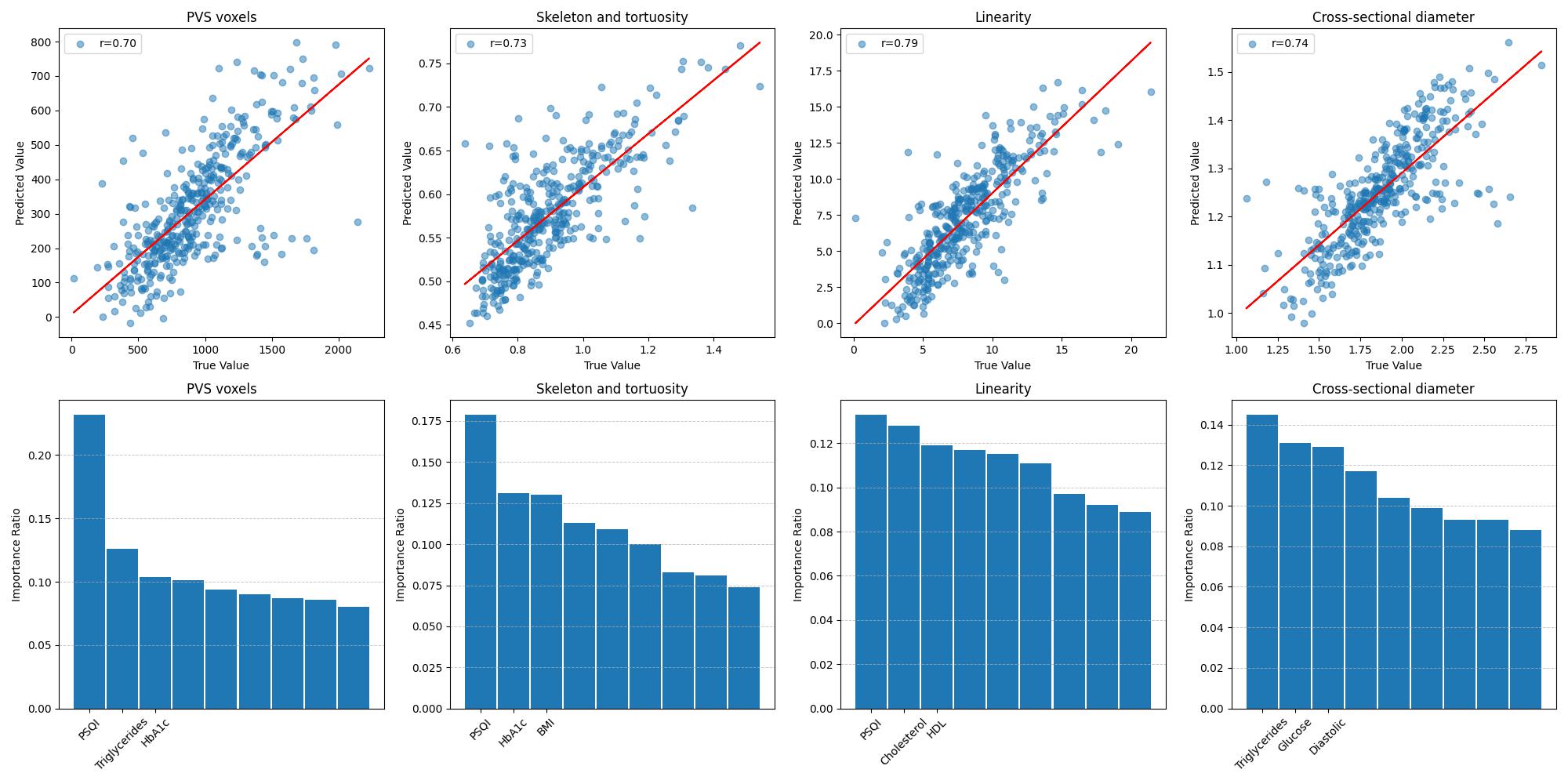
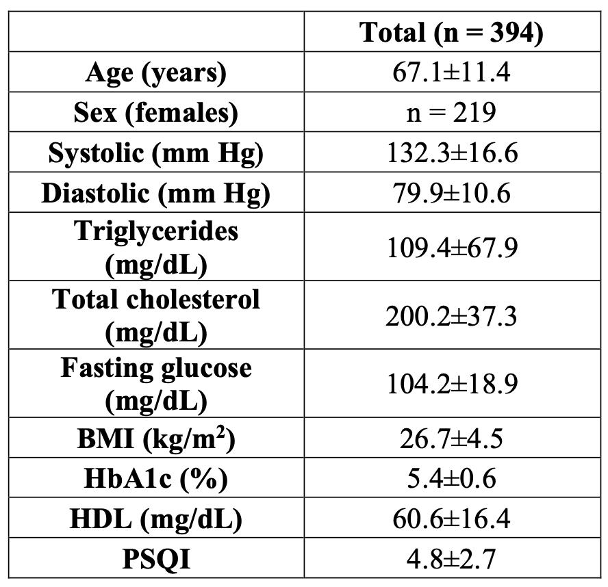
Table 1 Demographic and clinical characteristics of the studied subjects
BMI: body mass idex; HDL: high-density lipoprotein; PSQI: Pittsburgh Sleep Quality Index