0998
Test-retest reliability of coupling between cerebrovascularoscillations and cerebrospinal fluid flow fluctuations1Center for Magnetic Resonance Imaging Research & Key Laboratory of Brain-Machine Intelligence for Information Behavior (Ministry of Education and Shanghai), Shanghai International Studies University, Shanghai, China
Synopsis
Keywords: Neurofluids, Reproductive, gBOLD-CSF coupling, Glymphatics, Sleep deprivation
Motivation: The coupling strength of global blood-oxygen-level-dependent (gBOLD) signals and cerebrospinal fluid (CSF) inflow have been suggested to be an indicator of glymphatic system function. However, few studies have validated its test-retest reproducibility.
Goal(s): To assess the reproducibility of gBOLD-CSF coupling.
Approach: Thirteen adults of the sleep deprivation (SD) group and 14 adults of the control group underwent three fMRI. The reliability of gBOLD-CSF coupling was evaluated by the intraclass correlation coefficient (ICC).
Results: A higher ICC (0.525, P = 0.001) was observed in the control group compared with the SD group (0.137, P = 0.086).
Impact: The gBOLD-CSF coupling shows good reproducibility, but care should be taken when interpreting longitudinal changes of the gBOLD-CSF coupling that may be influenced by participants’ drowsiness. Sleep should be considered an important factor in future studies exploring the glymphatic system.
INTRODUCTION
The glymphatic system (GS) is a recently discovered brain-wide perivascular fluid transport system in the cerebral nervous system1. This fluid transport system facilitates the exchange of cerebrospinal fluid (CSF) and brain interstitial fluid, which ultimately results in fluid movement across the brain parenchyma to remove metabolic waste products2. It is commonly believed that hemodynamic oscillations in the brain facilitate CSF movement. A coherent pattern of low-frequency hemodynamic oscillations and CSF movement, expressed by the coupling of global blood-oxygen-level-dependent (gBOLD) and CSF signals, was recently found during non-rapid eye movement sleep via resting-state functional MRI (rfMRI)3. This finding raises a fundamental question: the test-retest reproducibility of the gBOLD-CSF coupling. This study aimed to quantify the test-retest reliability of gBOLD-CSF coupling.METHODS
We analyzed rfMRI data from 27 healthy adults (12 females, mean age = 34.86 ± 9.83 years) as a part of a strictly controlled in-laboratory sleep deprivation study4. Fourteen participants in the control group slept 8-9 hours every night in the laboratory. Thirteen participants in the SD group stayed awake all night on the second night and had 12 hours of recovery sleep on the third night. The in-laboratory study lasted 5 consecutive days and 4 nights. Each subject was scanned three times on the morning of days 2, 3, and 5.The rfMRI and high-resolution T1-weighted anatomic imaging (for more details, see 4) were performed using a 3T Siemens Trio system (Siemens Medical Systems, Erlangen, Germany). All rfMRI was prepressed using SPM 12. Nuisance regressions of global signal, CSF signal, and motion parameters were skipped to avoid loss of crucial hemodynamic information3. Consistent with previous studies3,5, the gBOLD signal was extracted from the gray matter region6 in the original space and the CSF signal was extracted manually from the bottom slice of the fMRI acquisition.
The maximal cross-correlation over (− 20 s to 20 s) time lags between the gBOLD signal and CSF signal was calculated for each participant, with the BOLD signal as the reference. The maximal anticorrelation was used to quantify the strength of gBOLD-CSF coupling. Voxel-wise cross-correlations were conducted to derive maximal cross-correlation coefficients (MCCC) maps7. Intra-class correlation coefficients (ICC) were calculated to quantify test-retest reliability across the scans.
RESULTS
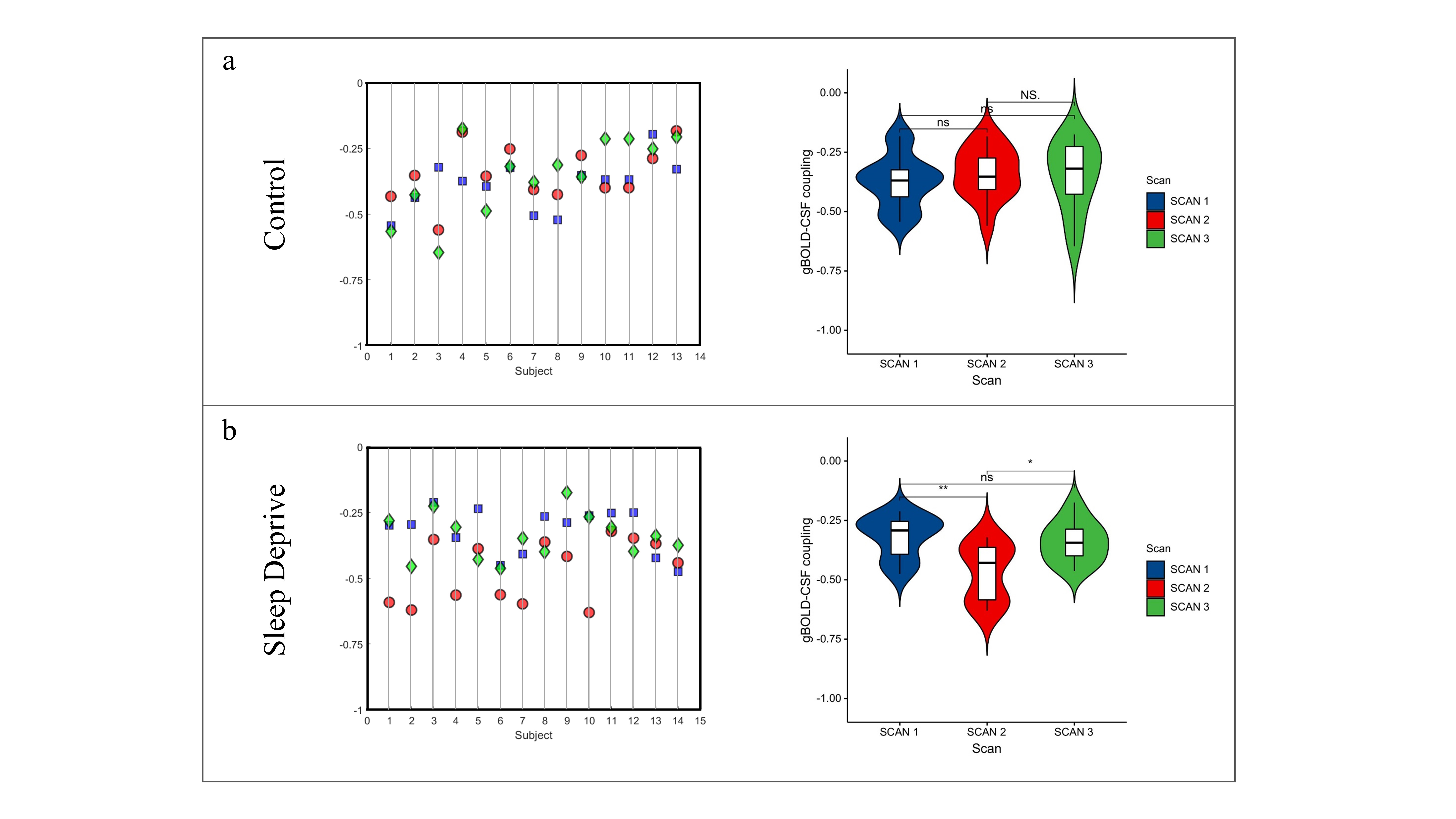
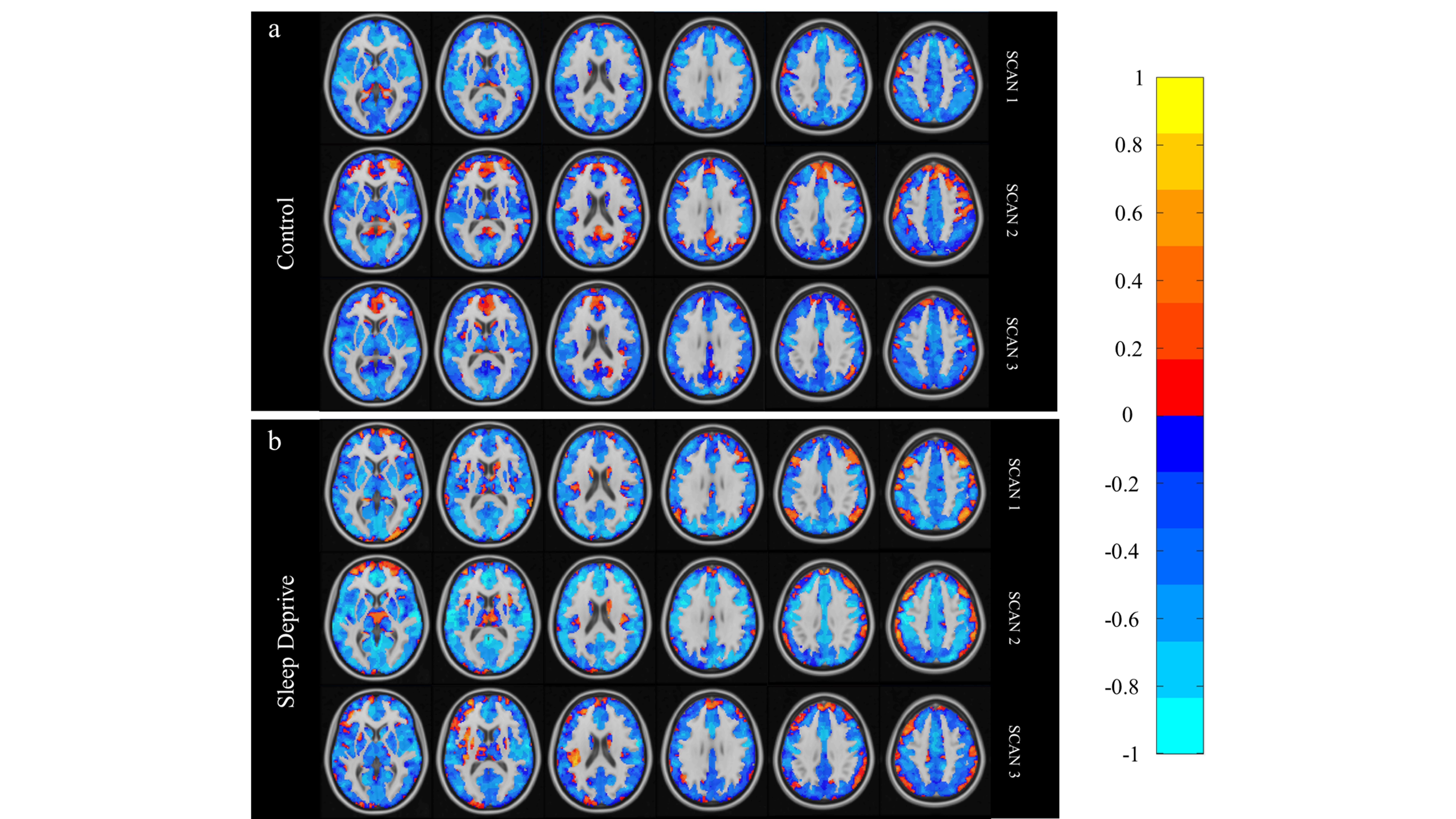
The gBOLD-CSF coupling in the control group demonstrated higher test-retest reliability (ICC = 0.525, P = 0.001) compared with the SD group (ICC = 0.137, P = 0.086). Figure 1 shows the individual and average results of gBOLD-CSF coupling. The gBOLD-CSF couplings were not different across three scans in the control group (P = 0.580). However, the coupling indexes of scan 2 were changed significantly compared to scan 1 and scan 3 in the SD group (P < 0.001).Figure 2 shows the voxel analysis of the MCCC maps. Lightbox view of whole brain MCCC shows that most of the gBOLD in the grey matter voxels were negatively correlated (blue) with CSF inflow fluctuation. The voxel analysis of the MCCC showed a similar trend of low test-retest reliability in the SD group, with the value of the voxels in the MCCC maps of scan 2 being higher than those of scan 1 and scan 3 (Figure 2b). These changes were not observed in the control group.
DISCUSSION
In this study, we evaluated the reproducibility of an emerging, noninvasive method for assessing glymphatic system function based on rfMRI (gBOLD-CSF coupling). The gBOLD-CSF coupling showed acceptable test-retest reliability with ICC values of 0.525. However, the reliability of gBOLD-CSF coupling was significantly reduced in the strictly controlled in-laboratory sleep deprivation group, with an ICC value of 0.137.Reduced test-retest reliability in SD group may stem from changes in the coupling results of scan 2, which was conducted after the subjects experienced a full night of sleep deprivation. Previous research found increases in the cortical interstitial space by more than 60% during sleep when compared with an awake condition, resulting in efficient convective clearance of β-amyloid and other compounds8. This highly sleep-dependent clearance was observed in both human and mouse models9,10. In our study, these significant changes in the gBOLD-CSF coupling index, which could be attributed to participants' drowsiness, were confirmed across three scans and may be responsible for the reduced reproducibility.
CONCLUSION
The gBOLD-CSF coupling shows good reproducibility, but care should be taken when interpreting longitudinal changes of the gBOLD-CSF coupling index that may be influenced by participants’ drowsiness. It would be of great interest to consider sleep as an important factor in future studies exploring the glymphatic system.Acknowledgements
No acknowledgement found.References
1. Iliff, J. J. et al. A paravascular pathway facilitates CSF flow through the brain parenchyma and the clearance of interstitial solutes, including amyloid β. Sci. Transl. Med. 4, 147 (2012).
2. Nedergaard, M. & Goldman, S. A. Glymphatic failure as a final common pathway to dementia. Science 370, 50–56 (2020).
3. Fultz, N. E. et al. Coupled electrophysiological, hemodynamic, and cerebrospinal fluid oscillations in human sleep. Science (80-. ). 366, 628–631 (2019).
4. Yang, F. N. et al. Sleep deprivation enhances inter-stimulus interval effect on vigilant attention performance. Sleep 41, (2018).
5. Han, F. et al. Decoupling of Global Brain Activity and Cerebrospinal Fluid Flow in Parkinson’s Disease Cognitive Decline. Mov. Disord. 36, 2066–2076 (2021).
6. Power, J. D. et al. Ridding fMRI data of motion-related influences: Removal of signals with distinct spatial and physical bases in multiecho data. Proc. Natl. Acad. Sci. U. S. A. 115, E2105–E2114 (2018).
7. Tong, Y., Bergethon, P. R. & Frederick, B. D. An improved method for mapping cerebrovascular reserve using concurrent fMRI and near-infrared spectroscopy with Regressor Interpolation at Progressive Time Delays (RIPTiDe). Neuroimage 56, 2047–2057 (2011).
8. Xie, L. et al. Sleep drives metabolite clearance from the adult brain. Science 342, 373–377 (2013).
9. Bateman, R. J. et al. Human amyloid-beta synthesis and clearance rates as measured in cerebrospinal fluid in vivo. Nat. Med. 12, 856–861 (2006).
10. Kang, J.-E. et al. Amyloid-beta dynamics are regulated by orexin and the sleep-wake cycle. Science (80-. ). 326, 1005–1007 (2009).
Figures