0997
Distinct Effects of Respiratory Depth and Frequency on CSF Flow1Graduate Program for Neuroscience, Boston University, Boston, MA, United States, 2Athinoula A. Martinos Center for Biomedical Imaging, Massachusetts General Hospital, Charlestown, MA, United States, 3Institute for Medical Engineering and Science, Electrical Engineering and Computer Science, Massachusetts Institute of Technology, Cambridge, MA, United States, 4Department of Biomedical Engineering, Boston University, Boston, MA, United States, 5Institute for Medical Engineering and Science, Electrical Engineering and Computer Science, Massachusetts General Hospital, Cambridge, MA, United States, 6Department of Radiology, Harvard Medical School, Cambridge, MA, United States, 7Division of Health Sciences and Technology, Massachusetts Institute of Technology, Cambridge, MA, United States
Synopsis
Keywords: Neurofluids, Neurofluids, Cerebrospinal Fluid Flow, CSF, phase contrast, velocity
Motivation: The flow of cerebrospinal fluid (CSF) is essential for maintenance of brain function.
Goal(s): We aimed to understand the effects of respiration on CSF flow dynamics by quantitatively testing the change in CSF flow across varying paced breathing frequencies.
Approach: Using flow-sensitive fMRI, phase contrast imaging, and physiological recordings, we measured changes in CSF flow and velocity during a visually guided paced breathing task.
Results: We examined CSF flow across breath frequencies ranging from 0.1 Hz to 0.25 Hz, and found that slower frequencies of breathing increase CSF flow, independent of breath depth.
Impact: Our results demonstrate that key features of human respiration, its timing and its depth, induce separate effects on CSF flow. Our identification of respiratory frequency as a modulator of CSF flow provides an accessible mechanism to modulate CSF flow.
INTRODUCTION
In awake humans, respiration has been identified as a driver of CSF flow1-5. While breathing depth is known to modulate flow, another potential modulator is the frequency of breathing, which varies across arousal and autonomic states. The precise effects of different breathing frequencies (i.e., slower vs. faster breathing) on CSF flow are not well understood3-6. When measured with phase contrast imaging, fast (0.17Hz) and slow (0.125Hz) breathing induce positive CSF velocities in the aqueduct during the inhale period of the breath cycle, with slow breathing having a higher increase1. However, those two frequencies do not reflect the full range of fast or slow breathing present across arousal states which can range from 0.1Hz to ~0.33Hz7. Recent data in our lab showed that respiratory paces between fast (0.25Hz) and slow (0.1Hz) frequencies increase CSF inflow signals in the 4th ventricle (measured via fMRI inflow signals) relative to free breathing, with slower frequencies having higher signal increases (Fig 2a)8. A critical question is whether changes in the depth of breathing could explain those frequency-dependent effects. To answer this question we expanded on our previous paced breathing study with phase contrast imaging to quantify flow velocities while recording respiration, expired CO2, and heartrate to assess changes in autonomic state and gas exchange. Additionally, we assessed the contribution of breathing depth to the changes in CSF inflow across frequencies.METHODS
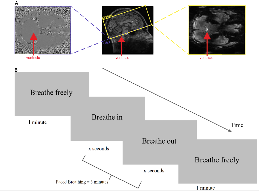
Fourteen subjects gave informed consent and were scanned on a 7T Siemens whole-body scanner with a custom-built 64-channel head coil array. Subjects completed 30 minutes of guided diaphragmatic breath training before the scan to practice minimizing motion. Sessions began with a 0.75 mm isotropic multi-echo MPRAGE. Functional MRI runs were acquired using a simultaneous multislice (SMS) gradient-echo EPI sequence (R=2 acceleration, MultiBand factor=4, 2 mm isotropic voxels, TR=499ms, TE=24 ms, echo-spacing=0.59 ms, flip angle=40°). Phase contrast (PC) MRI runs were acquired using a GRE sequence to collect a single axial slice (VENC=10cm/s, 2x2x5 mm voxels, TR=903ms, TE=5.9ms). The bottom edge of the acquisition volume was placed perpendicular to the base of the fourth ventricle, in order to measure CSF inflow signals and velocity using fMRI and PC-MRI respectively (Fig 1a). Stimuli consisted of a visual prompt that guided subjects through five-minute breathing tasks consisting of free, 0.25Hz, 0.17Hz, 0.125Hz, and 0.1Hz breathing frequencies (Fig 1b). Respiratory, heart rate, and CO2 data were collected to measure task compliance and peripheral physiology. Data were preprocessed using slice-timing correction and CSF signals were extracted from within the fourth ventricle from a manually drawn ROI. CSF flow, velocity, and respiratory depth responses to the breathing task were computed by binning the MR time courses by the phase of the breath cycle for a given pace. Group averages for respiratory-locked CSF flow were calculated using the 15th percentile of the MR time course as baseline, approximating a no-flow baseline. Statistical analyses used Levene’s test for variance to determine that nonparametric tests should be used, and then applied the Kruskal Wallis test to estimate whether flow increases were significant, and the Tukey-Kramer to test for differences between breathing frequencies.RESULTS
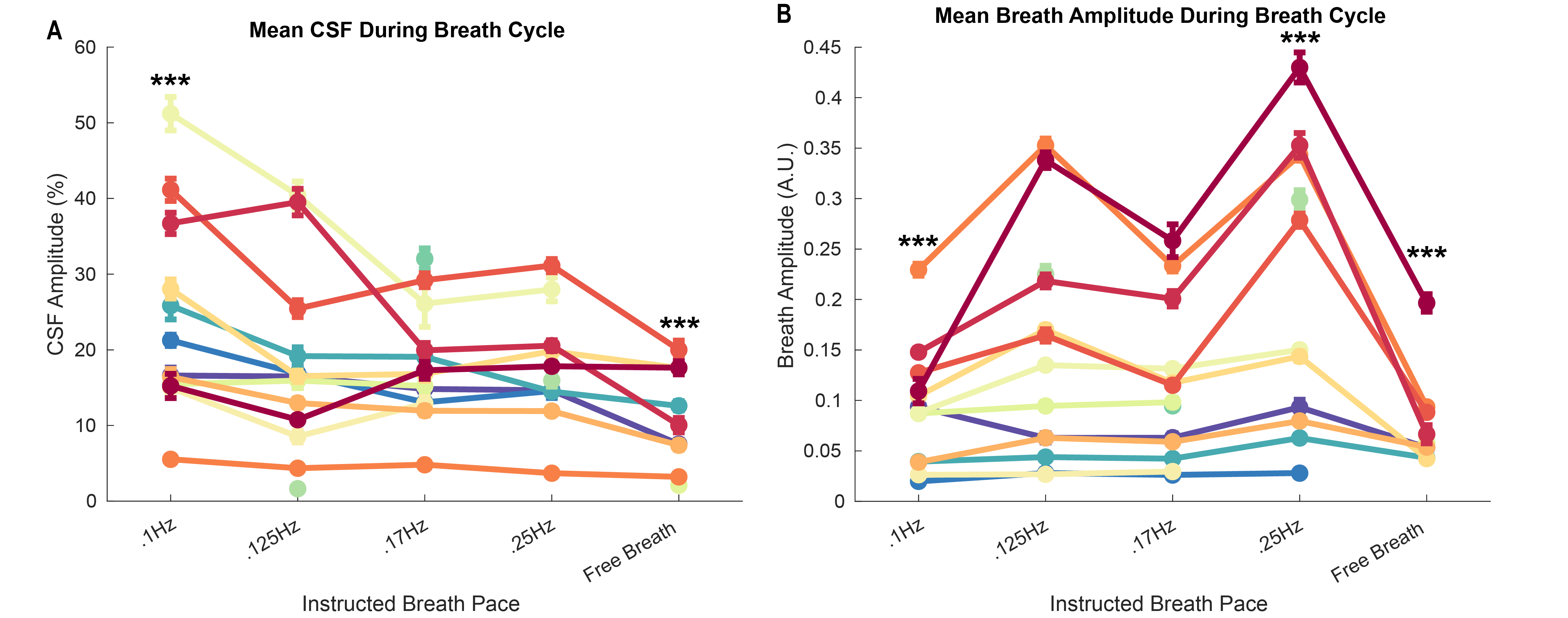
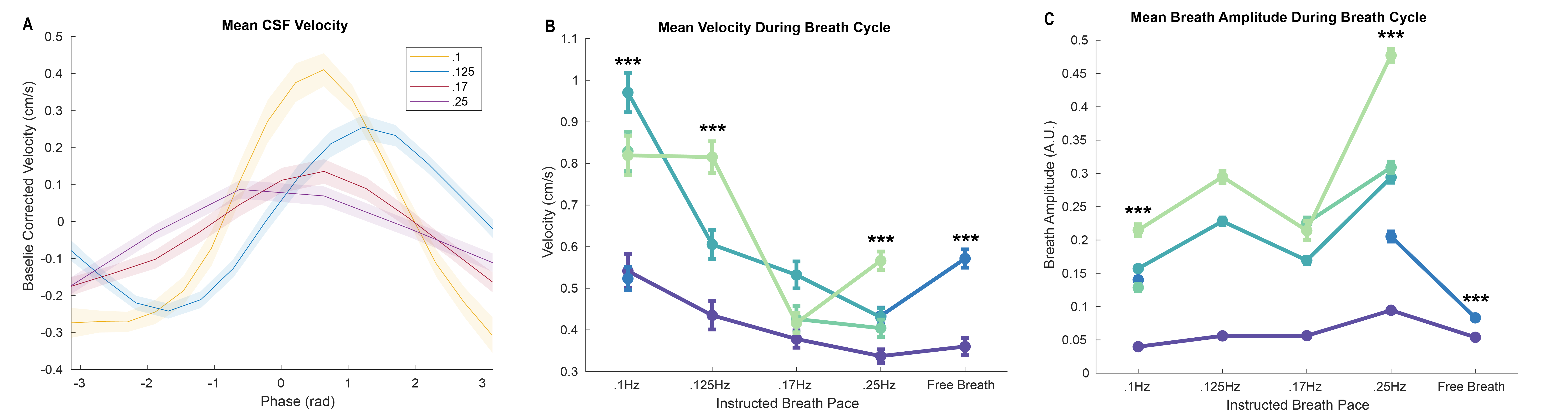
We first analyzed fMRI flow signals to obtain high temporal resolution measurement of how paced breathing affects CSF flow. All paced breathing frequencies significantly increased the mean CSF flow during each breath relative to free breathing, with 0.1 Hz inducing significantly larger flow than all other paces (p<.001, Fig 2a). The depth of respiration was significantly higher for paced breathing than free breathing, but with the opposite pattern: largest depth during 0.25 Hz breathing, and smallest depth during 0.1Hz breathing (p<.001, Fig 2b). Next, using quantitative measurement of velocity using phase contrast, we found the same pattern of velocity amplitudes and respiratory depths during paced breathing (p<.001, Fig 3, with slow breathing eliciting velocities as much as two times higher than fast breathing.DISCUSSION
These results show an opposing relationship between breath frequency and breath depth, suggesting that breath frequency can modulate fourth ventricle CSF flow independent of breath depth. Future work should explore potential mechanisms such as the altered CO2 levels or changes in autonomic state caused by slow paced breathing.CONCLUSION
We found that breath frequency can modulate 4th ventricle CSF flow, and this effect cannot be explained by increases in breath depth. Paced and especially slow breathing can significantly increase CSF flow in humans.Acknowledgements
This work was funded by NIH R01-AT011429, P41-EB030006, the Simons Collaboration on Plasticity in the Aging Brain (no. 811231), the Sloan Fellowship, the 1907 Trailblazer Award, and was made possible by the resources provided by Shared Instrumentation Grant S10-OD023637.References
1. Chen, L., Beckett, A., Verma, A., & Feinberg, D. A. (2015). Dynamics of respiratory and cardiac CSF motion revealed with real-time simultaneous multi-slice EPI velocity phase contrast imaging. Neuroimage, 122, 281-287.
2. Dreha-Kulaczewski, S., Joseph, A. A., Merboldt, K. D., Ludwig, H. C., Gärtner, J., & Frahm, J. (2015). Inspiration is the major regulator of human CSF flow. Journal of neuroscience, 35(6), 2485-2491
3. Aktas, G., Kollmeier, J.M., Joseph, A.A. et al. Spinal CSF flow in response to forced thoracic and abdominal respiration. Fluids Barriers CNS 16, 10 (2019). https://doi.org/10.1186/s12987-019-0130-0 4. Yildiz, S., Grinstead, J., Hildebrand, A., Oshinski, J., Rooney, W. D., Lim, M. M., & Oken, B. (2022). Immediate impact of yogic breathing on pulsatile cerebrospinal fluid dynamics. Scientific Reports, 12(1), 10894.
5. Kollmeier, J. M., Gürbüz-Reiss, L., Sahoo, P., Badura, S., Ellebracht, B., Keck, M., ... & Dreha-Kulaczewski, S. (2022). Deep breathing couples CSF and venous flow dynamics. Scientific reports, 12(1), 2568.
6. Fultz, N. E., Bonmassar, G., Setsompop, K., Stickgold, R. A., Rosen, B. R., Polimeni, J. R., & Lewis, L. D. (2019). Coupled electrophysiological, hemodynamic, and cerebrospinal fluid oscillations in human sleep. Science, 366(6465), 628-631.
7. Migliaccio, G.M., Russo, L., Maric, M., Padulo, J. Sports Performance and Breathing Rate: What Is the Connection? A Narrative Review on Breathing Strategies. Sports (Basel). 2023 May 10;11(5):103. doi: 10.3390/sports11050103. PMID: 37234059; PMCID: PMC10224217.
8. Banks, M., Gomez, E.P.D., Beldzik, E., Cicero, N., Napadow, V., Polimeni, J., Lewis, D.L. Respiratory Modulation of Cerebrospinal Fluid Flow During Paced Breathing [abstract]. ISMRM 2023; June 3-9; Toronto, CA.
Figures