0995
Imaging GBCA enhancement in the periosteal and meningeal layers of dura mater using high resolution MRI at 7T1F.M. Kirby Research Center for Functional Brain Imaging, Kennedy Kreiger Institude, Baltimore, MD, United States, 2Biomedical Engineering, Johns Hopkins University School of Medicine, Baltimore, MD, United States, 3Neurosection, Division of MRI Research, Russell H. Morgan Department of Radiology and Radiological Science, Johns Hopkins University School of Medicine, Baltimore, MD, United States, 4Department of Neurology, Johns Hopkins University School of Medicine, Baltimore, MD, United States
Synopsis
Keywords: Neurofluids, Neurofluids, CSF, dura mater
Motivation: The GBCA enhancement detected in the parasagittal dura (PSD) is hypothesized to originate from the dura mater blood vessels lacking a blood-brain-barrier. The dura mater consists of two layers: periosteal and meningeal dura mater. No prior study has examined the pattern of GBCA enhancement within these layers.
Goal(s): To visualize GBCA enhancement in the periosteal and meningeal layers of dura mater in the human brain.
Approach: High-resolution black-blood and FLAIR MRI were performed at 7T.
Results: The two layers and their GBCA-enhancement could be visualized. T1 and T2* values in each layer were measured.
Impact: This work allows us investigate the relationship between GBCA-enhancement in the dura mater and GBCA-induced-signal-changes in the PSD and meningeal-lymphatic-vessels. This is of importance given that GBCA-enhanced MRI has been a gold-standard technique for investigating human brain clearance.
Introduction
Although Gadolinium-based contrast agents (GBCAs) are generally considered intravascular contrast agents, various studies have demonstrated that intravenously administrated GBCAs can be detected at multiple extravascular locations in the human central nervous system (CNS). One main site is the parasagittal dura (PSD). It is hypothesized that the GBCA enhancement in the PSD primarily originates from the blood vessels in the dura mater since these vessels lack a blood-brain barrier (BBB). The dura mater is the outer layer of the meninges (above the arachnoid mater and pia mater) composed of fibrous connective tissue. The dura mater surrounding the brain can be sub-divided into two layers: the periosteal dura mater (outer layer that connects to the skull) and the meningeal dura mater (inner layer above the subdural cavity and arachnoid mater). The dural venous sinuses, situated between the two layers, drain to the internal jugular veins. To the best of our knowledge, no prior study has examined the pattern of GBCA enhancement within the two layers of dura mater. In this study, a first attempt is made using high-resolution MRI at 7T to visualize GBCA enhancement in the periosteal and meningeal layers of dura mater in the human brain. This is of importance given that GBCA-enhanced MRI has been a gold-standard technique for investigating human brain clearance.Methods
MRI: Eleven healthy volunteers were scanned on a Philips 7T scanner. GBCA (Gadoteridol) was administered intravenously (0.1 mmol/kg). The following scans were performed for each subject both before and after GBCA administration: 1) black-blood MRI: voxel=0.5x0.5x0.75mm3, fast-spin-echo, TR/TE/echo-spacing=1572/50/13.6ms; 2) T2 FLAIR: voxel=0.5mm isotropic, TR/TI/TE=8000/2200/300ms; 3) MP2RAGE to measure T1: voxel=0.5x0.5x1mm3, TR/TI1/TI1/TE=8250/1000/3300/2.3ms; 4) Multiple-gradient-echo to measure T2*: voxel=0.5x0.5x1mm3, TR=52ms, TE=5,10,15ms.Data analysis: The black-blood and FLAIR images were co-registered using SPM12. The periosteal and meningeal layers of dura mater surrounding two brain regions (a cortical region in the parietal lobe and the PSD) were manually delineated using ITK-snap on pre-GBCA FLAIR images. T1 values were calculated from the MP2RAGE data. T2* values were fitted using the multi-gradient-echo data.Results
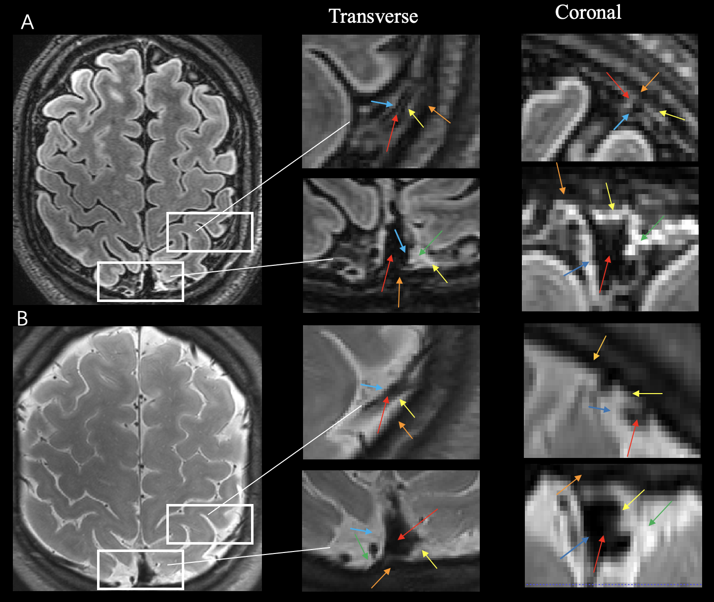
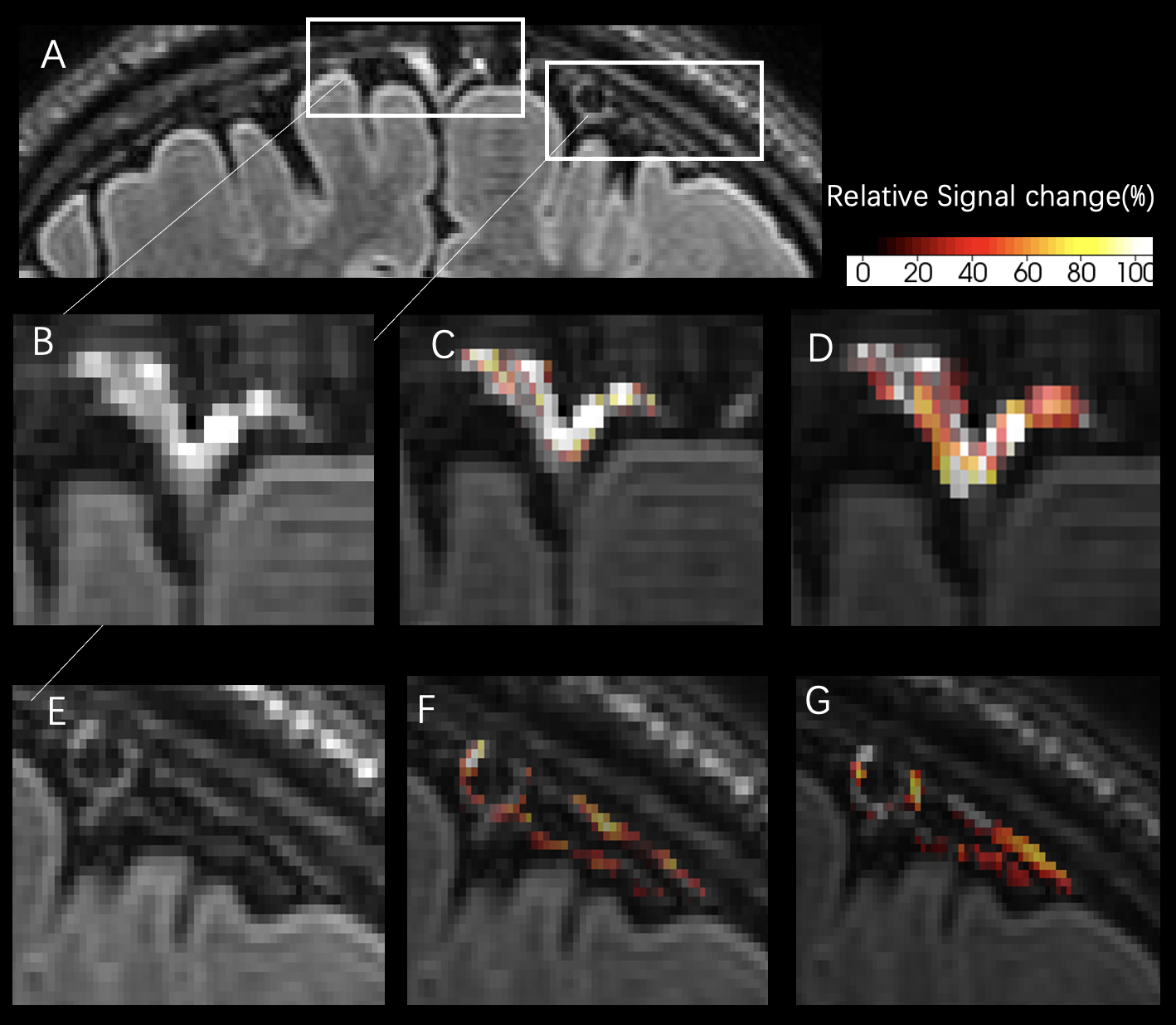
Fig.1 shows representative pre-GBCA black-blood and FLAIR images on which the periosteal and meningeal layers of dura mater surrounding a parietal region and the PSD can be identified. On both black-blood and FLAIR images, the dura mater layers showed hyper-intensities between the skull and subarachnoid space, both of which were hypo-intense. On the black-blood images, the dural venous sinuses between the two layers also were hypo-intense. Table 1 summarizes the T1 and T2* values measured in each layer. The periosteal layer showed shorter T1 than the meningeal layer (P<0.05). Fig.2 shows representative maps of GBCA enhancement in black-blood and FLAIR images in the periosteal and meningeal layers. Table 2 summarizes the quantitative results. Both the periosteal and meningeal layers showed significant GBCA-induced signal increase in black-blood and FLAIR images. No significant difference was found in the GBCA enhancement between the two layers (P>0.1). Interestingly, the dura surrounding the PSD showed greater (P<0.05) GBCA enhancement than the dura surrounding the parietal region.Discussion & Conclusion
By employing high-resolution black-blood and FLAIR MRI on 7T, the periosteal and meningeal layers of dura mater in the human brain can be identified on pre-GBCA images. GBCA enhancement can be detected in both layers after intravenous GBCA administration. The magnitudes of GBCA enhancement were similar between the two layers but varied around different brain regions. The dura surrounding the PSD showed more intense GBCA enhancement, possibly due to its rich vascular network. Subsequently, we will use a recently developed MRI method to investigate the relationship between GBCA enhancement in the two layers of dura mater and GBCA induced signal changes in the PSD and meningeal lymphatic vessels.Acknowledgements
Thank Mr. Joseph S. Gillen, Mr Ganji Sandeep, Mrs. Terri Lee Brawner, Ms. Kathleen A. Kahl, and Ms. Ivana Kusevic for assistance.References
1. Patel LD, Raghavan P, Tang S, Choi S, Harrison DM. Imaging of the meningeal lymphatic network in healthy adults: A 7T MRI study. Journal of Neuroradiology. 2023;50(4):369-376. doi:10.1016/j.neurad.2023.03.002
2. Ringstad G, Eide PK. Cerebrospinal fluid tracer efflux to parasagittal dura in humans. Nat Commun. 2020;11(1):354. doi:10.1038/s41467-019-14195-x
3. Martina Absinta, Seung-Kwon Ha, Govind Nair, Pascal Sati, Nicholas J Luciano, Maryknoll Palisoc, Antoine Louveau, Kareem A Zaghloul, Stefania Pittaluga, Jonathan Kipnis, Daniel S Reich (2017) Human and nonhuman primate meninges harbor lymphatic vessels that can be visualized noninvasively by MRI eLife 6:e29738
4. Cao, D, Sun, Y, Li, Y, et al. Concurrent measurement of perfusion parameters related to small blood vessels and cerebrospinal fluid circulation in the human brain using dynamic dual-spin-echo perfusion MRI. NMR in Biomedicine. 2023; 36(10):e4984. doi:10.1002/nbm.4984
5. Yushkevich PA, Piven J, Hazlett HC, et al. User-guided 3D active contour segmentation of anatomical structures: Significantly improved efficiency and reliability. NeuroImage. 2006;31(3):1116-1128. doi:10.1016/j.neuroimage.2006.01.015
Figures