0987
Evaluation of the glymphatic system activity during sleep-wake states through quantitative CSF measurement using a three-pool water model1Biomedical Engineering, Hankuk University of Foreign Studies, Yongin-si, Korea, Republic of
Synopsis
Keywords: Alzheimer's Disease, Alzheimer's Disease, Glymphatic system, CSF
Motivation: The glymphatic system plays a crucial role in brain waste clearance, and dysfunction has been linked to neurodegenerative diseases like Alzheimer's Disease. Understanding this system is essential for advancing our knowledge of brain health.
Goal(s): We aim to develop a non-invasive method to assess glymphatic activity in the human brain.
Approach: We utilize a quantitative CSF measurement technique with a three-pool model and multi-echo spin-echo images.
Results: Significant variations in glymphatic activity were observed across different brain regions and found to be influenced by sleep.
Impact: Our method can potentially reveal sleep-influenced glymphatic activity variations, enabling early Alzheimer's Disease diagnosis.
Introduction
The glymphatic system, which is a waste clearance pathway in the brain that relies on glial cells, has been acknowledged as an interstitial waste clearance system1. The presence of dysfunction in the glymphatic system has been observed to be linked to Alzheimer's Disease (AD). This dysfunction leads to a diminished ability to eliminate AD-related proteins, such as amyloid beta and tau.2 Sleep is also considered a potential biomarker for AD pathology and cognitive impairment.3 Recent studies have elucidated a correlation between cerebrospinal fluid (CSF) and the active removal of waste during sleep state4. However, the assessment of glymphatic system activity using MRI is constrained by the sluggish movement of CSF. Several studies have proposed indirect methodologies involving gadolinium-based contrast agents (GBCA), to examine glymphatic function in mice and the human brain.5-10 T1-mapping has demonstrated its potential for assessing dynamic glymphatic activity in the human brain11. Nevertheless, there is an increasing demand for developing non-invasive methods to measure glymphatic function through the visualization of CSF. In this study, we propose a non-invasive and direct method for quantitatively measuring cerebrospinal fluid (CSF) using a three-pool model and multi-echo spin-echo images to evaluate glymphatic activity.Methods
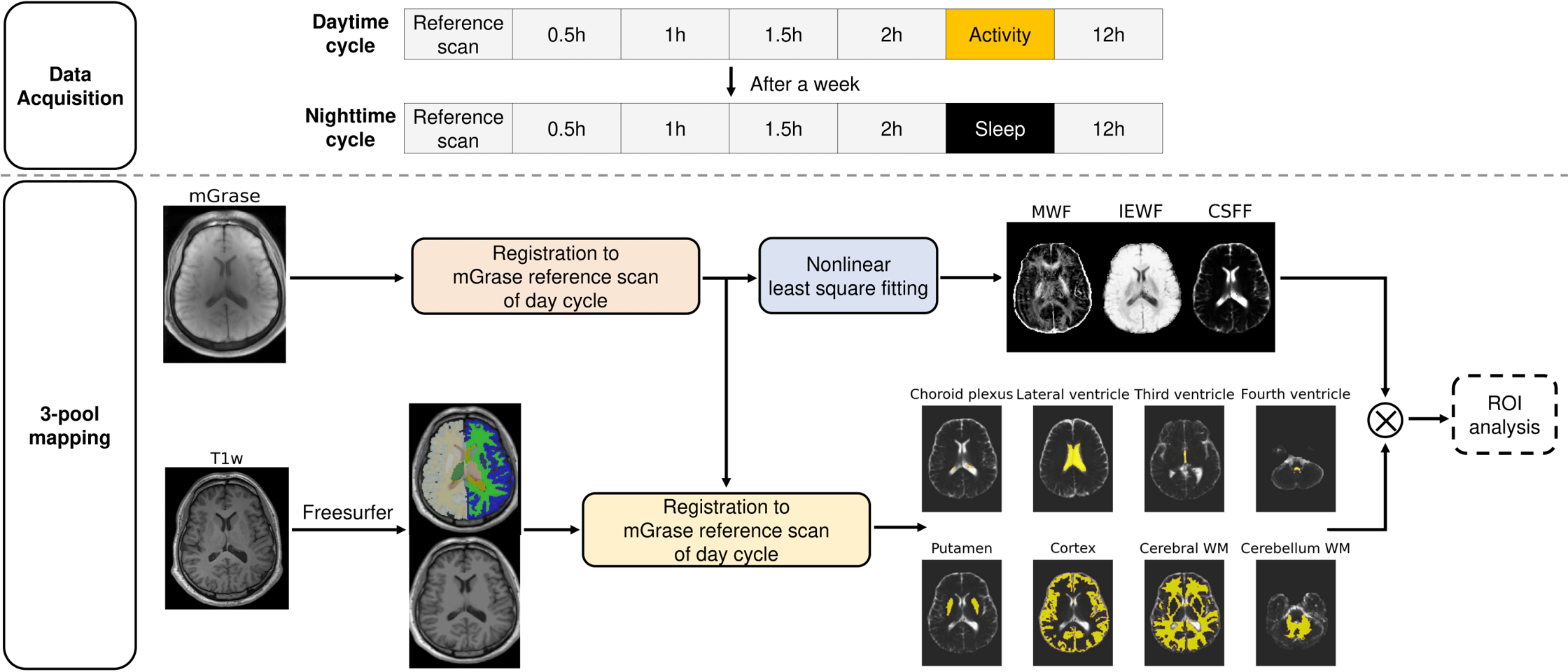
Data acquisitionTwenty-four healthy subjects (IRB-informed) aged 20-30 were scanned on 3T (Siemens Trio) . Figure 1 shows the overall study pipeline. The dataset consists of two cycles: (1) daytime and (2) nighttime. During each cycle, 3D T1w (TR/TE = 8.6/2.75 ms, 1.0 mm3 isotropic) and multi-echo gradient-spin-echo (mGRASE, TR/TE = 1000/10 ms, 32 echoes, 2 x 2 x 5 mm3) were acquired at 0 (= reference scan), 0.5, 1, 1.5, 2, and 12 hours. During the daytime cycle, the subjects had daily activities between 2 and 12 hours. In the nighttime cycle, the subjects had a regular sleep between 2 and 12 hours.
Image preprocessing
The 3D T1w was used to define eight ROIs (choroid plexus, lateral ventricle, third ventricle, fourth ventricle, cortex, cerebral white matter (WM), and cerebellum WM) using Freesurfer12. Affine registration was applied to match the T1w from the Freesurfer results to the reference scan of the daytime cycle. The transformation matrix derived from the registration of T1w was subsequently applied to the ROI mask by matrix multiplication.
3-pool mapping algorithm
Myelin water fraction (MWF), intra/extra-cellular fraction (IEWF), and CSF fraction (CSFF) maps were generated from the mGRASE of all time points and all cycles using a voxel-wise multi-exponential nonlinear least squares fitting by defining the following equations:
$$mGrase_{voxel}(t) = A_{MW} \times exp^\frac{-TE}{T2_{MW}} + A_{IEW} \times exp^\frac{-TE}{T2_{IEW}}+A_{CSF} \times exp^\frac{-TE}{T2_{CSF}}$$
,where mGRASEvoxel(t) is the acquisition signal in a voxel for 32 echo times, MW, IEW are myelin water, intra/extra-cellular water, respectively, and A is the amplitude of each water compartment.
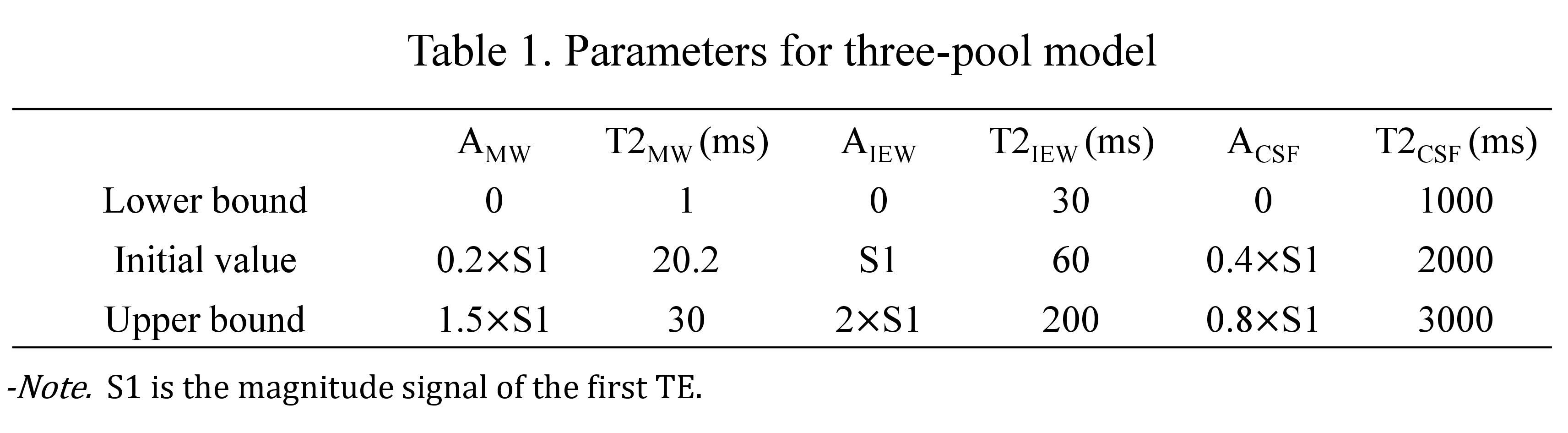
The parameters for the three-pool model were set as shown in Table 1. MWF, IEWF, and CSFF were calculated as the corresponding water compartment volume ratio to the total water volume.
ROI analysis
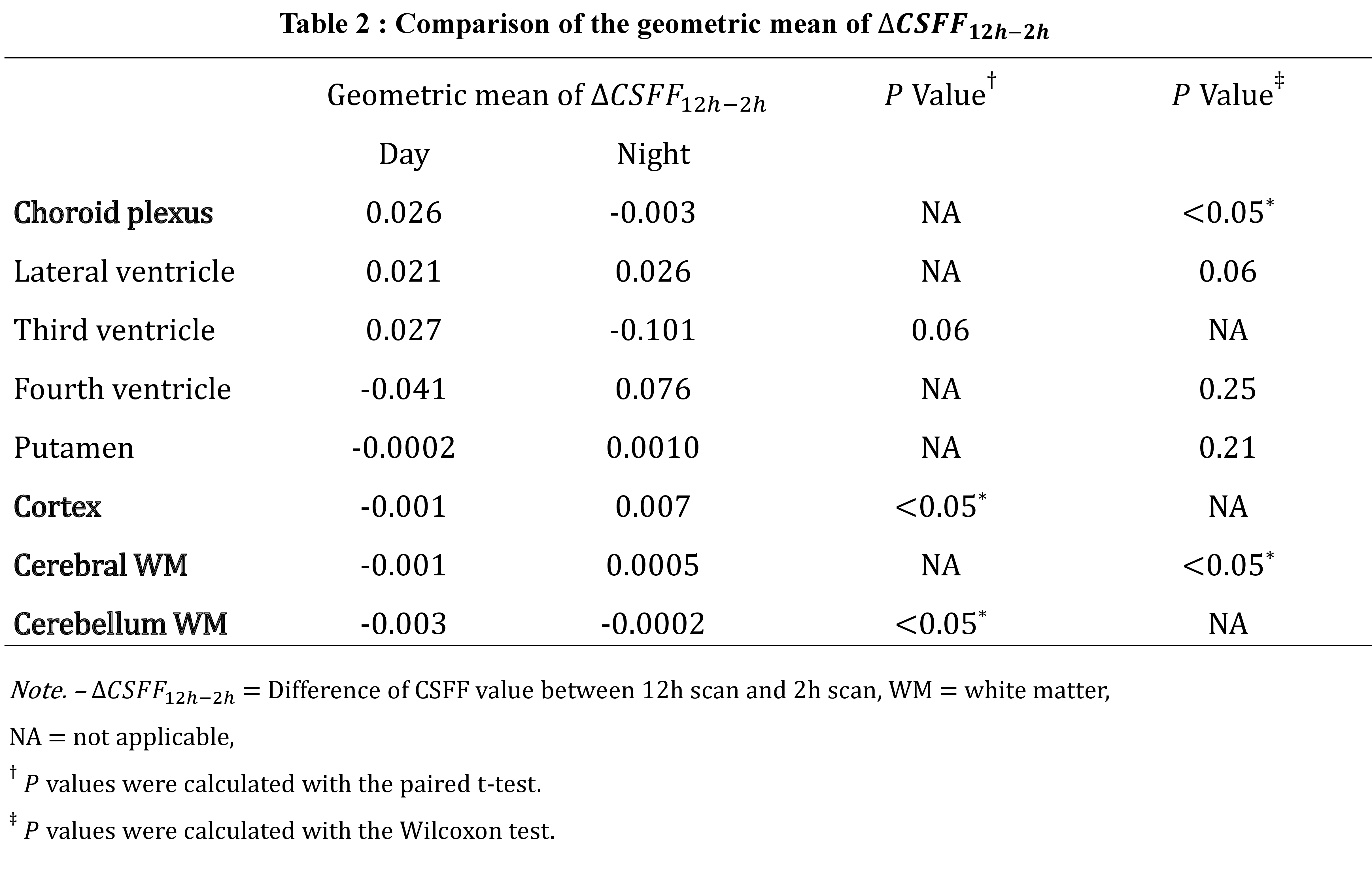
We compared the mean fraction change in CSF between the 2-hour scan (CSFF2h) and the 12-hour (CSFF12h) obtained from the daytime and nighttime cycles.
Results
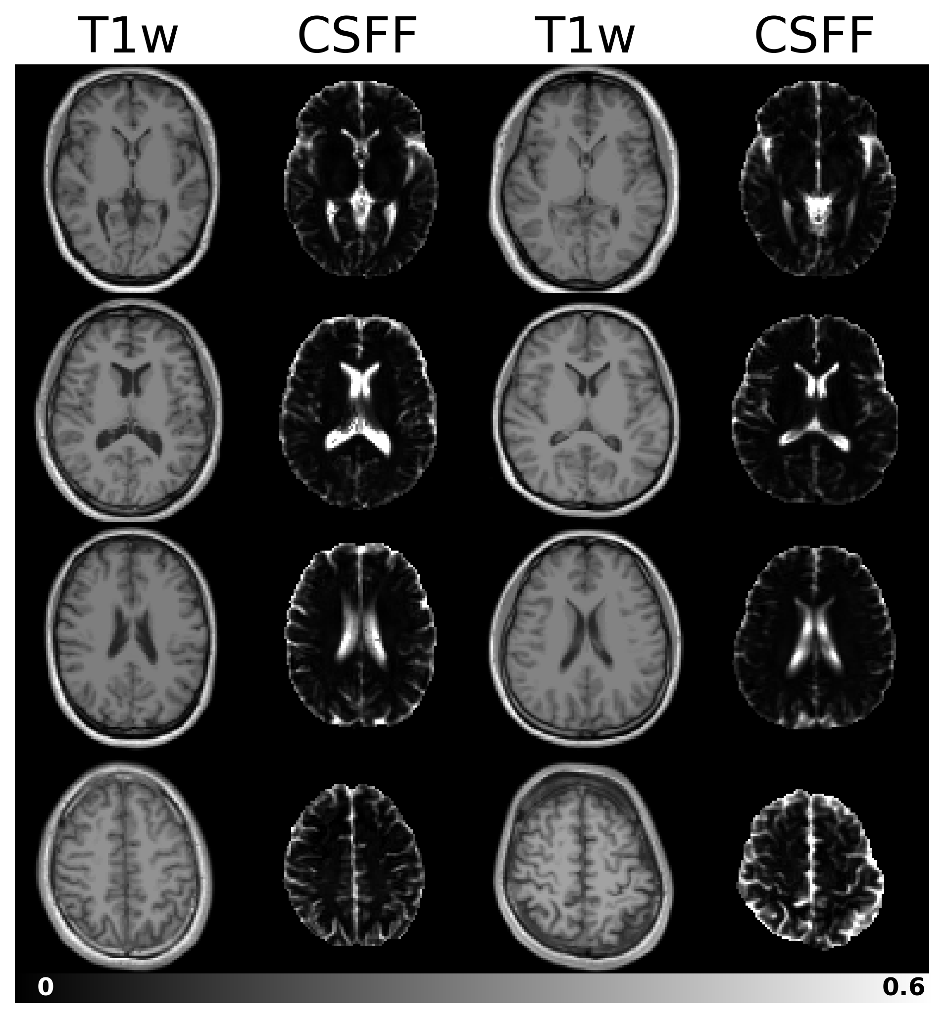
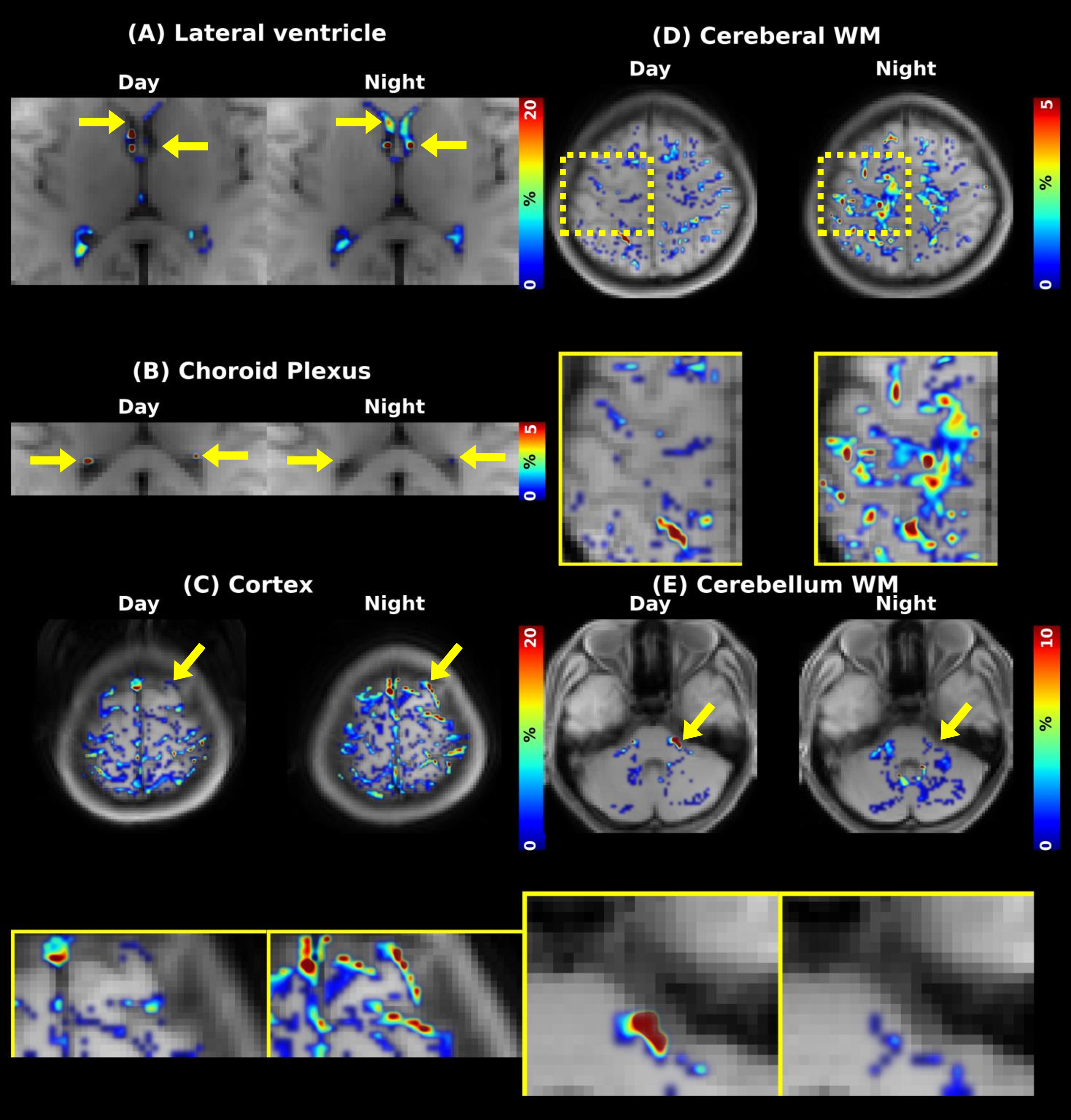
Figure 2 represents the typical CSFF maps with the corresponding T1w images. Figure 3 shows the difference map derived from a representative subject's comparison of CSFF2h and CSFF12h. Figure 3-(A) highlights increased CSFF in the lateral ventricle of the night scan. In Figure 3-(B), the choroid plexus shows decreased CSFF in the night scan. Figures 3-(C) and (D) demonstrate increased CSFF in the cortex and cerebral WM, while Figure 3-(E) reveals decreased CSFF in the cerebellum WM in the night scan. After verifying the normality using the Shaprio-Wilk test, a paired t-test was conducted for ROIs that followed normality and a Wilcoxon test for those that did not. Statistical analysis indicates significant differences between the daytime and nighttime cycles (p<0.05) in the choroid plexus, cortex, cerebral WM, and cerebellum WM. These variations in CSFF may be attributed to the influence of sleep.Discussion and Conclusion
We demonstrate a non-invasive quantitative CSF measurement method using the three-pool mapping model for assessing glymphatic activity in the human brain. Significant sleep-related effects on the CSFF difference in the choroid plexus, cortex, cerebral WM, and cerebellum WM were observed. These evidences imply it may help us better understand the glymphatic activity in the brain in a non-invasive way and provide clinically useful information. This is an initial investigation, and it serves as a starting point toward clinical scanning. Our method was exclusively evaluated on healthy subjects, but its application to patients with Alzheimer's Disease and other neurodegenerative disorders may offer valuable insights regarding their glymphatic activity.Acknowledgements
This work was supported by the National Research Foundation of Korea (NRF) grant funded by the Korea government (MSIT) (NRF-2023R1A2C1007292)References
1. Iliff JJ, Wang M, Liao Y, et al. A paravascular pathway facilitates CSF flow through the brain parenchyma and the clearance of interstitial solutes, including amyloid β. Science translational medicine. 2012;4(147):147ra11-ra11.2. Silva I, Silva J, Ferreira R, Trigo D. Glymphatic system, AQP4, and their implications in Alzheimer’' 's disease. Neurological research and practice. 2021;3:1-9.
3. Lucey BP, McCullough A, Landsness EC, et al. Reduced non–rapid eye movement sleep is associated with tau pathology in early Alzheimer’' 's disease. Science translational medicine. 2019;11(474):eaau6550.
4. Fultz NE, Bonmassar G, Setsompop K, et al. Coupled electrophysiological, hemodynamic, and cerebrospinal fluid oscillations in human sleep. Science. 2019;366(6465):628-31.
5. Kress BT, Iliff JJ, Xia M, et al. Impairment of paravascular clearance pathways in the aging brain. Annals of neurology. 2014;76(6):845-61.
6. Iliff JJ, Lee H, Yu M, et al. Brain-wide pathway for waste clearance captured by contrast-enhanced MRI. The Journal of clinical investigation. 2013;123(3):1299-309.
7. Taoka T, Jost G, Frenzel T, Naganawa S, Pietsch H. Impact of the glymphatic system on the kinetic and distribution of gadodiamide in the rat brain: observations by dynamic MRI and effect of circadian rhythm on tissue gadolinium concentrations. Investigative Radiology. 2018;53(9):529-34.
8. Eide PK, Ringstad G. Delayed clearance of cerebrospinal fluid tracer from entorhinal cortex in idiopathic normal pressure hydrocephalus: a glymphatic magnetic resonance imaging study. Journal of Cerebral Blood Flow & Metabolism. 2019;39(7):1355-68.
9. Watts R, Steinklein J, Waldman L, Zhou X, Filippi C. Measuring glymphatic flow in man using quantitative contrast-enhanced MRI. American Journal of Neuroradiology. 2019;40(4):648-51.
10. Deike-Hofmann K, Reuter J, Haase R, et al. Glymphatic pathway of gadolinium-based contrast agents through the brain: overlooked and misinterpreted. Investigative radiology. 2019;54(4):229-37.
11. Lee S, Yoo R-E, Choi SH, et al. Contrast-enhanced MRI T1 mapping for quantitative evaluation of putative dynamic glymphatic activity in the human brain in sleep-wake states. Radiology. 2021;300(3):661-8.
12. Fischl B. FreeSurfer. Neuroimage. 2012;62(2):774-81.
Figures