0986
Detection of vasomotion in the human brain using Fourier decomposition of T1-weighted Cine-FLASH MRI (FD-FLASH)1Siemens Medical Solutions USA Inc, Malvern, PA, United States, 2Neurology, Perelman School of Medicine, University of Pennsylvania, Philadelphia, PA, United States, 3Radiology, Perelman School of Medicine, University of Pennsylvania, Philadelphia, PA, United States
Synopsis
Keywords: Neurofluids, Neurofluids
Motivation: Vasomotion has been hypothesized as a mechanism for brain paravascular clearance, which could be impaired in multiple neurodegenerative conditions.
Goal(s): To measure vasomotion in vivo in human brains using MRI.
Approach: A single-shot T1-weighted Cine-FLASH sequence was optimized to provide high temporal resolution in two adults, as well as during a visual stimulation paradigm in one. We performed a Fourier decomposition of the signal in brain vascular structures.
Results: An ultra-low-frequency signal was observed consistent with vasomotion in posterior cerebral arteries while no such signal could be identified in the parenchyma. A substantial amplification of this signal could be observed during visual stimulation.
Impact: This method could help studying brain waste clearance and its dysfunction in humans in vivo.
Introduction
Vasomotion, defined as an ultra-low frequency (≈0.08 Hz) spontaneous oscillation of blood vessels, has been hypothesized as a mechanism for resting-state BOLD functional connectivity1 and for driving paravascular waste clearance in the brain2. While vasomotion oscillations have been successfully detected in the rodent brain where they can be entrained by functional stimuli modulated at near the vasomotion frequency1, direct localized measurement in humans has not been demonstrated.We propose here a proof-of-concept for measuring vasomotion in the human brain by Fourier decomposition of high frame-rate Cine-FLASH data.
Methods
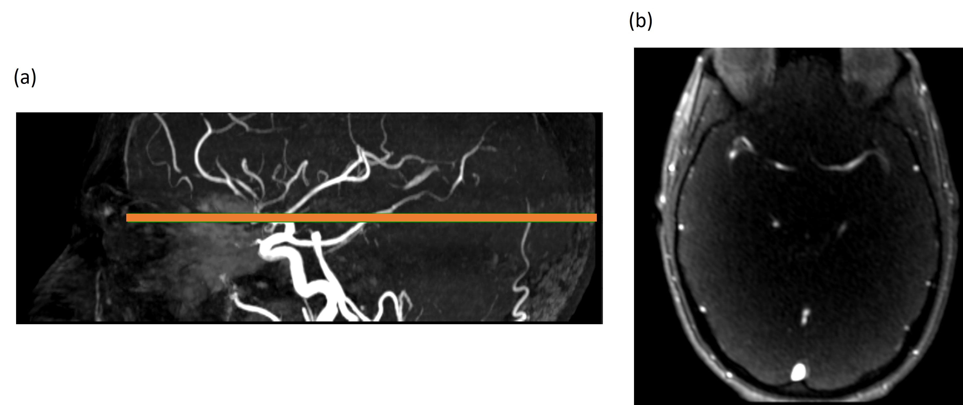
Concept: In a Cine-FLASH sequence, stationary spins get saturated because of shortly repetition time RF pulses especially if the flip-angle is above Ernst angle3. In a region of interest encompassing a pulsating structure, the total signal in that ROI will be modulated by the pulsation frequency. Therefore, if the temporal resolution is sufficient to respect Nyquist criteria (leading to a sampling rate of at least twice the frequency of the signal to be detected), vascular oscillations could potentially be detected by performing a Fourier decomposition4 of a temporal series acquired in a slice containing a vascular region of interest.Experiments: Two healthy adults (54 and 64 yo males) were scanned at 3T (MAGNETOM Prisma, Siemens Healthineers, Erlangen, Germany) using a 64-ch head/neck coil. After localizers, we acquired a 3D Time-of-Flight angiogram (TOF-MRA) accelerated with Compressed Sensing5 to localize a slice of interest (0.4mm isotropic, R=10.3), containing branches of both middle cerebral arteries (MCA) and posterior cerebral arteries (PCA). Once the spatial location was selected, an axial single slice, single-shot Cine-FLASH sequence was acquired continuously for 2.7min (512 phases) as illustrated in Figure 1. In order to increase temporal resolution as much as possible, we used parallel imaging (GRAPPA6 with R=2), phase Partial Fourier (6/8) as well as an asymmetric echo (TE = 2.06ms). With a matrix of 160x160 and FOV = 180x180mm2, bandwidth = 947Hz/pixel, we reached a minimum TR = 315ms for a 3-mm thick slice. We used a flip angle alpha = 35 degrees to saturate parenchymal signal (assuming T1 = 0.8/1.6s in white and gray matter7). In one of the of the volunteers, we presented a visual stimulation consisting of 16 Hz flicker with a modulation frequency of 0.1Hz as an attempt to amplify the vasomotion using neural stimulation. Starting in total dark, we acquired Cine-FLASH data for 512 phases (2.7min) prior to switching on the visual stimulation for the same amount of time.
Processing: Cine time series were saved as DICOM images and processed offline. After realignment of the time series using mcflirt (FSL)8, a 512-points discrete Fast Fourier Transform (FFT) was performed in MATLAB on a time-series spatially averaged in a manually defined ROI, leading to a spectral resolution of 0.0062Hz. A 3-points sliding window averaging was used for noise reduction.
Results and Discussion
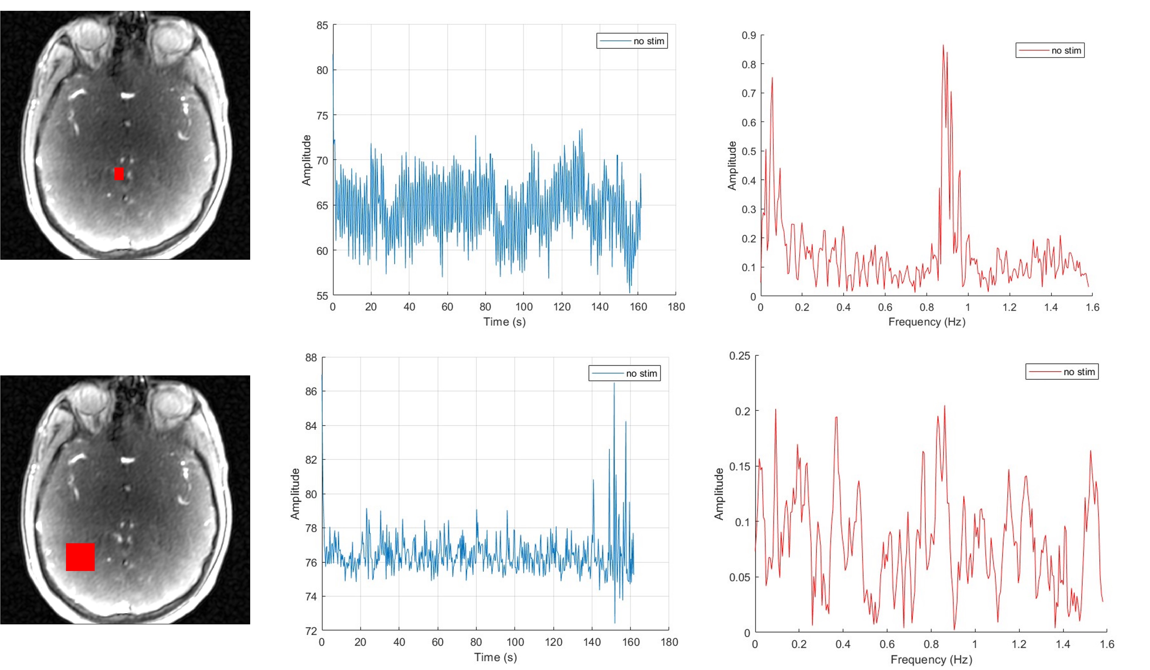
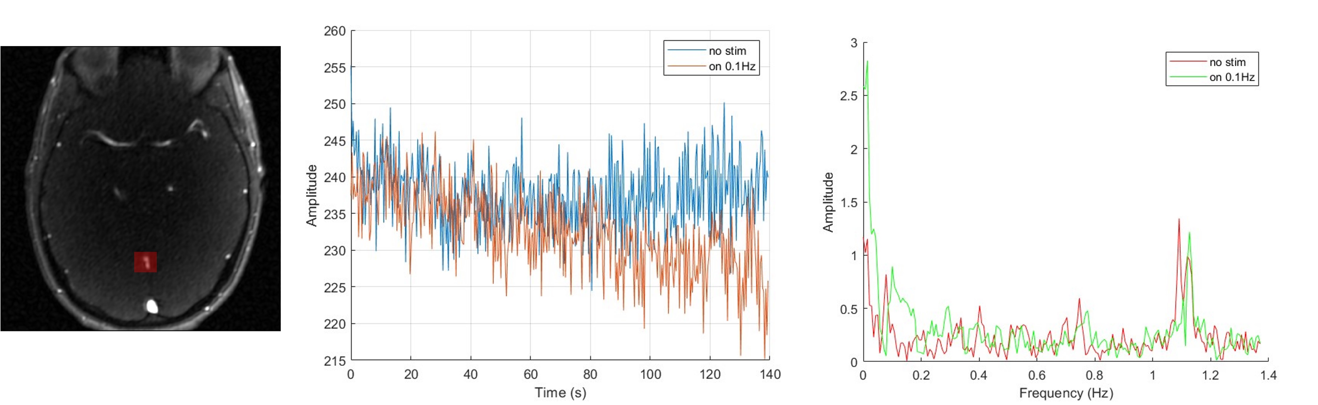
An example of the raw time series is shown in two different ROIs placed in the parenchyma and PCA. When looking at their respective Fourier decomposition, while the parenchymal ROI does not highlight any specific feature, the one from the PCA shows specific peaks reflective of cardiac pulsation at 0.9Hz (corresponding to RR=1111ms, HR=54bpm), but also a peak in the ultra-low frequency range ≈ 0.05-0.1 Hz consistent with vasomotion.When looking at the FD-FLASH with and without visual stimulation, an increase in the power spectrum in the 0.1Hz frequency range, with a shift from 0.08 to 0.1Hz with amplification by 154% of the 0.1Hz signal is observed (Figure 3). Interestingly, we could also observe a different signal time course with a signal drift and shift of the cardiac peak in the frequency spectrum suggesting systemic modification of hemodynamic parameters during visual stimulation.
Conclusions
In this preliminary pilot work, an ultra-low frequency signal suggestive of vasomotion could be detected in vivo in the human brain using a Fourier decomposition of Cine-FLASH T1-weighted MRI, as well as potential sign of vasomotion modulation with functional stimulation. Future work will be geared towards improving acquisition SNR and temporal resolution using for example Non-Cartesian radial trajectories. Additionally, modulation of the visual stimulation frequency will also be explored to assess whether we can detect associated vasomotion modulations.Acknowledgements
No acknowledgement found.References
1. Mateo, C., Knutsen, P. M., Tsai, P. S., Shih, A. Y. & Kleinfeld, D. Entrainment of Arteriole Vasomotor Fluctuations by Neural Activity Is a Basis of Blood-Oxygenation-Level-Dependent “Resting-State” Connectivity. Neuron 96, 936-948.e3 (2017).
2. van Veluw, S. J. et al. Vasomotion as a Driving Force for Paravascular Clearance in the Awake Mouse Brain. Neuron 105, 549-561.e5 (2020).
3. Ernst, R. R. & Anderson, W. A. Application of Fourier Transform Spectroscopy to Magnetic Resonance. Rev. Sci. Instrum. 37, 93–102 (1966).
4. Bauman G et al. Non‐contrast‐enhanced perfusion and ventilation assessment of the human lung by means of fourier decomposition in proton MRI. Magn. Reson. Med. 62, 656–664 (2009).
5. Lustig, M., Donoho, D. & Pauly, J. M. Sparse MRI: The application of compressed sensing for rapid MR imaging. Magn. Reson. Med. 58, 1182–1195 (2007).
6. Griswold, M. A. et al. Generalized autocalibrating partially parallel acquisitions (GRAPPA). Magn. Reson. Med. 47, 1202–1210 (2002).
7. Stanisz, G. J. et al. T1, T2 relaxation and magnetization transfer in tissue at 3T. Magn. Reson. Med. 54, 507–512 (2005).
8. Jenkinson, M., Beckmann, C. F., Behrens, T. E., Woolrich, M. W. & Smith, S. M. Fsl. Neuroimage 62, 782–90 (2012).
Figures