0983
Non-contrast MRI assessment of CSF drainage into superior sagittal sinus1Department of Radiology, Johns Hopkins School of Medicine, Baltimore, MD, United States, 2Department of Biomedical Engineering, Johns Hopkins University, Baltimore, MD, United States, 3Department of Biomedical Engineering, Johns Hopkins School of Medicine, Baltimore, MD, United States, 4Department of Biomedical Engineering, Zhejiang University, Hangzhou, China, 5Department of Neurology, Johns Hopkins School of Medicine, Baltimore, MD, United States
Synopsis
Keywords: Neurofluids, Neurofluids
Motivation: The absorption of CSF into the superior-sagittal-sinus (SSS) is an important CSF drainage pathway and has been implicated in brain disorders like Alzheimer’s disease and idiopathic-intracranial-hypertension. However, we still lack non-invasive and non-contrast techniques to evaluate this CSF-venous drainage system.
Goal(s): To develop a novel non-contrast MRI technique to assess the CSF drainage into the SSS.
Approach: We propose to magnetically label the CSF spins and use a control-label subtraction to isolate signals from the CSF spins that have been absorbed into SSS.
Results: In all subjects, we observed considerable signals from absorbed CSF spins in frontal SSS. Test-retest experiments demonstrated good reproducibility.
Impact: We have proposed a novel non-contrast MRI technique to assess the drainage of CSF into the superior-sagittal-sinus, which may address an important technical gap in evaluating the circulation of CSF.
INTRODUCTION
The cerebrospinal-fluid (CSF) provides hydromechanical protection to the brain and maintains the brain’s fluid homeostasis1. CSF also plays a critical role in the brain’s waste clearance system2,3. CSF is mainly produced by choroid plexuses in the ventricles and circulates in the subarachnoid-space3. A major drainage pathway of CSF is its absorption through arachnoid villi or granulations into dural venous sinuses, including the superior-sagittal-sinus (SSS)1. Impairment of this CSF-venous drainage pathway has been linked to brain disorders such as Alzheimer’s disease and idiopathic-intracranial-hypertension3,4. However, assessment of the CSF-venous drainage pathway has primarily relied on injection of dye, contrast agent or radiative tracers1,3, and we still lack non-invasive and non-contrast techniques to evaluate this drainage system. In this work, we propose a novel MRI technique, dubbed CSF-Absorption-into-SSS-by-T2-Labeling (CASTL), to assess the CSF drainage into the SSS without contrast agent.METHODS
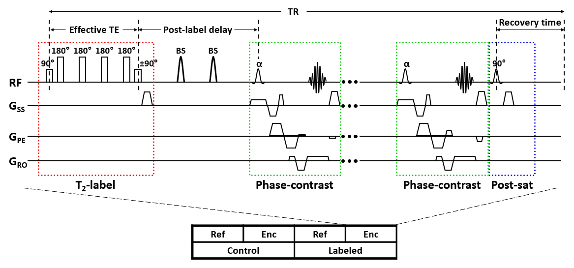
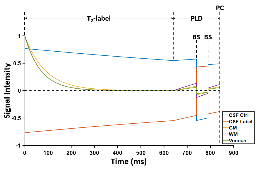
Pulse sequence: Briefly, CASTL MRI magnetically labels the CSF spins and uses a control-label subtraction to isolate signal from the CSF spins that have entered the SSS. Figure 1 illustrates the CASTL pulse sequence. To exclusively label the CSF spins, we use a non-selective T2-label module (red box) with ultra-long effective-TE (640ms). At the end of the T2-label module, for the control scan, the magnetization is flipped to the +z axis by a −90° pulse; while for the label scan, the magnetization is flipped to the -z axis by a +90° pulse. Note that because of the ultra-long effective-TE, only magnetization from the CSF spins (T2≈2000ms5) remains while the magnetization from spins in brain tissues or venous blood (typically T2<150ms6,7) decays to 0. After a post-label-delay (PLD), phase-contrast acquisition (green boxes) is employed to isolate pure blood signal in the SSS, eliminating partial volume contamination from surrounding brain tissues and CSF that has not been absorbed into SSS. To enhance the SNR, two background-suppression pulses were played during the PLD to suppress the background brain tissue and venous blood signal. Figure 2 depicts the simulated signal evolutions.Study 1: Feasibility and reproducibility: Six healthy subjects (3M/3F, age 30±7) were scanned on a 3T Siemens Prisma scanner. CASTL used the following parameters: 2D single slice in the mid-sagittal plane, field-of-view=200×200mm2, slice thickness=10mm, reconstructed in-plane resolution=0.8×0.8mm2, velocity-encoding (VENC)=20cm/s in anterior-posterior direction, recovery-time=6300ms, PLD=200ms, 10 averages, and scan time=5.1min. The CASTL scan was repeated once to evaluate the reproducibility. For quantification purposes, we also acquired an M0 scan with recovery-time=10000ms and scan time=21s.
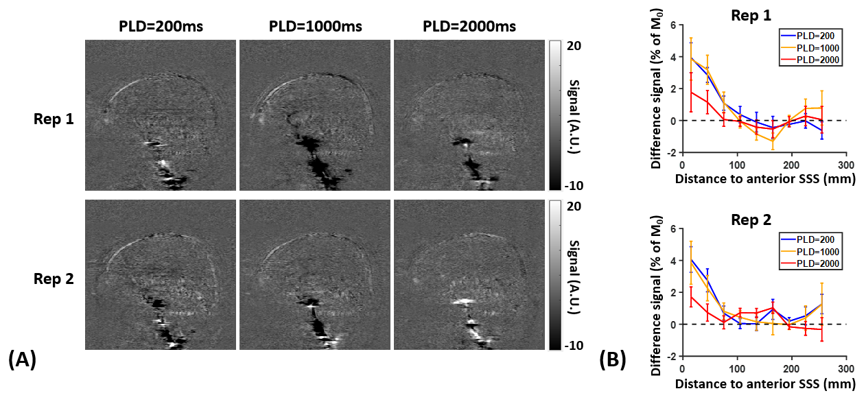
Study 2: Multi-PLD experiment: To examine the dependence of the CASTL signal on the PLD, we acquired three PLDs (200ms, 1000ms and 2000ms) with two repetitions on one subject (female, 25 years old).
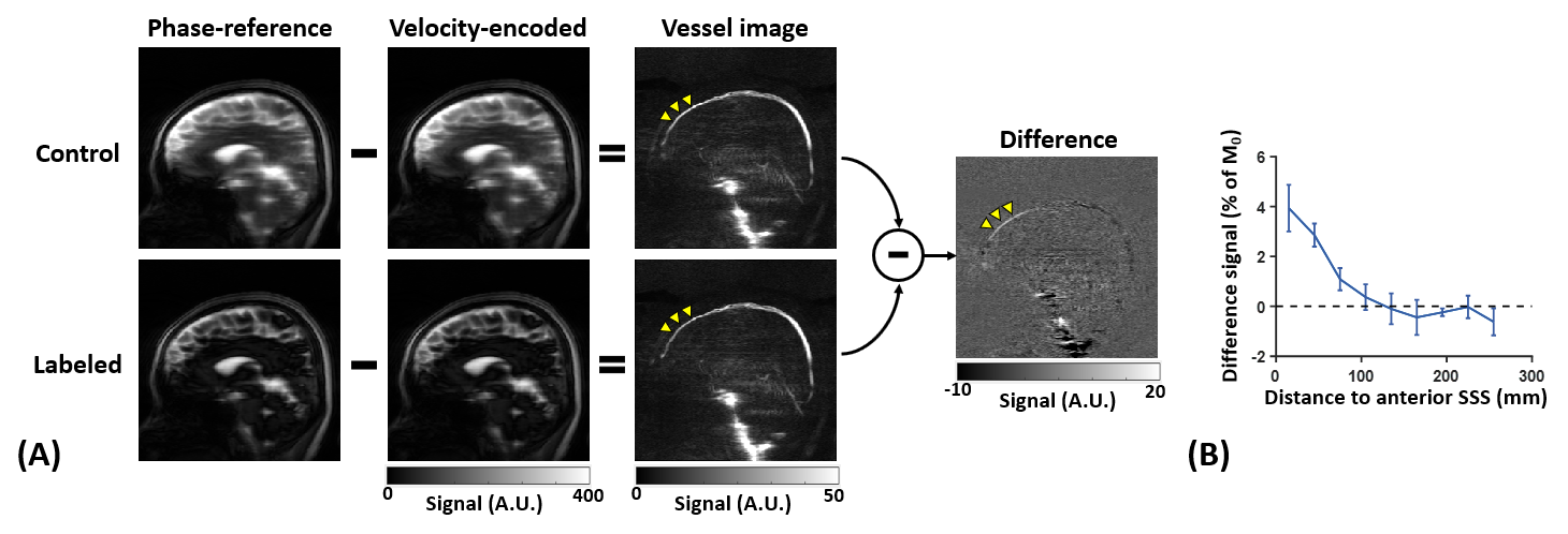
Data analysis: As illustrated in Figure 3, CASTL acquires a phase-reference image and a velocity-encoded image for both control and labeled scans. Complex subtraction between phase-reference and velocity-encoded images yielded vessel images with pure blood signal in the SSS. Further subtraction between the control and labeled vessel images generated the final difference image showing signals of the CSF spins that have entered SSS. To evaluate the spatial pattern of the difference signal, we divided the SSS into 30-mm segments, and calculated the difference signal of each segment as a percentage of the M0 signal of the same segment (Figure 3B).
RESULTS AND DISCUSSION
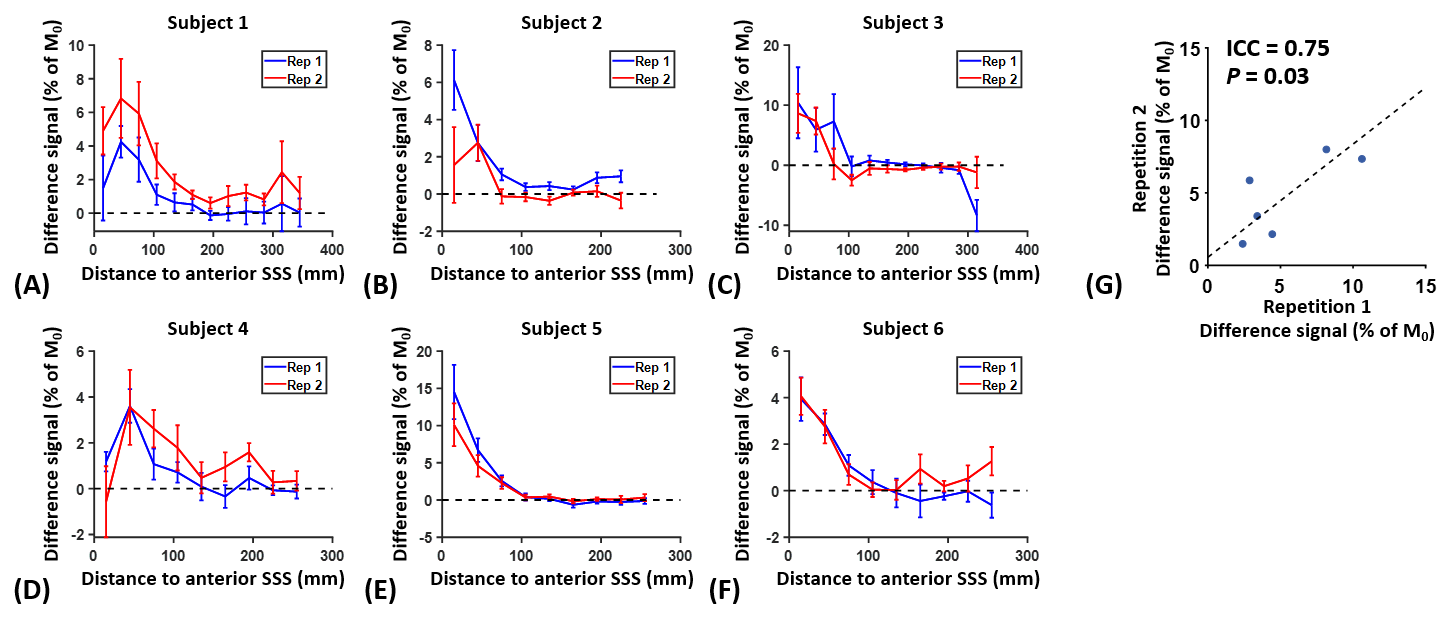
Study 1: As demonstrated by the representative data in Figure 3, we observed considerable CASTL difference signals in the frontal SSS (yellow arrowheads). The difference signal gradually decreased to nearly 0 after 100mm from the anterior end of SSS. Possible cause of this anteriorly dominant pattern may be that subarachnoid CSF is more abundant in the frontal lobe compared to parietal and occipital lobes8, resulting in a larger bolus of labeled CSF spins.Figure 4A-F depicts the CASTL signal profiles in all six subjects, showing a good consistency between the two repetitions. We computed the averaged difference signals in the two anteriormost segments of SSS and found a significant correlation between the two repetitions (intraclass-correlation-coefficient=0.75, P=0.03, Figure 4G), suggesting good reproducibility of CASTL MRI.
Study 2: As illustrated in Figure 5, the CASTL signal at PLD=1000ms was similar to that at PLD=200ms, which may reflect the competing effects of T1-decay and the arrival of more labeled spins from subarachnoid CSF into the SSS. The difference signal at PLD=2000ms exhibited considerable decay compared to the other two PLDs. This interesting temporal pattern was consistent between two repetitions. Further experiments with more PLDs and a larger sample size are needed to elucidate the temporal evolution of the CASTL signal.
CONCLUSION
We proposed a novel non-contrast MRI technique to assess the CSF-venous drainage system.Acknowledgements
No acknowledgement found.References
1. Proulx ST. Cerebrospinal fluid outflow: a review of the historical and contemporary evidence for arachnoid villi, perineural routes, and dural lymphatics. Cell Mol Life Sci 2021;78:2429-2457.2. Iliff JJ, Lee H, Yu M, Feng T, Logan J, Nedergaard M, Benveniste H. Brain-wide pathway for waste clearance captured by contrast-enhanced MRI. J Clin Invest 2013;123:1299-1309.
3. Agarwal N, Lewis LD, Hirschler L et al. Current Understanding of the Anatomy, Physiology, and Magnetic Resonance Imaging of Neurofluids: Update From the 2022 "ISMRM Imaging Neurofluids Study group" Workshop in Rome. J Magn Reson Imaging 2023. doi: 10.1002/jmri.28759.
4. Mondejar V, Patsalides A. The Role of Arachnoid Granulations and the Glymphatic System in the Pathophysiology of Idiopathic Intracranial Hypertension. Curr Neurol Neurosci Rep 2020;20:20.
5. Jiang D, Gou Y, Wei Z, Hou X, Yedavalli V, Lu H. Quantification of T(1) and T(2) of subarachnoid CSF: Implications for water exchange between CSF and brain tissues. Magn Reson Med 2023;90:2411-2419.
6. Lu H, Nagae-Poetscher LM, Golay X, Lin D, Pomper M, van Zijl PC. Routine clinical brain MRI sequences for use at 3.0 Tesla. J Magn Reson Imaging 2005;22:13-22.
7. Lu H, Xu F, Grgac K, Liu P, Qin Q, van Zijl P. Calibration and validation of TRUST MRI for the estimation of cerebral blood oxygenation. Magn Reson Med 2012;67:42-49.
8. Qin Q. A simple approach for three-dimensional mapping of baseline cerebrospinal fluid volume fraction. Magn Reson Med 2011;65:385-391.
Figures