0976
High-resolution diffusion tensor imaging shows cortical microstructure changes in multiple sclerosis across the lifespan1Biomedical Engineering, University of Alberta, Edmonton, AB, Canada, 2Campbell Family Mental Health Research Institute, Toronto, ON, Canada, 3Neurology, University of Alberta, Edmonton, AB, Canada, 4Pediatric Neurology, University of Alberta, Edmonton, AB, Canada, 5Computer Science, Université de Sherbrooke, Sherbrooke, QC, Canada, 6Radiology and Diagnostic Imaging, University of Alberta, Edmonton, AB, Canada
Synopsis
Keywords: Gray Matter, Multiple Sclerosis
Motivation: Patterns of cortical microstructural damage in multiple sclerosis (MS) can be examined in vivo with high-resolution diffusion tensor imaging (DTI).
Goal(s): To assess cortical diffusion changes in MS across the lifespan.
Approach: High-resolution DTI from controls (5-74 years) and MS participants (13-72 years) were segmented using an only-DTI-based method. Thickness, standard DTI metrics and radiality were evaluated in the entire cortex in MS against normative development/aging.
Results: Cortical changes were observed in ~1/3 of MS participants versus controls over the entire lifespan, such as thinning, higher mean (MD), axial (AD) and radial (RD) diffusivities, and lower radiality.
Impact: This study highlights microstructural abnormalities in the cortex of multiple sclerosis (MS) patients throughout the lifespan. These findings will help to understand in vivo cortical pathology in MS that might precede atrophy and that could be linked with disease progression/phenotypes.
Introduction
Cortical pathology is key aspect of multiple sclerosis (MS) at all stages and is related to cognitive disability and disease progression [1,2], but it is underestimated by conventional MRI. Diffusion tensor imaging (DTI), if acquired with sufficient spatial resolution, can characterize the cytoarchitectural properties of cortex in vivo, which may differ in ‘normal-appearing cortex’ and MS lesions. Although not as pronounced as in white matter, water diffusion in the cortex is anisotropic, with the primary diffusion direction typically perpendicular to the cortical surface (i.e., radial direction) [3]. Two high-resolution (1.5 mm isotropic) DTI studies showed elevated mean diffusivity (MD) over the ‘normal-appearing cortex’ in MS in early onset adults [4] and more extensive brain coverage with longer disease duration [5]. A post-mortem study in MS showed a correlation of the primary diffusion direction (radiality) versus histology, which identified microstructural properties such as mini-columnar and axon bundle organization [6]. The purpose here is to identify cortical diffusion changes in a cross-sectional MS cohort (n=57, 13-72 years) compared to a normative population (n=190, 5-74 years) [7] factoring for age.Methods
Whole-brain DTI was acquired on a 3T Siemens Prisma scanner equipped with a 64 channel RF coil using: single-shot EPI, 90 1.5 mm axial/oblique slices, 1.5x1.5 mm2 in-plane 2x zero-filled to 0.75x0.75 mm2, GRAPPA R=2, partial Fourier 6/8, TR 4700 ms, TE 64 ms, 6 b=0 and 30 b=1000 s/mm2, scan time 3.3 minutes. The two cohorts included 190 normative controls (108 females, 5-74 years) and 57 MS participants (49 females, 13-72 years, EDSS 3.5±2.0), 46 relapsing remitting (RRMS), 9 secondary progressive (SPMS), and 2 primary progressive MS (PPMS). DTI processing included denoising, eddy current and motion correction, tensor calculation, and segmentation of the inner, outer, and mid-cortical surfaces based only on the diffusion images/maps (not T1) using an in-house developed package [8]. Cortical thickness was measured between the inner and outer surfaces, and five diffusion metrics were sampled from the mid-surface: fractional anisotropy (FA), MD, axial (AD) and radial (RD) diffusivity, and radiality reflecting the direction of the primary eigenvector relative to the mid-cortex surface (1 perpendicular to 0 parallel) [10]. These metrics over the entire cortex were fit versus age in the normative population using linear, quadratic, cubic, Poisson, and exponential decreasing models that were selected using Akaike Information Criteria. 95% confidence intervals were used for normative prediction bands across age, except for exponential models that used ± one standard deviation. Cortical metrics of the MS population were then compared to these normative ranges.Results
Automated cortex segmentation performed well at various ages in both cohorts (Figure 1). Over the entire cortex of the normative cohort, all diffusion measures showed significant age relationships over 5-74 years that differed from the exponential decrease in cortical thickness (Figure 2). MD, AD, and RD were best fit by quadratic curves with minima ~30-40 years, FA by a Poisson curve with a minimum ~20-35 years (steep FA decrease during childhood/adolescence which subsequent increases), and radiality by a cubic curve decreasing in childhood/adolescence, flat and then decreasing further >60 years.The total cortex metrics were outside the normative range in a substantial portion of the 57 MS participants including 26% with lower cortical thickness, 36% with lower radiality, 32% with elevated MD, 29% with elevated AD, and 31% with elevated RD; in contrast FA was similar to controls. The abnormal cortical metrics covered the full age span from 13 to 72 years. Focusing on radiality, a larger proportion were affected in the progressive MS participants including 100% (2/2) of PPMS and 44% (4/9) of SPMS versus 33% (15/46) of RRMS. Representative cortical maps demonstrated widespread areas affected with a larger MD, AD, and RD, and lower radiality in MS (Figure 3).
Discussion
In addition to confirming previous findings of elevated MD in normal appearing cortex of MS [4,5], the new investigation of radiality highlighted microarchitectural disorganization of the cortex as a whole, particularly in a greater proportion of progressive MS participants. Notably, even 3/6 of the children/adolescents with MS had abnormally low radiality suggesting effects early in the disease. Human histology studies of MS brain implicate that these diffusion changes are the result of alterations of the mini-column microcircuit organization [6]. The regional distribution of the diffusion changes (lobes, lesions) was not examined yet, but may provide insight into the other 2/3 of MS participants that did not differ from age-relevant controls over the entire cortex. High-resolution DTI has revealed extensive cortex changes in MS across the lifespan.Acknowledgements
No acknowledgement found.References
- Calabrese, M., Magliozzi, R., Ciccarelli, O., Geurts, J. J. G., Reynolds, R. & Martin, R. Exploring the origins of grey matter damage in multiple sclerosis. Nat. Rev. Neurosci. 16, 147–158 (2015).
- Mainero, C., Treaba, C. A. & Barbuti, E. Imaging cortical lesions in multiple sclerosis. Curr. Opin. Neurol. 36, 222–228 (2023).
- Heidemann, R. M., Anwander, A., Feiweier, T., Knösche, T. R. & Turner, R. k-space and q-space: Combining ultra-high spatial and angular resolution in diffusion imaging using ZOOPPA at 7T. Neuroimage 60, 967–978 (2012).
- Granberg, T., Fan, Q., et al. In vivo characterization of cortical and white matter neuroaxonal pathology in early multiple sclerosis. Brain 140, 2912–2926 (2017).
- Solana, E., Martinez-Heras, E., et al. Regional grey matter microstructural changes and volume loss according to disease duration in multiple sclerosis patients. Sci. Rep. 11, 1–11 (2021).
- McKavanagh, R., Torso, M., et al. Relating diffusion tensor imaging measurements to microstructural quantities in the cerebral cortex in multiple sclerosis. Hum. Brain Mapp. 40, 4417–4431 (2019).
- Acosta-Franco, J. A., Little, G. & Beaulieu, C. High resolution diffusion tensor imaging shows non-linear trajectories in the human cortex over the healthy lifespan. in Proc. Int. Soc. Magn. Reson. Med. 3357. Toronto, Ontario. (2023).
- Little, G. T. & Beaulieu, C. Automated cerebral cortex segmentation based solely on diffusion tensor imaging for investigating cortical anisotropy. Neuroimage 237, 118105 (2021).
- Van Essen, D. C., Glasser, M. F., Dierker, D. L., Harwell, J. & Coalson, T. Parcellations and hemispheric asymmetries of human cerebral cortex analyzed on surface-based atlases. Cereb. Cortex 22, 2241–2262 (2012).
- McNab, J. A., Polimeni, J. R., et al. Surface based analysis of diffusion orientation for identifying architectonic domains in the in vivo human cortex. Neuroimage 69, 87–100 (2013).
Figures
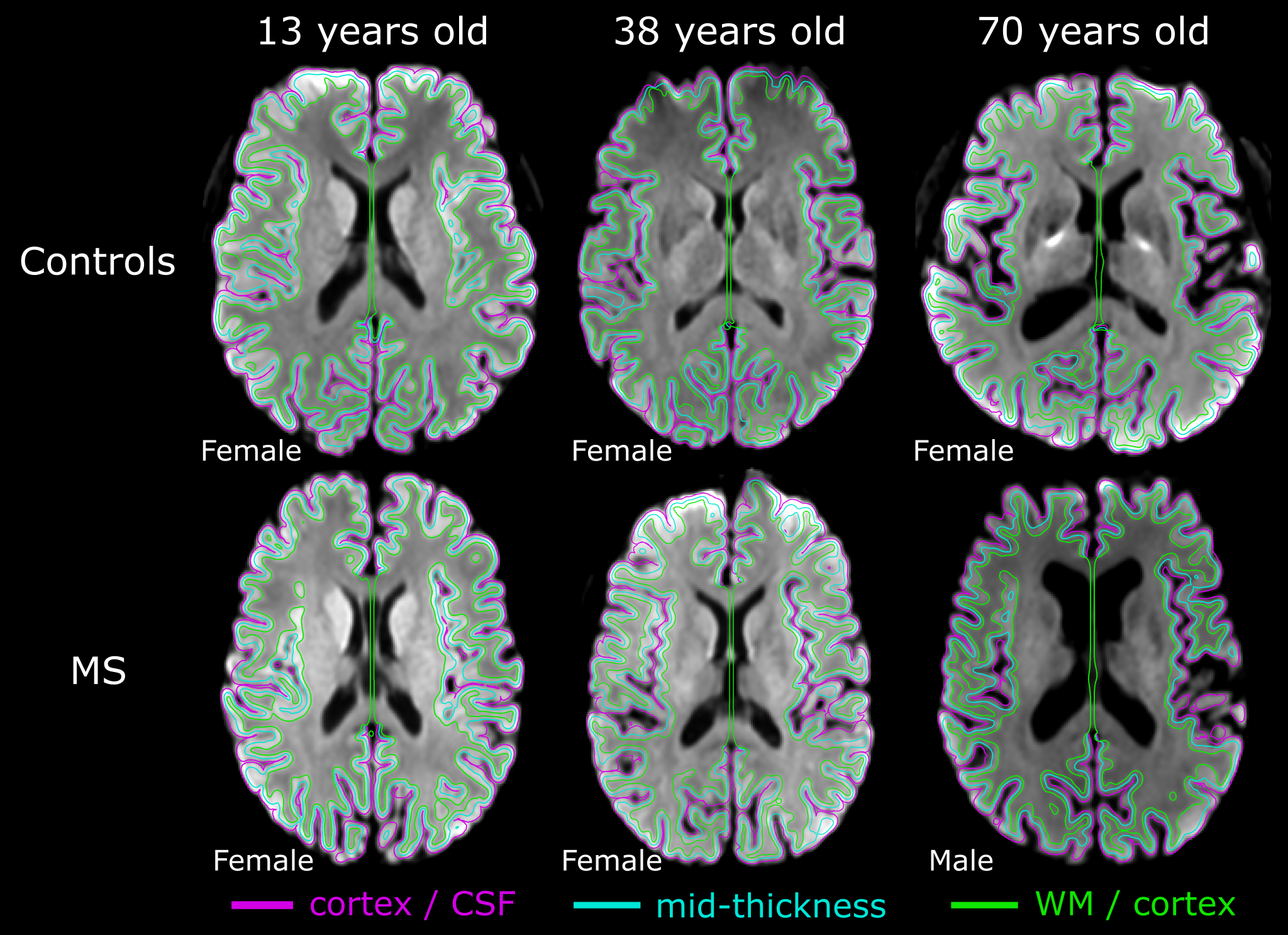
Figure 1. Examples of automated cortex segmentation directly on the 1.5 mm isotropic diffusion images (mean DWI b1000 shown) in 3 sets of age-paired participants from the controls and MS cohorts. Diffusion measurements were extracted based on the mid-thickness surface, and thickness was calculated as the distance between the cortex/CSF and WM/cortex surfaces.
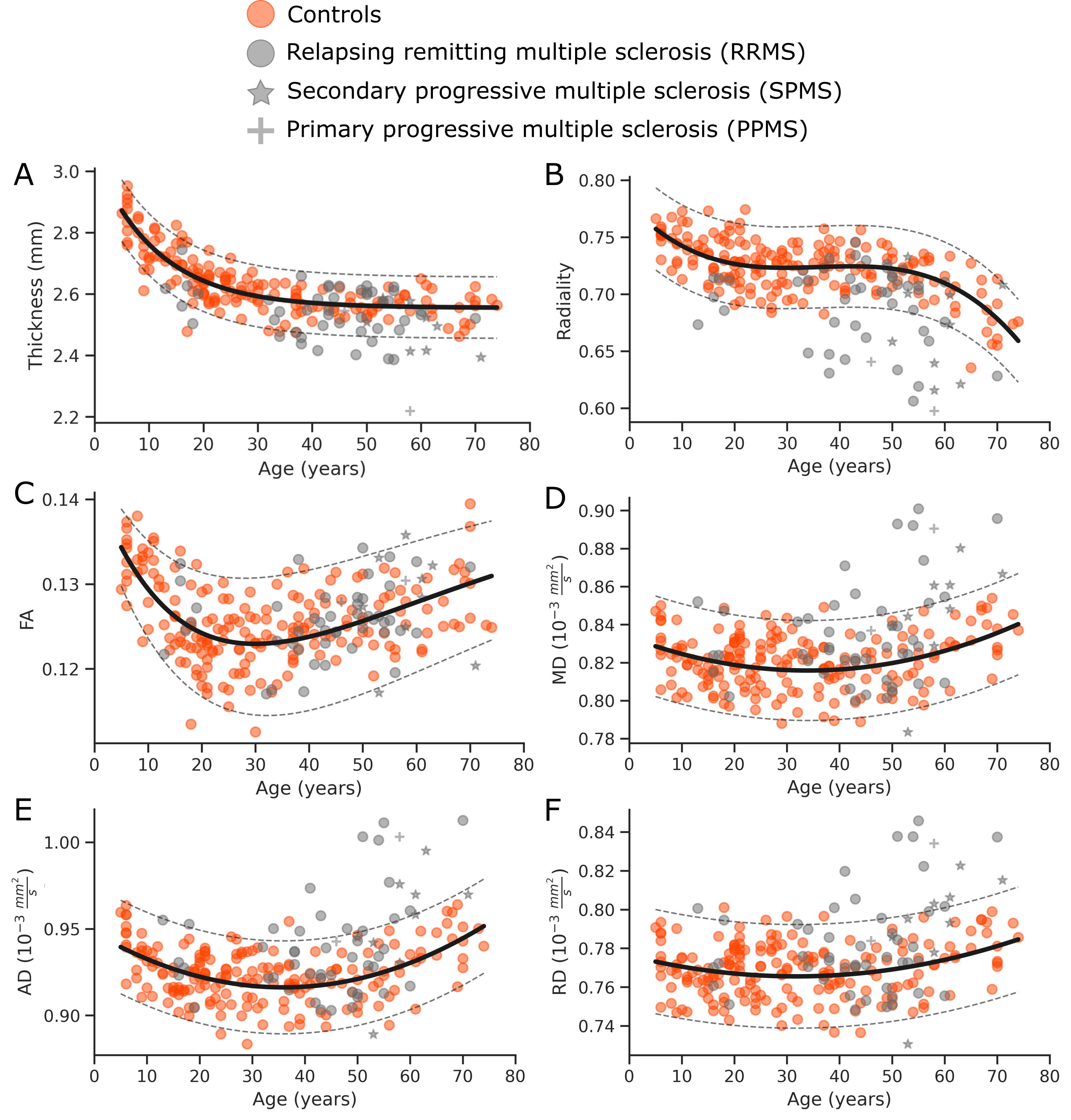
Figure 2. Developmental and aging trajectories of cortical metrics in healthy controls (n=190) (orange dots) and MS (n=57) (gray symbols) across the entire cortex with best fit models and prediction bands. The MS group (~1/3 of them) show higher MD, AD and RD, and lower radiality suggesting extensive cortical alterations. The different DTI-derived metrics have unique age trajectories compared to the cortical thickness exponential trajectory, implicating microstructural changes beyond cortical thinning.
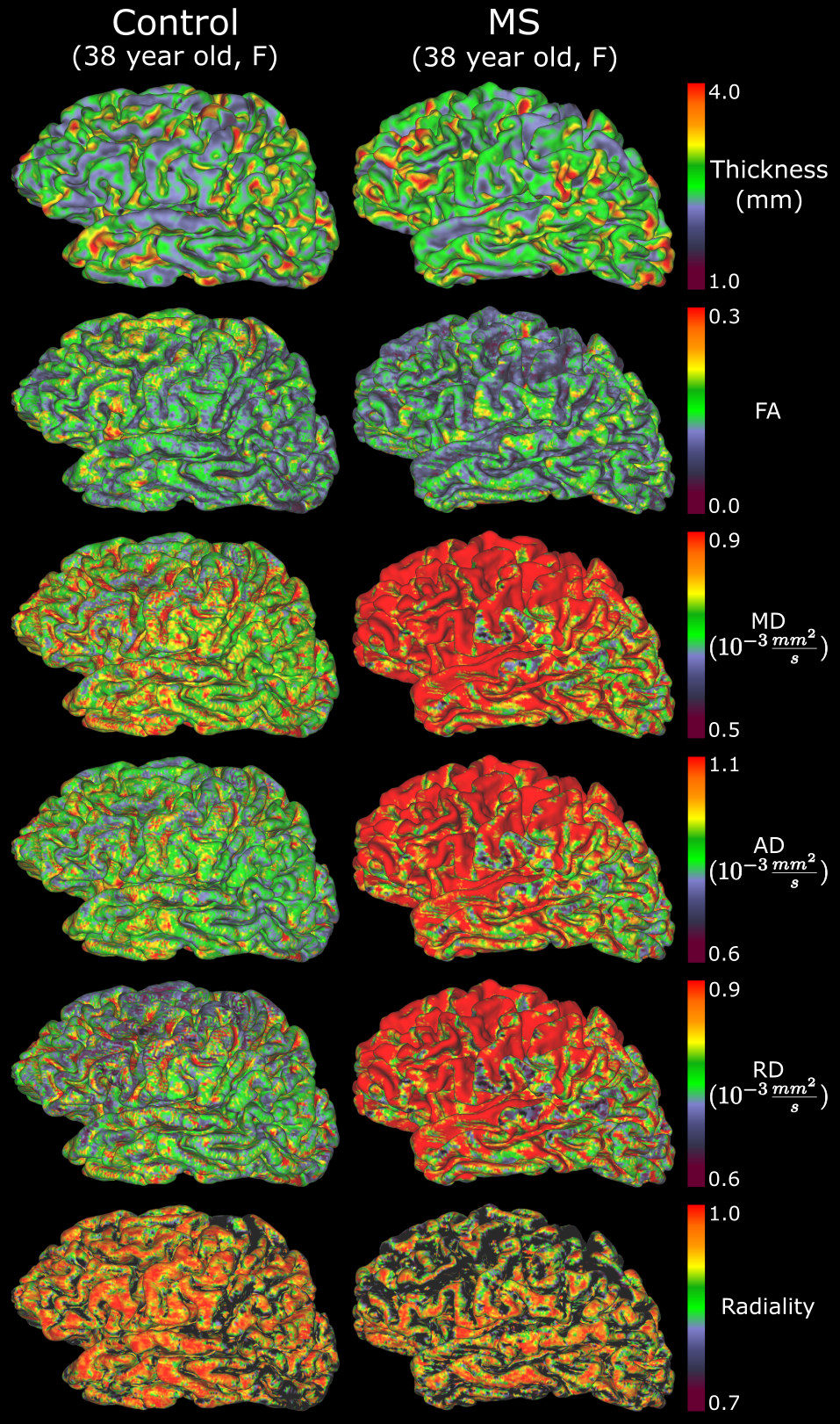
Figure 3. Cortical DTI maps from two 38-year-old female representative participants from both cohorts. The RRMS participant (11 years since onset time of MS, EDSS score 4.5) demonstrated widespread regions of higher MD, AD and RD, as well as lower radiality, primarily in the frontal lobe of the shown left hemisphere. Similar trends were observed in the right hemisphere (not shown).