0972
Brain Region Mapping and Quantification of White Matter Hyperintensity: Estimating the Direct and Indirect impact of WMH Load on Cognition1Department of Biological Sciences, Indian Institute of Science Education and Research Berhampur, Berhampur, India
Synopsis
Keywords: Aging, Ischemia
Motivation: Understanding the magnitude and threshold of PVWMH and DWMH that disrupt cognitive abilities in MCI and AD, compared to healthy aging.
Goal(s): To investigate white matter hyperintensity distribution & its impact on cognitive functions.
Approach: Neuroanatomic segmentation & quantification of Periventricular WMH and DeepWMH, with mediation analysis assessing their impact on cognitive functions
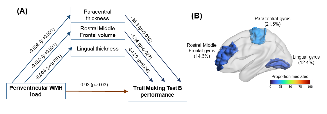
Results: WMH load accrues vascular insult to brain structures, which in-turn mediates impaired cognitive functions, specifically motor and executive functions. WMH load in periventricular region abrogates the information processing and processing speed indirectly mediated through paracentral gyrus thickness, rostral middle frontal volume and lingual gyrus thickness.
Impact: Periventricular white matter hyperintensity progresses faster compared to DeepWMH with Aging. We establish that the Regional Distribution of DeepWMH load is distinct for CN, MCI and AD. High WMH load impairs Executive memory and Motor Memory via specific structural atrophy.
Introduction
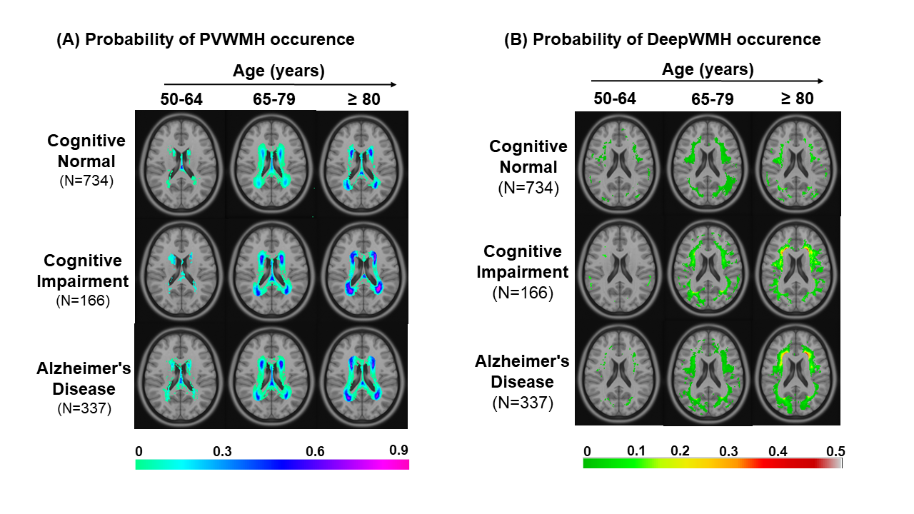
Cerebral Small Vessel Disease (CSVD) is a prevalent aging brain pathology, characterized by small arterial infarcts, causing chronic ischemia and White Matter Hyperintensity(WMH)1 in white matter regions visible on T2-FLAIR MR images. The kinetics of WMH accumulation across Periventricular (PVWMH) and Deep white matter hyperintensity(DWMH) with aging and its impact on cognition remains unexplored. What is the magnitude and threshold of PVWMH and DWMH that perturbs cognitive abilities need to be quantified to unravel the role of PVWMH and DWMH in MCI and AD compared to healthy aging? Further, we have also explored whether WMH load directly mediates its impact on cognition or is it mediated by a set of brain structures?Here, we have quantified total WMH, PVWMH and DWMH in cognitively normal(CN), cognitively impaired(CI), and AD subjects using the National Alzheimer’s Coordinating Center (NACC) cohort to understand the effect of WMH magnitude and kinetics on cognition. To decipher WMH spatial distribution, Probability maps were established to elucidate brain-wide probabilistic insights into the prevalence and localization of WMH, enabling insights into evolving cerebrovascular pathology with age and cognitive status.Methods
T2-FLAIR and T1-weighted(T1w) MRI from subjects with normal cognition(CN,N=734), with cognitive impairment (CI,N=173), and subjects with etiological diagnosis of Alzheimer's disease (CI-AD,N=330) were segmented for quantification of WMH. A cluster-based pipeline based on k-nearest neighbors (k-NN) algorithm, UBO Detector2 was established for segmentation and measurement of PVWMH and DWMH. The quantity of WMH load in various cognitive groups was stratified across three age groups: early(50-64 years), intermediate(65-79 years), and late age group(>79 years). For probability map, Continuous WMH masks were thresholded into binary masks, added to a matrix, followed by normalization by the number of subjects in each group. Mediation analysis was conducted to examine the relationship between WMH load(X) and neuropsychological test scores(Y) in 695 CN subjects. Significant neuroanatomic regions(M) in a simple mediation analysis were identified and integrated into a parallel mediation model for the best fit, using 95% confidence intervals and 5000 bootstrap samples.Results
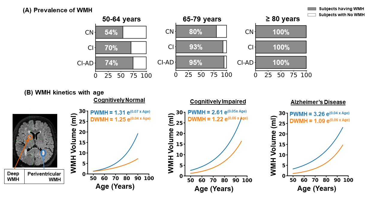
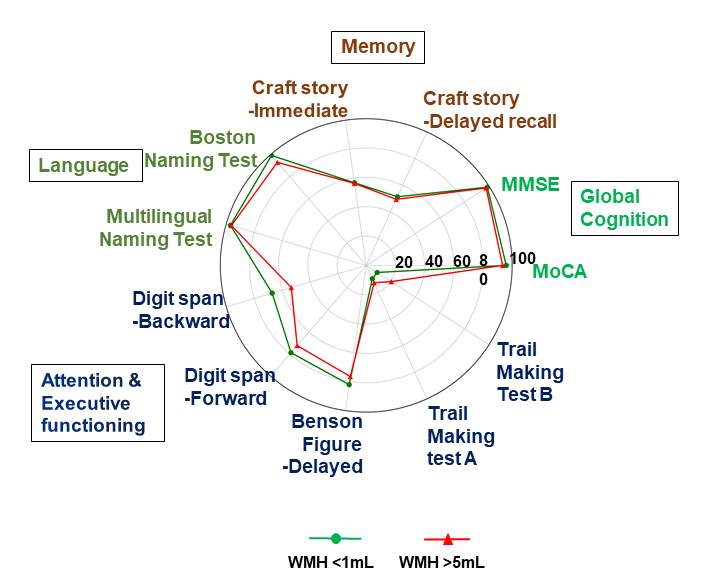
Age-related disparities in WMH prevalence are evident across cognitive states, with a higher WMH load in the CI and CI-AD subjects at all the age groups (Fig 1A). WMH burden increased exponentially with age across cognitive groups, with faster progression rate in CI and CI-AD vs. CN(Fig 1B). The magnitude of periventricular WMH load was distinct across intermediate and late age groups for CN, CI and CI-AD cohort. Deep WMH was significantly elevated in Frontal and parietal lobes compared to temporal and occipital brain regions. Voxel-wise probability mapping in the periventricular region showed higher WMH likelihood in the frontal and occipital horns, increasing with age, especially in cases of CI and AD (Fig 2A). In the deep white matter, WMH lesions initially appear in different areas but become more likely near the periventricular region with age(Fig 2B).Various neuropsychological tests assessed cognitive performance across domains: global cognition, memory, language, attention, and executive function. Test scores were age-normalized. Subjects with low WMH (<1mL) outperformed those with high WMH (>5mL) in attention and executive function tests (Digit Span Test, Benson Complex Figure, and Trail Making Test)(Fig 3). Our parallel mediation model demonstrated a significant direct effect of PVWMH load on TMTB Performance (c'=0.93,p=0.03). Mediation analysis identified three neuroanatomic mediators: Paracentral thickness, Rostral Middle Frontal volume, and Lingual thickness accounting for 48.5% of the total effect(Fig4). Supramarginal atrophy mediated decline in benson complex figure performance due to occipital WMH load.Discussion
WMH magnitude increases exponentially with age, notably accelerated in CI and CI-AD. Periventricular WMH load caused atrophy of structures involved in visual processing (lingual gyrus) and executive functioning (RMFG) alongwith paracentral gyrus, which has roles in motor abilities. Similar results were observed in case of occipital WMH, affecting supramarginal area (involved in perception of space and limbs location) giving insight as WMH may affect fine motor movements to complete the tasks using hand movements, affecting cognitive score.Conclusion
The rate of progression of WMH is significantly faster in cognitively impaired subjects suggestive of increased vascular insults on neuroanatomic health owing to WMH accumulation. WMH load across brain regions have distinct regional patterns among cognitive states. The temporal and spatial accumulation of periventricular and deep white matter hyperintensity with aging are the potential vascular indicators of brain health and cognitive abilities. Decline in cognitive flexibility due to periventricular WMH load is mediated by atrophy of paracentral, RMFG, and lingual gyrus.Acknowledgements
The study was funded by Indian Council of Medical Research (ICMR). The MRI and cognitive data were obtained from National Alzheimer’s Coordinating Centre (NACC) database funded by NIA/NIH Grant U24 AG072122References
Li Q, Yang Y, Reis C, et al., Cerebral Small Vessel Disease. Cell Transplant. 2018 Dec;27(12):1711-1722. doi: 10.1177/0963689718795148. Epub 2018 Sep 25. PMID: 30251566; PMCID: PMC6300773
Jiang J, Liu T, et al., UBO Detector - A cluster-based, fully automated pipeline for extracting white matter hyperintensities. Neuroimage. 2018 Jul 1;174:539-549. doi: 10.1016/j.neuroimage.2018.03.050. Epub 2018 Mar 22. PMID: 29578029.
Figures