0968
Detecting the pathogenic threshold of neuromelanin accumulation in Parkinson’s disease and prodromal Parkinson’s disease patients1Paris Brain Institute – ICM, MOVIT team, Sorbonne Université, Inserm U1127, CNRS 7225, Hôpital Pitié-Salpêtriere, Paris, France, 2Paris Brain Institute – ICM, Data Analysis Core, Sorbonne Université, Inserm U1127, CNRS 7225, Hôpital Pitié-Salpêtriere, Paris, France, 3Paris Brain Institute – ICM, Centre de NeuroImagerie de Recherche – CENIR, Sorbonne Université, Inserm U1127, CNRS 7225, Hôpital Pitié-Salpêtriere, Paris, France, 4Paris Brain Institute – ICM, Centre d'Investigation Clinique (CIC), Sorbonne Université, Inserm U1127, CNRS 7225, Hôpital Pitié-Salpêtriere, Paris, France, 5Neurodegenerative Diseases Research Group, Vall d’Hebron Research Institute (VHIR)-Network Center for Biomedical Research in Neurodegenerative Diseases (CIBERNED), Barcelona, Spain
Synopsis
Keywords: Parkinson's Disease, Parkinson's Disease
Motivation: Neuromelanin is a pigment that accumulates specifically in neurons population that are vulnerable in Parkinson's disease. The role of neuromelanin in pathogenesis is still unclear.
Goal(s): We tested the hypothesis that there is a pathogenic threshold of neuromelanin accumulation that triggers neurodegeneration in Parkinson's disease patients.
Approach: We performed longitudinal neuromelanin-MRI imaging of the substantia nigra of Parkinson's disease, prodromal Parkinson's disease (iRBD) patients, and healthy volunteers.
Results: We confirmed accelerated decrease of neuromelanin-MRI signal in patients with Parkinson's disease, which started from the maximum of healthy volunteer, in line with hypothetic pathogenic threshold. iRBD patients showed similar trajectory delayed by 5 years.
Impact: Results support the hypothesis of a pathogenic threshold of neuromelanin. Its role in Parkinson's disease pathogenesis needs more investigations. Late reach of this threshold in prodromal patients results in delayed age of Parkinson's disease onset, suggesting different progression pattern.
Purpose
Neuromelanin (NM) is a pigment accumulating through human lifespan in the dopaminergic neurons of the substantia nigra (SN) and noradrenergic neurons of the Locus Coeruleus (LC)1. These specific populations of neurons are vulnerable in Parkinson’s disease (PD)2. In a previous work, we demonstrated that NM accumulation in the SN of rats triggered PD phenotype with neurodegeneration, Lewy bodies accumulation, and motor symptoms3. We hypothesized that intracellular NM accumulation over a threshold elicited neurodegeneration. In fact, PD patients showed increased intracellular NM concentration compared to healthy volunteers (HV)3.To further investigate this hypothesis, we present longitudinal NM-MRI on a large cohort of PD patients and patients with isolated rapid eye movement sleep behavior disorder (iRBD), considered as prodromal PD. We hypothesized that NM-MRI would be able to detect a pathogenic threshold of NM concentration and that PD patients would reach that threshold at younger age than HV.
Methods
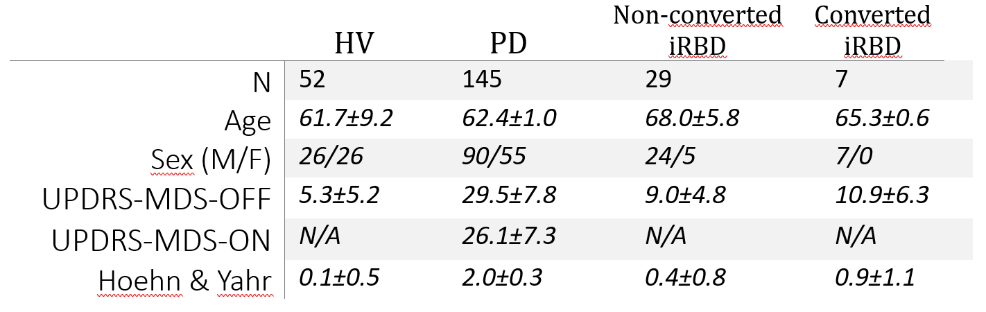
Participants: Participants from the HV, PD and iRBD were included from the ICEBERG study4. Participants were followed longitudinally up to 5 years with 2 to 3 imaging sessions. Table 1 summarized clinical data of the cohort.Imaging: T1-weighted whole brain (3D MP2RAGE, TR/TE=5000/2.98 ms, Resolution = 1x1x1 mm3, acquisition time = 08:12) and NM-sensitive of the SN (2D TSE, TR/TE=890/13 ms, in-plane resolution = 0.4x0.4 mm2, 48 slices, slice thickness = 3 mm, acquisition time=06:55 min) were acquired on a 3T Siemens PRISMA system.
Image analysis: NM-MRI image was registered to the anatomical T1-weighted image of each participant. SN was then manually segmented on NM-MRI image by two trained raters blind to the groups. Normalized Signal Intensity (NSI) was computed as the mean signal in the SN normalized by the mean signal in the background region, which included the tegmentum and superior cerebral peduncle.
Results
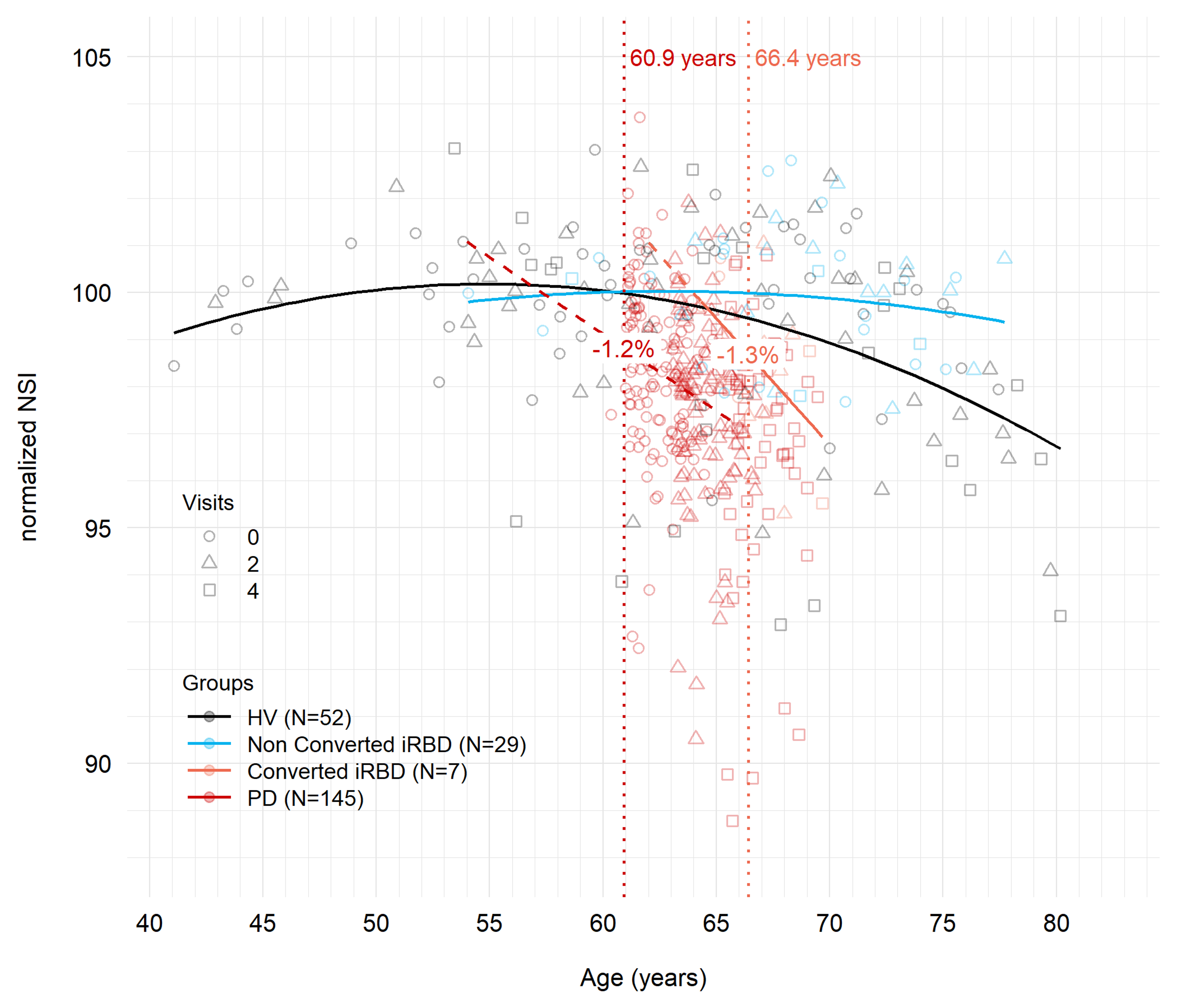
Longitudinal follow-up of HV showed parabolic trajectory of the NM signal reaching a maximum at 56 years of age (Figure 1, black line). PD patients showed reduced NM-MRI SNR in the SN compared with HV and accelerated decrease with time (Figure 1, red line). At the time of onset, 60.9 years old on average, PD patients showed 1.2% NSI reduction compared to HV. HV reached similar NSI 10 years later.iRBD patients who did not convert into PD during the study showed similar parabolic trajectory than HV, with a maximum reached around 63 years of age (Figure 1, blue line). iRBD patients who converted during the study showed accelerated decrease of SNR similar to PD patients (Figure 1, orange line). In this group, the onset of PD was delayed by 5.5 years compared to idiopathic PD patients. At the time of onset, converting iRBD patients showed 1.3% reduction of NSI compared to non-converting iRBD patients.
Discussion
NM-MRI in HV (Figure 1, black line) displayed a parabolic trajectory that was consistent with previous study with a maximum reached between 50 and 60 years5. The increase in NM-MRI signal at younger age can be attributed to the accumulation of intracellular NM and the subsequent decrease to neurodegeneration5. iRBD patients that did not convert into PD during the study (Figure 1, blue line) showed a similar parabolic trajectory of NM-MRI with age, delayed by 9 years compared to HV.PD patients and iRBD patients who converted to PD presented accelerated and early decrease in the NM signal suggesting neuronal loss.
Interestingly, the average NSI in PD and converting iRBD patients crossed the parabolic curves in HV and non-converting iRBD at their maximum, respectively. These results are in line with the hypothesis of PD patients reaching a deleterious threshold of NM concentration and showing vulnerability to NM-induced toxicity. iRBD patients reached this threshold 9 years older than HV, resulting in later onset of PD in converting iRBD patients.
Conclusion
This longitudinal NM-MRI study confirmed the parabolic trajectory of NM-MRI NSI in the SN of HV and compared this trajectory with those in PD and iRBD patients. PD patients showed accelerated decrease in NSI with significant difference with HV at time of onset. These results are consistent with the existence of a pathogenic threshold of NM-MRI in PD. Non-converting iRBD patients presented a trajectory similar to that of HV delayed by 9 years, suggesting that this pathogenic threshold was reached at an older age in iRBD patients. The origin of this difference in age at the pathogenic threshold between PD and iRBD remains to be investigated but could be related to differences in the propagation of the disease process6.Acknowledgements
This project was funded by Agence Nationale de la Recherche (ANR JPND NIPARK). The ICEBERG study was funded by grants from the Investissements d'Avenir, IAIHU-06 (Paris Institute of Neurosciences – IHU), ANR-11-INBS-0006, Fondation d’Entreprise EDF, Biogen Inc., Fondation Thérèse and René Planiol, Fondation Saint Michel, Unrestricted support for Research on Parkinson’s disease from Energipole (M. Mallart), M.Villain and Société Française de Médecine Esthétique (M. Legrand).References
1. Sasaki, M. et al. Neuromelanin magnetic resonance imaging of locus ceruleus and substantia nigra in Parkinson’s disease. NeuroReport 17, 1215–1218 (2006).
2. Braak, H., Ghebremedhin, E., Rüb, U., Bratzke, H. & Del Tredici, K. Stages in the development of Parkinson’s disease-related pathology. Cell Tissue Res. 318, 121–134 (2004).
3. Carballo-Carbajal et al. Brain tyrosinase overexpression implicates age-dependent neuromelanin production in Parkinson’s disease pathogenesis. Nat. Commun. 10, 973 (2019).
4. Biondetti, E. et al. Spatiotemporal changes in substantia nigra neuromelanin content in Parkinson’s disease. Brain J. Neurol. 143, 2757–2770 (2020).
5. Xing, Y., Sapuan, A., Dineen, R. A. & Auer, D. P. Life span pigmentation changes of the substantia nigra detected by neuromelanin-sensitive MRI. Mov. Disord. Off. J. Mov. Disord. Soc. 33, 1792–1799 (2018).
6. Horsager, J. et al. Brain-first versus body-first Parkinson’s disease: a multimodal imaging case-control study. Brain J. Neurol. 143, 3077–3088 (2020).
Figures