0967
Volumetric MR, Blockface Imaging, and Histology Deliver High Fidelity Coregistered MR-Histology1Department of Bioengineering, Stanford University, Stanford, CA, United States, 2Department of Radiology, Stanford University, Stanford, CA, United States, 3Athinoula A. Martinos Center for Biomedical Imaging, Harvard Medical School, Boston, MA, United States, 4Department of Pathology, Stanford University, Stanford, CA, United States, 5Forschungszentrum Jülich, Jülich, Germany, 6Department of Electrical & Computer Engineering, University of Florida, Gainesville, FL, United States
Synopsis
Keywords: Alzheimer's Disease, Neurodegeneration
Motivation: Validating pathological findings from ultra-high-resolution ex-vivo MRI through histology is significant but challenging due to nonlinear 3D deformations between MRI and histological samples.
Goal(s): Addressing the challenge of accurately quantifying complex neurodegenerative diseases by improving the alignment of post-mortem MRI data with histological images.
Approach: We built a novel pipeline integrating advanced imaging techniques with innovative registration algorithms, linking high-resolution MRI with blockface imaging and histological sections.
Results: Our methodology successfully generated blockface volumes with minimal distortion and artifacts, accomplished precise alignment between MRI and blockface volumes, and achieved an accurate 2D correspondence between MRI and histology slides.
Impact: This study introduces an advanced correlative MRI-histology pipeline with robust 2D and 3D coregistration methods, promising to enhance our understanding of neurodegenerative diseases and contribute to the evolution of MRI-based biomarkers for the disease.
Introduction
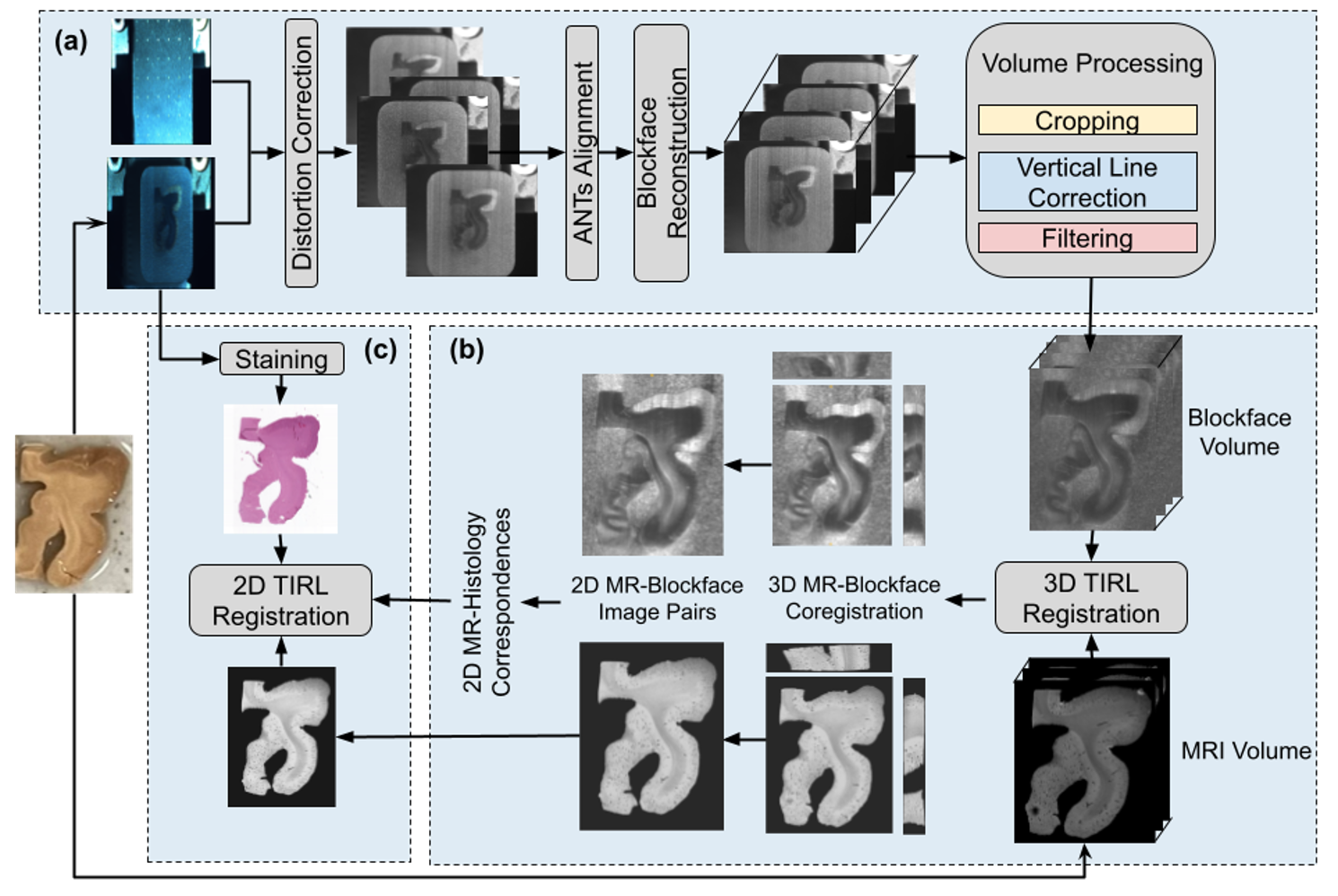
MRI holds potential for the noninvasive detection of micropathology, pivotal for understanding neurodegenerative disorders such as Alzheimer's disease. Defining the relationship between imaging findings and neuropathology is essential for this translation. Thus, a precise registration with multimodal histology is critical. Current methods face significant challenges in aligning MRI with paraffin-embedded histological images1-3, including 3D nonlinear deformation during the paraffin-embedding process and 2D nonlinearities during tissue mounting. We built a novel pipeline combining advanced imaging techniques with robust registration algorithms to tackle this challenge. We begin with high-resolution MRI to capture detailed tissue structure before paraffin embedding, followed by Brewster’s angle telecentric paraffin blockface imaging to provide accurate surface maps for each section. These maps are reconstructed into a 3D volume and serve as a registration intermediate to stained histologic sections. Using the Tensor Image Registration Library (TIRL)4 with Modality Independent Neighbourhood Descriptor (MIND)5, we perform a precise alignment of 3D MRI with the 3D blockface, then register the corresponding 2D MRI slices with the corresponding 2D stained sections.Methods
Image acquisition: A human hippocampal specimen was dissected from a formalin-fixed human brain and divided into hippocampal head and tail sections. We obtained high-resolution multi-echo gradient echo ex-vivo MRI (0.1mm isotropic) of each using a 7.0T Bruker scanner.Blockface imaging: The specimens were embedded in paraffin and placed in a microtome with a home-locked position. An optical image of the tissue block's surface was captured before each section was cut using a PL-D775 5.0MP rolling shutter USB camera and a 0.0128X bi-telecentric lens to reduce barrel distortion. The camera, equipped with a polarized light filter, was positioned at Brewster’s angle (55° from the normal for the paraffin-air boundary), opposite an LED light source also at 55°, to image the polarized light reflected by the blockface for both depth information elimination and contrast enhancement. There is a 1-to-1 correspondence between each blockface image and histology section. Histology sections were stained with H&E.
Blockface reconstruction and processing: We used a calibration grid to correct residual left-right distortion (Fig.1a). Serial 2D blockface images were aligned using ANTs6 to account for vertical shifts during sectioning. The reconstructed 3D volume was cropped and filtered to minimize imaging artifacts.
Blockface and MRI registration: Deformable registration of 3D MRI to the reconstructed 3D blockface volume in Fig.1b is performed using either ANTs or TIRL including Rigid, isotropic scaling, Affine, and Nonlinear transformation.
MRI and Histology registration: Utilizing the 3D MR-Blockface coregistration and the pre-established 2D Histology-Blockface image of the same section, we were able to extract the specific 2D MRI slice that corresponds to our stained histology slide. Subsequently, we executed a 2D deformable registration using either ANTs or TIRL.
Registration quantification: We manually and separately segmented MRI, blockface and histology of the entire hippocampal volume into three distinct categories using ITK-SNAP7: white matter, grey matter, and the dentate gyrus. We employed a Dice similarity coefficient8 between the segmentations after transformation to the same space.
Results
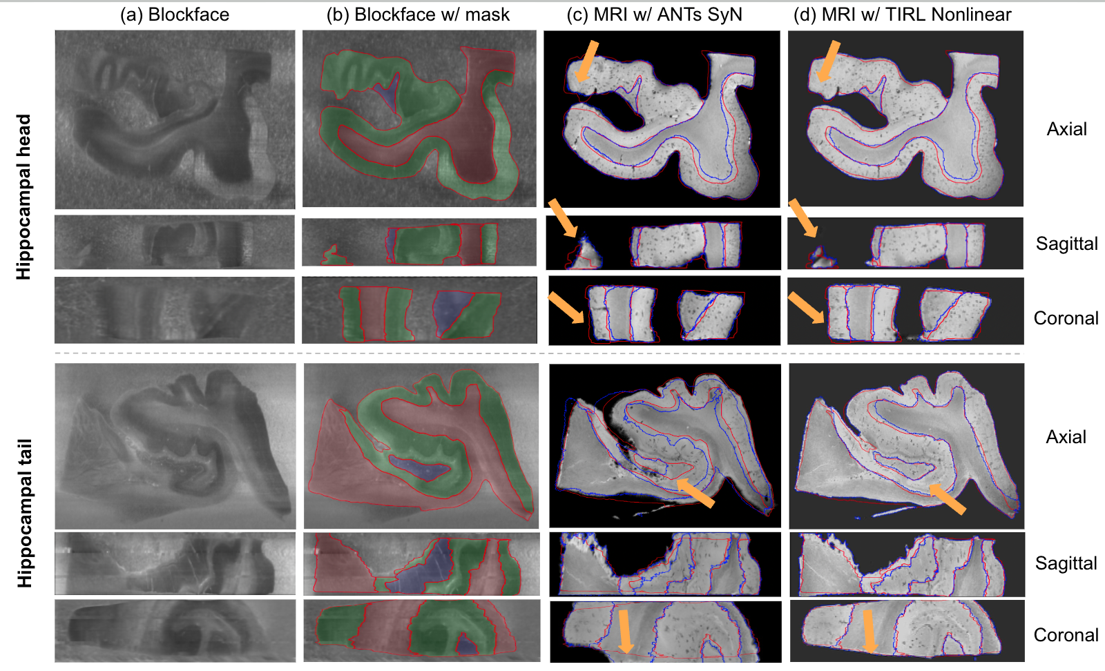
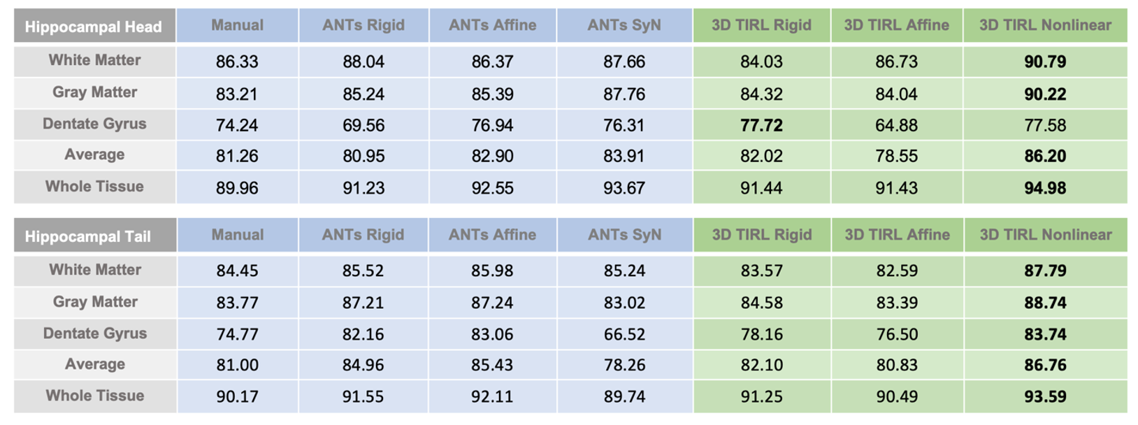
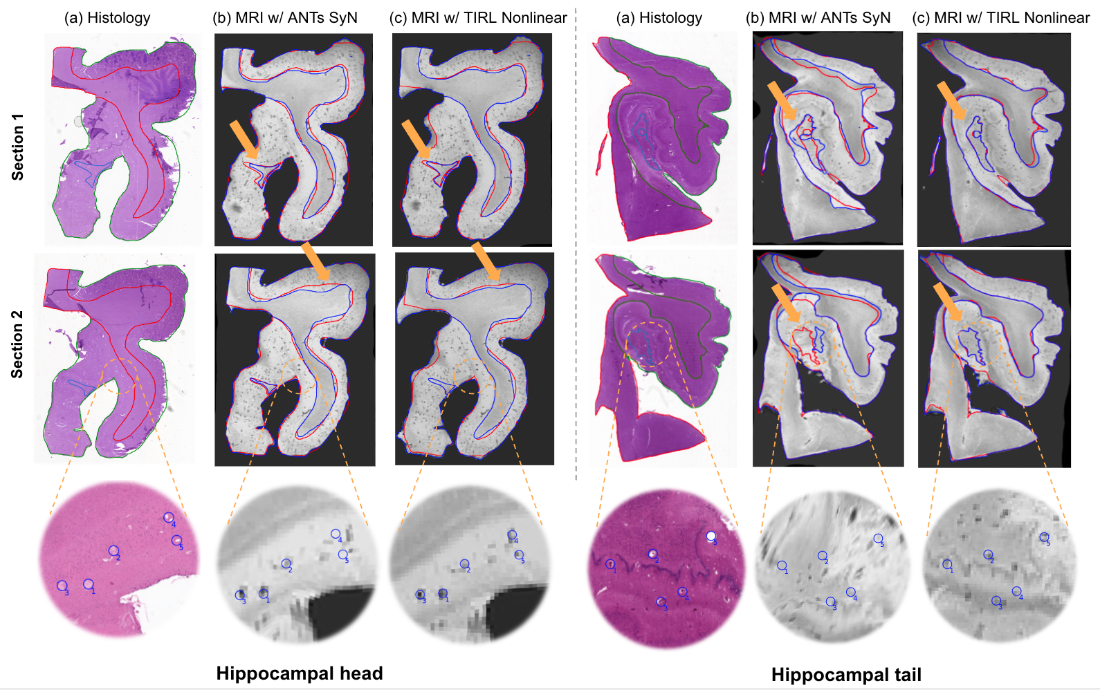
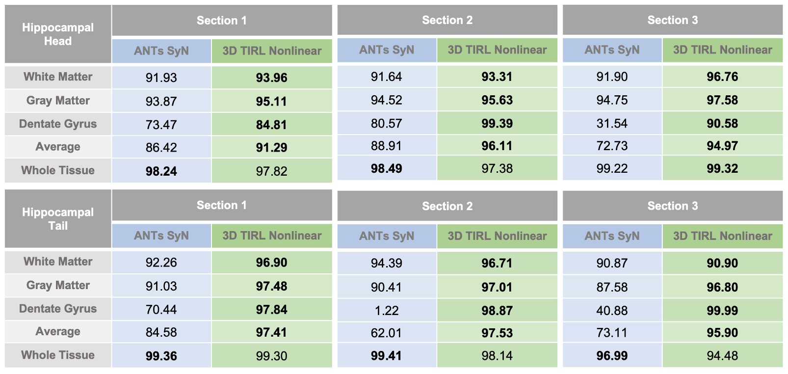
3D MR-Blockface coregistration: The pipeline achieved a close overlap with the MRI segmentation at the 3D blockface level (Fig. 2). The red lines represent the blockface boundaries, while the blue lines represent MRI boundaries after coregistration to the blockface images. In the hippocampal head, both techniques visually performed well, with mildly improved tissue boundaries for TIRL, but the improvement with TIRL was more conspicuous for the hippocampal tail, in particular the dentate gyrus (orange arrows). A comparison of Dice coefficients showed a superior overlap for TIRL, particularly when finely delineated at the subfield level (Table 1).2D MR-Histology coregistration: Our final goal of registering the 2D histology with MRI was achieved to a micron level scale (Fig. 3), with individual features matching between histology sections and the transformed MRI, in particular with the TIRL registration. Table 2 shows high overlap scores across sections. ANTs had higher overlap over the whole section, but TIRL outperformed ANTs at the more detailed level of the subregions and showed less MR section distortion (e.g., hippocampal tail inset).
Discussion and Conclusion
Our integrated pipeline has the potential to provide micron-level registration of MR with neuropathology. The utilization of polarized optics at Brewster’s angle in blockface imaging significantly enhances contrast and reduces obfuscation of the blockface volume by out-of-plane tissue. TIRL’s MIND-based coregistration is synergistic with these high-quality tissue volumes. The robust coregistration with thorough evaluation provides a reliable framework for correlating histological findings with MRI-detected anomalies in Alzheimer’s disease and beyond.Acknowledgements
The present work was supported by the National Institutes of Health (NIH), award numbers R01AG061120-01.References
1. Iglesias, J. E., Modat, M., Peter, L., Stevens, A., Annunziata, R., Vercauteren, T., ... & Alzheimer’s Disease Neuroimaging Initiative. (2018). Joint registration and synthesis using a probabilistic model for alignment of MRI and histological sections. Medical image analysis, 50, 127-144.
2. Pichat, J., Iglesias, E., Nousias, S., Yousry, T., Ourselin, S., & Modat, M. (2017). Part-to- whole registration of histology and mri using shape elements. In Proceedings of the IEEE International Conference on Computer Vision Workshops (pp. 107-115).
3. Casero, R., Siedlecka, U., Jones, E. S., Gruscheski, L., Gibb, M., Schneider, J. E., ... & Grau, V. (2017). Transformation diffusion reconstruction of three-dimensional histology volumes from two-dimensional image stacks. Medical image analysis, 38, 184-204.
4. Huszar, I. N., Pallebage-Gamarallage, M., Bangerter-Christensen, S., Brooks, H., Fitzgibbon, S., Foxley, S., ... & Jenkinson, M. (2023). Tensor image registration library: Deformable registration of stand‐alone histology images to whole‐brain post‐mortem MRI data. NeuroImage, 265, 119792.
5. Heinrich, M. P., Jenkinson, M., Bhushan, M., Matin, T., Gleeson, F. V., Brady, M., & Schnabel, J. A. (2012). MIND: Modality independent neighbourhood descriptor for multi-modal deformable registration. Medical image analysis, 16(7), 1423-1435.
6. Avants, B. B., Tustison, N., & Song, G. (2009). Advanced normalization tools (ANTS). Insight j, 2(365), 1-35.
7. Paul A. Yushkevich, Joseph Piven, Heather Cody Hazlett, Rachel Gimpel Smith, Sean Ho, James C. Gee, and Guido Gerig. User-guided 3D active contour segmentation of anatomical structures: Significantly improved efficiency and reliability. Neuroimage 2006 Jul 1;31(3):1116-28.
8. Dice LR. Measures of the amount of ecologic association between species. Ecology. 1945; 26:297–302.
Figures