0959
Multi-slab whole-brain in vivo 0.35 mm human brain at 7 T with low undersampling to validate future acceleration & denoising1Department of Cognitive Neuroscience, Maastricht Univesity, Faculty of Psychology and Neuroscience, Maastricht, Netherlands, 2Brain Innovation, Maastricht, Netherlands, 3Department of Radiology, Center for Magnetic Resonance Research, University of Minnesota, Minneapolis, Netherlands, 4Scannexus, Maastricht, Netherlands, 5School of Electrical Engineering and Computer Science, The University of Queensland, Brisbane, Australia
Synopsis
Keywords: Data Acquisition, Brain, Mesoscopic
Motivation: Our previous work provided 0.35×0.35×0.35 mm3 voxel resolution T2* dataset where the intracortical angioarchitecture details were captured and analyzed (Gulban et al. 2022). However, this work only covered a third of the brain while requiring two scanning sessions.
Goal(s): Our aim here is to explore reducing the scanning time while expanding the brain coverage to get similar quality data for vascular analyses.
Approach: Our approach consisted of exploring further acceleration for T2* imaging and boosting the SNR of T1 images though denoising instead of multi-run averaging.
Results: Our results suggest that we can reduce the scanning time five-fold while accomplishing whole brain overage.
Impact: We provide a low undersampling 0.35 mm in vivo human brain dataset and a scanning protocol (including 7 T T2*, T1 contrasts) for cortical angioarchitecture studies while delivering a reference dataset to test further acceleration and denoising.
Introduction
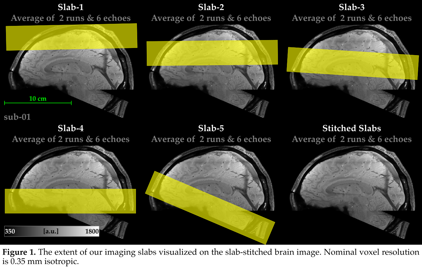
In vivo brain imaging at mesoscopic scale (0.1 to 0.5 mm) reveals the intricate details of the brain angioarchitecture [1]. However, there are three major constraints to advance the study of human angioarchitecture in vivo: (I) imaging the mesoscopic intracortical vessels in practicable scanning durations, (II) accurate and precise gray matter segmentation, (III) analysis methods to localize and quantify the vascular details. While some work outlined the path [references within 2, 3]; recently we progressed in all three aspects by providing a 0.35 mm T2* dataset where the intracortical angioarchitecture details are visible, developing and providing the analyses to start quantifying the mesoscopic intracortical vessels [3]. However, this work only covered a third of the brain while the total scanning lasted 3 hours/subject. Here, we provide two major advancements to increase the brain coverage while decreasing the overall duration:- We concatenate partial brain slabs to achieve whole-brain coverage while keeping each scan time around 10 minutes. We “slab-stitch” in the post-processing to have whole-brain images.
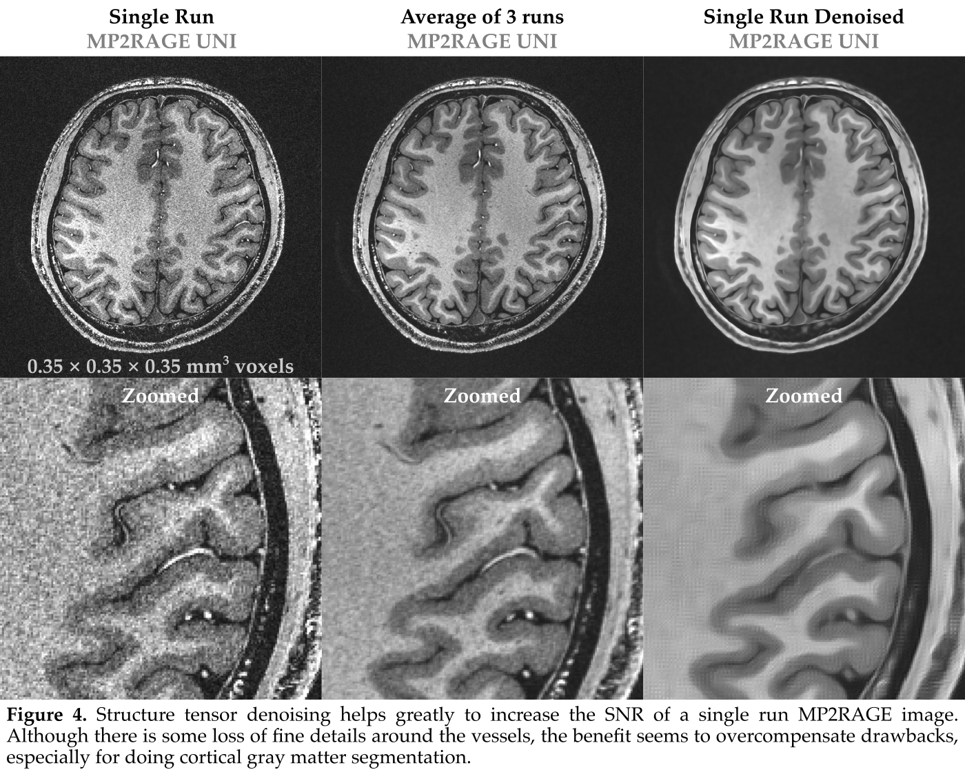
- We apply structure tensor denoising [4] to a single run of 0.35 mm MP2RAGE to achieve SNR similar to averaging 6 runs (used in [6]).
Methods
Data was acquired as described in [3] using ME GRE protocol [5] at 7 T with pTx [6]. Briefly, 6 bipolar echoes at 0.35 mm isotropic resolution were obtained using a low undersampling factor. For the first participant, in 5 sessions totalling 7.5 hours, we piloted:- Different slab positioning and angulations for our partial brain coverage slabs (Figure 1).
- Number of averages required for high SNR T2*-weighted contrast in our ME GRE images.
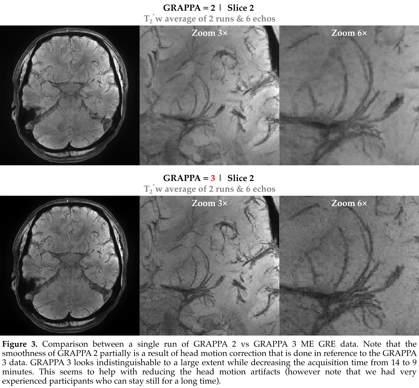
- The effect of GRAPPA 3 compared to 2 in ME GRE images.
- The effect of structure tensor denoising compared to multi-run MP2RAGE averaging.
- Instead of 4 ME GRE runs (used in [6]), 2 runs were deemed sufficient for depicting the angioarchitecture.
- Instead of GRAPPA 2, GRAPPA 3 was deemed satisfactory also for angioarchitecture visualization.
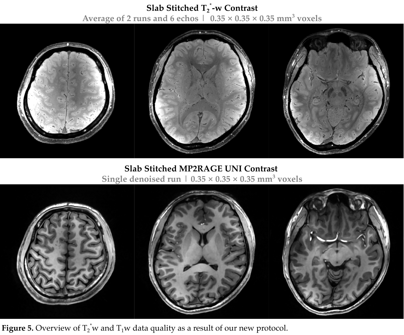
- Instead of 6 MP2RAGE runs (used in [6]), 1 run with structure tensor denoising was deemed sufficient for cortical gray matter segmentation.
The partial brain coverage slabs were stitched by zero filling a central slab and then performing overlap-mask-based affine registration (using ITKSNAP [7]). This step is semi automatic, requiring careful quality control by the user (takes an hour for stitching 5 slabs). Motion correction and averaging across echoes speeds up the process.
Slab-stitching of MP2RAGE images was done using the same method after the UNI contrast non brain tissue noise is suppressed [8] and structure-tensor-denoised (using `segmentator_filters` program implemented within Segmentator [9]).
Results
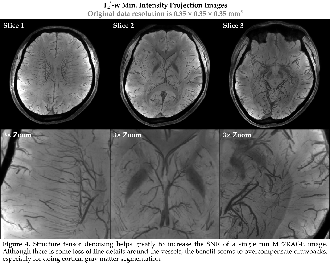
- Figure 2 depicts the intricate vascular details easily visible in our T2* weighted images.
- Figure 3 demonstrates that GRAPPA 3 data is to a very large extent indistinguishable from GRAPPA 2 data.
- Figure 4 illustrates that structure tensor denoising helps greatly to increase the SNR of a single run MP2RAGE image. Although there is some loss of fine details around the vessels, the benefit seems to substantially overcompensate the drawbacks, especially for doing cortical gray matter segmentation.
- Figure 5 shows the image quality after post-processing and slab-stitching.
Discussion & Conclusion
The results demonstrate excellent data quality despite reducing the total scanning time required five-fold. However, the in-plane undersampling factor of 3 without partial Fourier acquisition is still rather conservative since our additional aim is to provide a reference T2* dataset for future studies employing higher acceleration approaches (e.g. transformed domain NORDIC [10, 11]).Acknowledgements
OFG and RG have financial interests tied to Brain Innovation company. DHYT is employed by Scannexus.References
[1] Duvernoy, H.M., Delon, S., Vannson, J.L., 1981. Cortical blood vessels of the human brain. Brain research bulletin 7, 519–79. https://doi.org/10.1016/0361-9230(81)90007-1
[2] Bollmann, S., Mattern, H., Bernier, M., Robinson, S.D., Park, D., Speck, O., Polimeni, J.R., 2022. Imaging of the pial arterial vasculature of the human brain in vivo using high-resolution 7T time-of-flight angiography. eLife 11, 2021.06.09.447807. https://doi.org/10.7554/eLife.71186
[3] Gulban, O.F., Bollmann, S., Huber, L. (Renzo), Wagstyl, K., Goebel, R., Poser, B.A., Kay, K., Ivanov, D., 2022. Mesoscopic in vivo human T2* dataset acquired using quantitative MRI at 7 Tesla. NeuroImage 264, 119733. https://doi.org/10.1016/j.neuroimage.2022.119733
[4] Weickert, J., 1998. Anisotropic diffusion in image processing. Teubner Stuttgart.
[5] Eckstein, K., Dymerska, B., Bachrata, B., Bogner, W., Poljanc, K., Trattnig, S., Robinson, S.D., 2018. Computationally Efficient Combination of Multi-channel Phase Data From Multi-echo Acquisitions (ASPIRE). Magnetic resonance in medicine 79, 2996–3006. https://doi.org/10.1002/mrm.26963
[6] Tse, D.H.Y., Wiggins, C.J., Ivanov, D., Brenner, D., Hoffmann, J., Mirkes, C., Shajan, G., Scheffler, K., Uludağ, K., Poser, B.A., 2016. Volumetric imaging with homogenised excitation and static field at 9.4 T. Magnetic Resonance Materials in Physics, Biology and Medicine 29, 333–345. https://doi.org/10.1007/s10334-016-0543-6
[7] Yushkevich, P.A., Piven, J., Hazlett, H.C., Smith, R.G., Ho, S., Gee, J.C., Gerig, G., 2006. User-guided 3D active contour segmentation of anatomical structures: significantly improved efficiency and reliability. NeuroImage 31, 1116–28. https://doi.org/10.1016/j.neuroimage.2006.01.015
[8] Gulban, O.F., Schneider, M., Marquardt, I., Haast, R.A.M., De Martino, F., 2018. A scalable method to improve gray matter segmentation at ultra high field MRI. PloS one 13, e0198335. https://doi.org/10.1371/journal.pone.0198335
[9] O’Brien, K.R., Kober, T., Hagmann, P., Maeder, P., Marques, J., Lazeyras, F., Krueger, G., Roche, A., 2014. Robust T1-weighted structural brain imaging and morphometry at 7T using MP2RAGE. PLoS ONE 9. https://doi.org/10.1371/journal.pone.0099676
[10] Vizioli, L., Moeller, S., Dowdle, L., Akçakaya, M., De Martino, F., Yacoub, E., Uğurbil, K., 2021. Lowering the thermal noise barrier in functional brain mapping with magnetic resonance imaging. Nat Commun 12, 5181. https://doi.org/10.1038/s41467-021-25431-8
[11] Moeller, S. et al. Locally low-rank denoising in transform domains. in (2023).
Figures