0954
Fast volumetric Cartesian MRI with auto-calibrated local B0 coil array – a reproducing kernel Hilbert space perspective1High-Field MR center, Max Planck Institute for Biological Cybernetics, Tuebingen, Germany, 2Institute of Biomedical Imaging, Graz University of Technology, Graz, Austria, 3Department for Biomedical Magnetic Resonance, University of Tuebingen, Tuebingen, Germany
Synopsis
Keywords: Image Reconstruction, Image Reconstruction, nonlinear gradient
Motivation: The local B0 coil array has been shown to speed up 2D Cartesian MRI and provides a platform for investigating the most efficient B0 encoding fields. Nevertheless, optimizing the rapid modulations for accelerating volumetric scans without introducing additional artifacts becomes more challenging.
Goal(s): We explore distinct nonlinear modulation B0 fields and reconstruct artifact-free accelerated images.
Approach: With a recent RKHS framework, the k-space efficiency maps for various modulation fields are analyzed, and a novel auto-calibration reconstruction method is introduced.
Results: Our k-space analysis provides insights validating optimal modulation fields, and the ex-vivo and in-vivo scans demonstrate the robustness of the proposed reconstruction technique.
Impact: We demonstrate the RKHS formalism as a valuable tool for understanding 3D MRI scans encoded with nonlinear modulation fields. Our auto-calibration reconstruction, analogous to GRAPPA in parallel imaging, offers a promising approach for image acceleration with rapid B0 modulation.
Introduction
MRI sequentially samples object signals in Fourier domain1,2, which inherently leads to prolonged scan time, and therefore, has driven long-standing interests in developing image acceleration methods. Nonlinear gradients3–8, in addition to the well-established parallel imaging technique9–11, have demonstrated potential to speed up MRI acquisitions; however, a rigorous analysis comparing the encoding capability of various nonlinear and linear fields requires more in-depth investigations12.Early nonlinear (i.e., B0) gradient methods spatially encode spins using sinusoidal3, quadrupolar4,6, or quadratic5 magnetic fields for phase and frequency encoding. Later, sinusoidal modulation of 2nd/3rd-order gradient coils7,13 was applied during signal readout to accelerate scans, similar to bunched phase encoding14 or wave-CAIPI15, but with nonlinear modulation fields. To further investigate optimal field shapes for image acceleration, we developed a local B0 coil array to provide more flexible spatial-temporal modulation patterns to enhance the speed of MRI8,12.
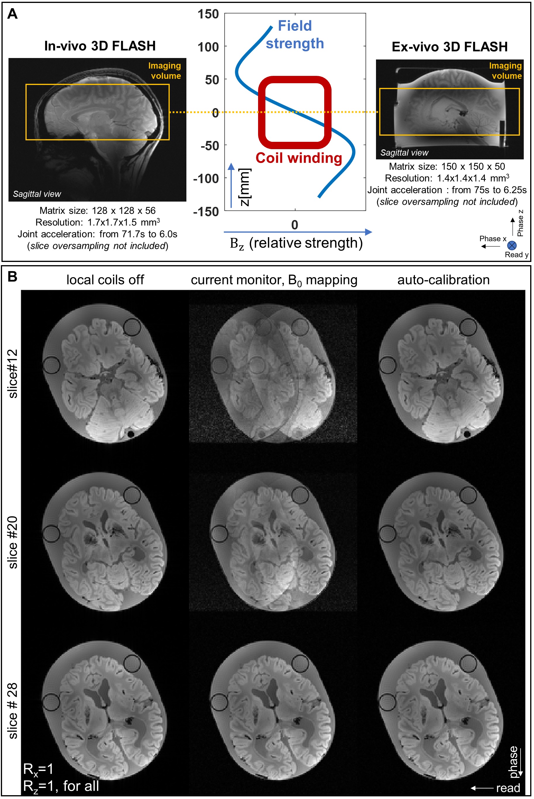
For accelerated 2D Cartesian scans12, the optimal modulation field by our 8-channel local coil setup converges to a linear gradient regardless of whether parallel imaging is combined or not, with the k-space sampling efficiency visualized in a novel reproducing kernel Hilbert Space (RKHS) framework16,12. This time, we apply the same principles12 to accelerate volumetric Cartesian MRI, and investigate the optimal modulation field shapes given a specific coil-patient position and readout axis, as in Figure 1(A).
Additionally, to enhance image reconstruction for 3D accelerated scans, we propose a robust auto-calibration algorithm to address field calibration challenges arising from potentially larger phase errors induced by eddy currents in 3D, as well as stronger motion due to longer acquisition time.
Methods
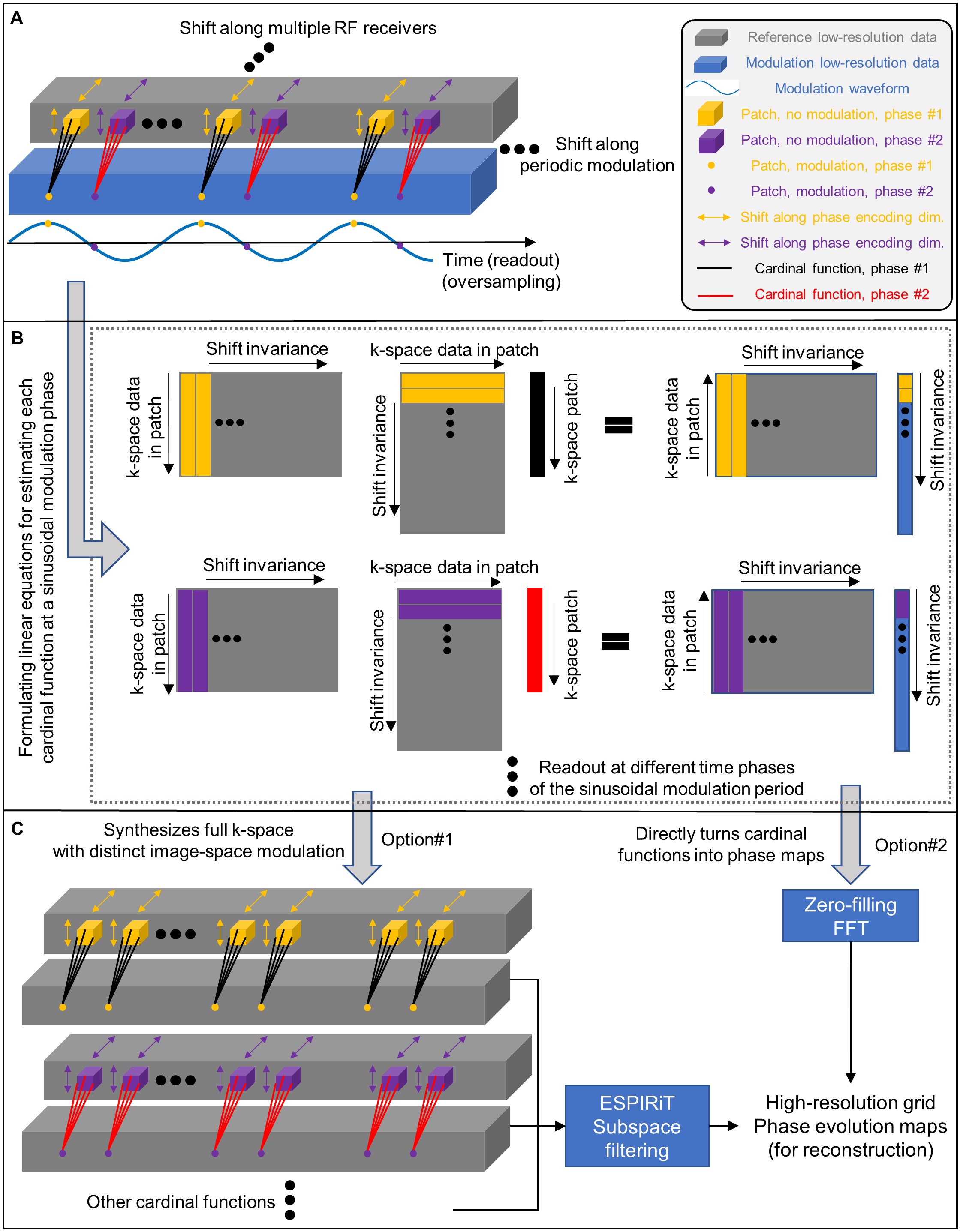
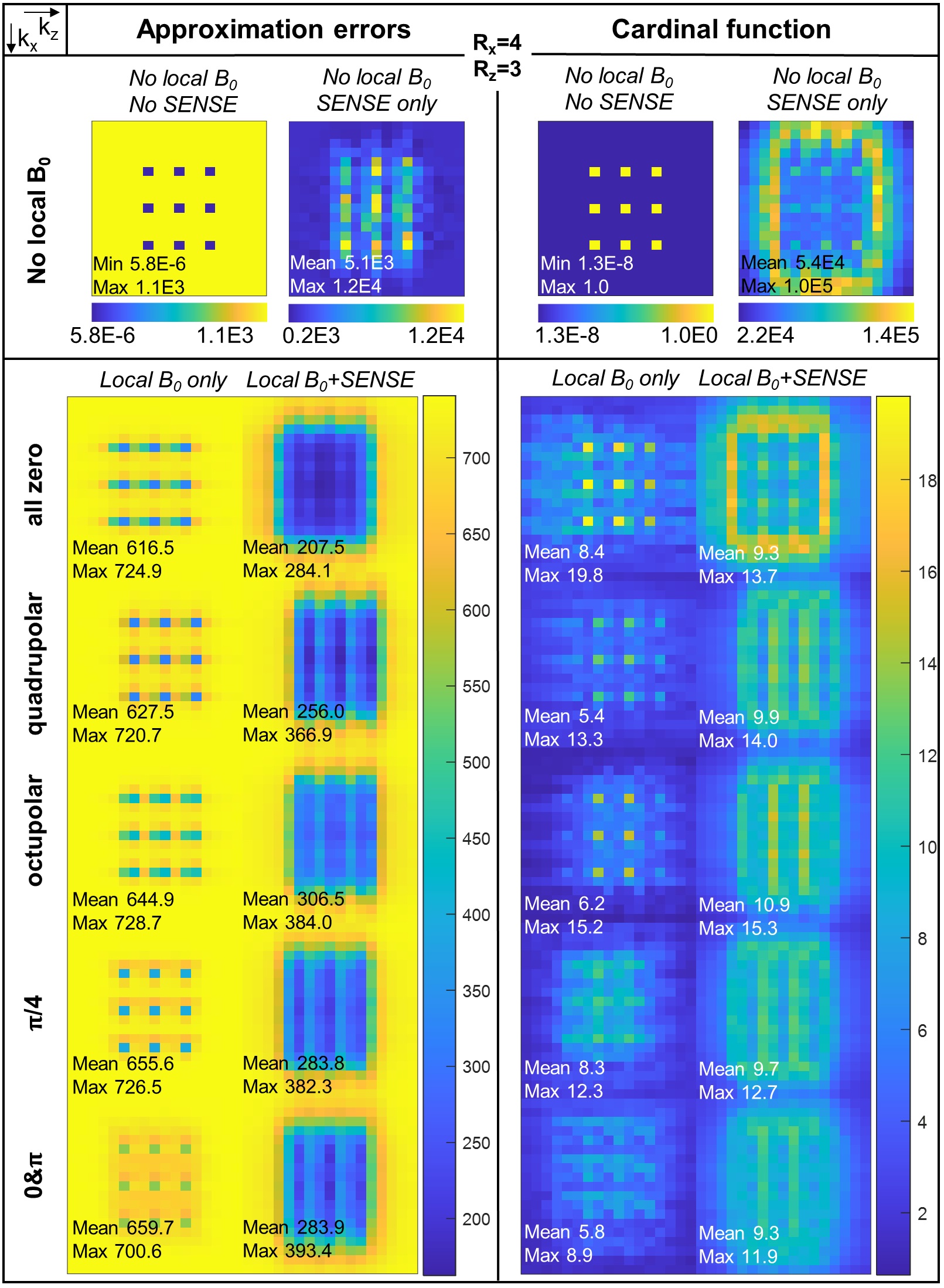
During signal readout of 3D FLASH scans in a Siemens 9T human scanner, sinusoidal currents in 9.26kHz/42Apeak-to-zero (ex. 9.80kHz/37Apeak-to-zero for in-vivo scans) with distinct phase offsets are applied to the 8 local B0 coils to accelerate acquisitions, corresponding to various modulation field shapes. In addition to the G-map, the k-space efficiency maps12 (i.e., cardinal and power functions) for visualizing noise amplifications and approximation errors have been computed and compared for different field patterns.In Figure 2, a novel algorithm is presented for extracting the additional spin phase evolution caused by local B0 modulations without field mapping scans, as a crucial step for the robust reconstruction of 3D accelerated scans. In section A, we formulate linear equations in k-space. Here, a single data point acquired with B0 modulations can be understood as a linear combination of a local region within low-resolution reference data without modulation (i.e., typically obtained for parallel imaging10,11,17), given the interpolation weights as a cardinal function12,16. By setting the sinusoidal modulation period (e.g., 108 μs) as an integer multiple of the scanner ADC dwell time (e.g., 3 μs), the cardinal functions undergo a periodic change in shape along the readout modulation. This results in independent linear equations obtained by shifting the interpolating position among readout time instants for the identical sinusoidal modulation phase, and across dimensions of phase encoding and RF receivers.
Hence, in section B, the local cardinal functions are estimated by solving the linear system. In section C, the cardinal functions can undergo zero-filling and Fourier transform to directly yield the estimated phase evolution maps in image space. Alternatively, they can be exploited to synthesize low-resolution k-space data with distinct image space modulation, which is used to produce phase evolution maps through ESPIRiT17 for additional subspace filtering.
Results
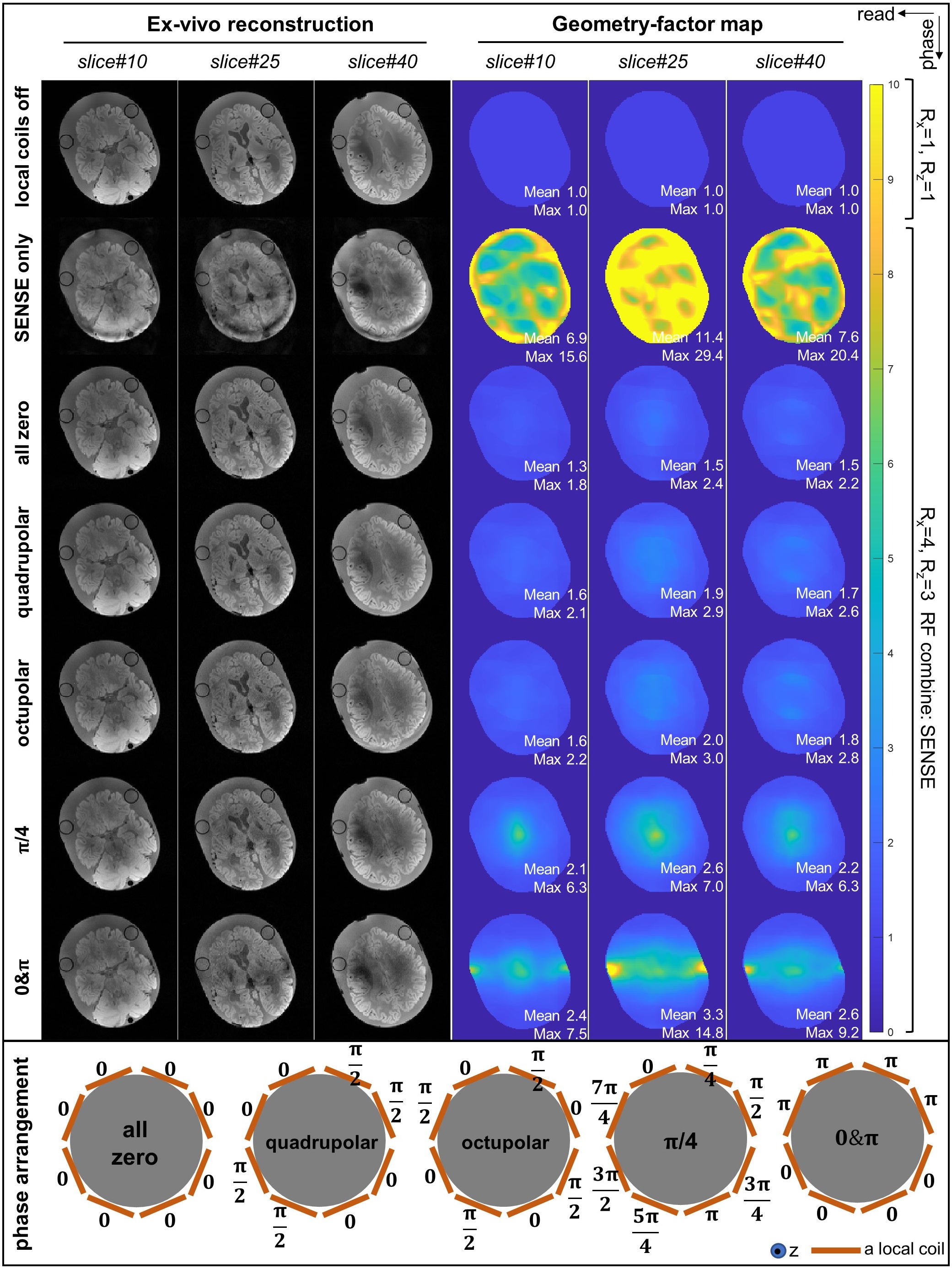
In Figure 1, the forward model reconstruction of ex-vivo phantom18,19 scans using auto-calibrated maps exhibits no noticeable artifacts, whereas the previous approach12 that involves field mapping scans and current monitors results in slice-dependent residue artifacts due to mis-calibration in three-dimension.In Figure 3, ex-vivo scans with distinct phase offsets (i.e., modulation field shapes) are jointly reconstructed with SENSE10,15,20, with a retrospective undersampling factor 4x4. Accelerated scans with phase arrangements of “all zero” and “quadrupolar” show no apparent difference except for very mild SNR loss compared to the reference image, which matches the G-map10 calculation.
In Figure 4, the superior encoding efficiency by the “all zero” configuration can be well explained by the k-space efficiency maps12, since this coil phase arrangement leads to strong modulation along the z dimension, which is usually insufficiently encoded by RF receivers' sensitivity10.
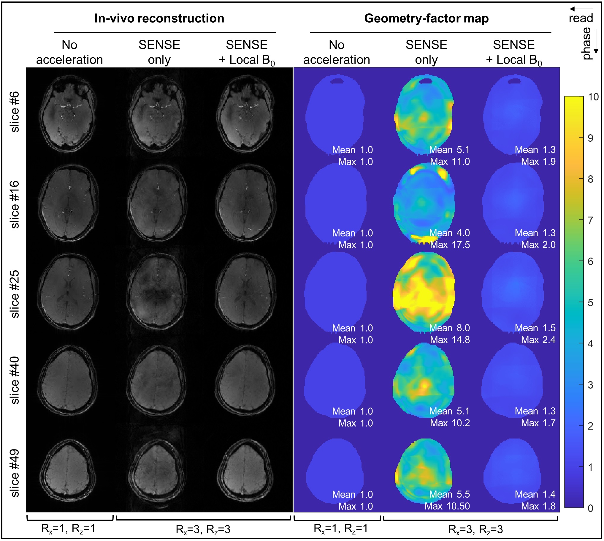
In Figure 5, the in-vivo scans with joint reconstruction of SENSE & B0 modulations show negligible artifacts given a retrospective undersampling factor of 3x3, which substantially outperforms the accelerated scans with SENSE alone.
Discussion/Conclusion
Our proposed robust reconstruction algorithm successfully enables 3D image acceleration by the local B0 coils, and the recently developed RKHS framework12 serves as a valuable tool for guiding the exploration of optimal field modulations.Acknowledgements
This study is supported by ERC Advanced Grant No 834940.
The ex-vivo brain phantom was with courtesy of the Institute of Clinical Anatomy and Cell Analysis, Department of Anatomy, Eberhard Karls University of Tübingen. The first author thanks Dr. Thomas Shiozawa (Institute of Clinical Anatomy and Cell Analysis) for assistance with sample preparation, and Dr. Gisela Hagberg for assistance in scanning this phantom.
The first author would also like to thank Stefan Plappert for guidance in programming the ADwin high-speed processor, and Pavel Povolni & Dr. Georgiy Alekseevich Solomakha for assistance in RF system testing.
References
1. Ljunggren S. A simple graphical representation of fourier-based imaging methods. J Magn Reson 1969. 1983;54(2):338-343. doi:10.1016/0022-2364(83)90060-4
2. Twieg DB. The k-trajectory formulation of the NMR imaging process with applications in analysis and synthesis of imaging methods. Med Phys. 1983;10(5):610-621. doi:10.1118/1.595331
3. Patz S, Hrovat MI, Pulyer YM, Rybicki FJ. Novel encoding technology for ultrafast MRI in a limited spatial region. Int J Imaging Syst Technol. 1999;10(3):216-224. doi:10.1002/(SICI)1098-1098(1999)10:3<216::AID-IMA3>3.0.CO;2-Q
4. Hennig J, Welz AM, Schultz G, et al. Parallel imaging in non-bijective, curvilinear magnetic field gradients: a concept study. Magn Reson Mater Phys Biol Med. 2008;21(1-2):5. doi:10.1007/s10334-008-0105-7
5. Stockmann JP, Ciris PA, Galiana G, Tam L, Constable RT. O-space imaging: Highly efficient parallel imaging using second-order nonlinear fields as encoding gradients with no phase encoding. Magn Reson Med. 2010;64(2):447-456. doi:10.1002/mrm.22425
6. Gallichan D, Cocosco ChrisA, Dewdney A, et al. Simultaneously driven linear and nonlinear spatial encoding fields in MRI. Magn Reson Med. 2011;65(3):702-714. doi:10.1002/mrm.22672
7. Wang H, Tam LK, Constable RT, Galiana G. Fast rotary nonlinear spatial acquisition (FRONSAC) imaging. Magn Reson Med. 2016;75(3):1154-1165. doi:10.1002/mrm.25703
8. Scheffler, K, Loktyushin, A, Bause, J, Aghaeifar, A, Steffen, T, Schölkopf, B. Spread-spectrum magnetic resonance imaging. Magn Reson Med. 2019; 82: 877–885. https://doi.org/10.1002/mrm.27766
9. Sodickson DK, Manning WJ. Simultaneous acquisition of spatial harmonics (SMASH): Fast imaging with radiofrequency coil arrays. Magn Reson Med. 1997;38(4):591-603. doi:10.1002/mrm.1910380414
10. Pruessmann KP, Weiger M, Scheidegger MB, Boesiger P. SENSE: Sensitivity encoding for fast MRI. Magn Reson Med. 1999;42(5):952-962. doi:10.1002/(SICI)1522-2594(199911)42:5<952::AID-MRM16>3.0.CO;2-S
11. Griswold MA, Jakob PM, Heidemann RM, et al. Generalized autocalibrating partially parallel acquisitions (GRAPPA). Magn Reson Med. 2002;47(6):1202-1210. doi:10.1002/mrm.10171
12. Tian R, Uecker M, Davids M, et al. Accelerated 2D Cartesian MRI with an 8-channel local B0 coil array combined with parallel imaging. Magn Reson Med. 2023;1-23. doi:10.1002/mrm.29799
13. Dispenza NL, Littin S, Zaitsev M, Constable RT, Galiana G. Clinical Potential of a New Approach to MRI Acceleration. Sci Rep. 2019;9(1):1912. doi:10.1038/s41598-018-36802-5
14. Moriguchi H, Duerk JL. Bunched phase encoding (BPE): A new fast data acquisition method in MRI. Magn Reson Med. 2006;55(3):633-648. doi:10.1002/mrm.20819
15. Bilgic B, Gagoski BA, Cauley SF, et al. Wave-CAIPI for highly accelerated 3D imaging: Wave-CAIPI for Highly Accelerated 3D Imaging. Magn Reson Med. 2015;73(6):2152-2162. doi:10.1002/mrm.25347
16. Athalye V, Lustig M, Martin Uecker. Parallel magnetic resonance imaging as approximation in a reproducing kernel Hilbert space. Inverse Probl. 2015;31(4):045008. doi:10.1088/0266-5611/31/4/045008
17. Uecker M, Lai P, Murphy MJ, et al. ESPIRiT-an eigenvalue approach to autocalibrating parallel MRI: Where SENSE meets GRAPPA. Magn Reson Med. 2014;71(3):990-1001. doi:10.1002/mrm.24751
18. Hagberg GE, Shiozawa-Bayer T, Mirkes C, et al. MRI of whole brain formalin-fixed samples at 9.4T: influence of the fixation agent and its dielectric properties on image quality. In Proceedings of the joint annual meeting of ISMRM-ESMRMB, Paris, France, June 2018. Abstract #3310.
19. Nazemorroaya A, Aghaeifar A, Shiozawa T, et al. Developing formalin-based fixative agents for post mortem brain MRI at 9.4 T. Magn Reson Med. 2022;87(5):2481-2494. doi:10.1002/mrm.29122
20. Pruessmann KP, Weiger M, Börnert P, Boesiger P. Advances in sensitivity encoding with arbitrary k-space trajectories. Magn Reson Med. 2001;46(4):638-651. doi:10.1002/mrm.1241
Figures