0953
MR Fingerprinting with Dynamic Transmit Shims1School of Biomedical Engineering and Imaging Sciences, King's College London, London, United Kingdom, 2London Collaborative Ultra high field System, London, United Kingdom, 3MR Research Collaborations, Siemens Healthcare Limited, Camberley, United Kingdom, 4Centre for the Developing Brain, London, United Kingdom
Synopsis
Keywords: MR Fingerprinting, MR Fingerprinting
Motivation: Transmit-inhomogeneities at ultra-high-field cause realized/effective flip-angles to strongly deviate from the nominal. In quantitative MRI, retrospective correction is possible but leads to spatially varying efficiency for parameter estimation. Parallel-transmit enables spatio-temporal modulation of excitation pulses in MR Fingerprinting (PTX-MRF), potentially improving encoding power by filling in regions of poor precision achieved with a static configuration.
Goal(s): Investigate an MRF prototype sequence with temporal modulation of parallel-transmit shims.
Approach: We sequentially apply different transmit-shims to modulate realized flip-angles and employ temporal low-rank for reconstruction of singular-component-images.
Results: We explored the potential and key requirements for PTX-MRF, showing that parallel-transmit could prove advantageous for MRF.
Impact: This work is an explorative step towards addressing transmit-field inhomogeneities at ultra-high-field. Its impact is mainly indicating new directions worthwhile investigating, supported by evidence from phantom experiments.
Introduction
Imaging at UHF enables high resolution because of increased SNR. However, image quality suffers from transmit-inhomogeneities, causing spatially varying contrast. With quantitative MRI, realized flip-angles can be incorporated into the signal model to correct for these1,7,8. While this addresses biases due to $$$B_1^+$$$, it does not address the problem that realized flip-angles $$$\alpha_{rea}$$$ deviate from nominal $$$\alpha_{nom}$$$, which are usually designed to maximize encoding efficiency. Hence, efficiency is spatially non-uniform2.In MR Fingerprinting3, sequence parameters can be changed dynamically, and parameters are estimated by matching measured signals to a dictionary of simulated signals. Cloos et al2 proposed to dynamically change the transmit shim and demonstrated feasibility in a special case, alternating between two complementary shim-modes. The underlying idea is that one shim enhances efficiency in areas where the other produces a suboptimal flip-angle evolution.
We worked towards generalizing this concept: as a thought experiment, imagine a flashlight illuminating different parts of the field-of-view at different times, potentially homogenizing efficiency. The main problem is reconstruction: for arbitrary transmit-shims, each voxel experiences a different flip-angle evolution, which means each voxel would need a different dictionary. Such approach exceeds any realistic time and memory limitations9.
Herein, we explore the use of parallel-transmit4 (PTX) in MRF as a new degree of freedom.
Methods
We conducted our experiments on a 7T scanner (MAGNETOM Terra, Siemens Healthcare, Erlangen, Germany; 8-TX 32-RX head coil, Nova Medical, USA), and used a homogeneous spherical gel phantom (FUNSTAR, Gold Standard Phantoms, UK).Transmit-fields were measured as channel-wise $$$B_1^+$$$-maps using CP-mode Actual Flip-angle Imaging5 and low flip-angle channel-wise GRE scans, see Fig. 1. These together with the applied transmit-shims determine realized flip-angle evolutions.
For exploration of PTX-MRF, we applied five shims sequentially, each for a duration of around 100ms: CP-mode and four pairs of consecutive channels with phases equal to those of CP-mode, see first and third row of Fig. 2.
Avoiding undersampling errors, a 35min stack-of-stars scan was done, with 1mm isotropic resolution, matrix-size 192x192x192, and $$$T_R=5$$$ms, measuring four radial spokes in each partition and for each of the 499 $$$T_R$$$ in the shim-schedule. As a reference, an equivalent 1Tx MRF-scan with Cramer-Rao-bound optimized flip-angles was performed10.
Using the voxelwise realized flip-angle evolutions, we simulated a dictionary of size 10GB, coarsely sampling $$$T_1=[400\!:\!20\!:\!800]$$$ms and $$$T_2=[22\!:\!2\!:\!40;\,45\!:\!5\!:\!60;\,70\!:\!10\!:\!120]$$$ms, around the expected values of the phantom ($$$T_1=520$$$ms, $$$T_2=33$$$ms). From this dictionary we derived a temporal compression matrix for temporal low-rank reconstruction using SVD6, see Fig. 4.
Results
Compared to MRF with static transmit-shimming, we find that around twice the temporal rank is required to achieve the same level of approximation. Here we use a rank of 10, with truncation error 1%.Fig 2. demonstrates how the shim illuminates different parts of the field of over time, and how signals change accordingly. The lower row in Fig. 2 indicates that for a representative selection of voxels $$$\alpha_{rea}$$$ and corresponding signals are considerably different.
Fig. 3. shows reconstructed images corresponding to MRF temporal low-rank components, using the basis in Fig. 4. Impressively, each component is imprinted with its own spatial non-uniformity, corresponding to different modes excited by the varying transmit-shims. Further, there are more high-magnitude components compared to 1Tx MRF; Fig. 3 demonstrates this experimentally, whereby the 10 components with high SNR can be reconstructed from the PTX-MRF scan while only 2 have this property for 1Tx. This could indicate that more information is available in the components of PTX-MRF, potentially translating to enhanced precision in estimation of relaxation times.
Discussion
Our explorations indicate potential benefits of MRF with dynamically varying transmit-shims over statically-shimmed experiments for addressing the problem of transmit-field inhomogeneities at ultra-high field. This includes the ability to achieving spatially uniform encoding efficiency by using complementary transmit shim-modes as well as changing the way information about tissue parameters is encoded into temporal low-rank components.For example, Cramer-Rao-bound optimizations10 often produce a period of low flip-angle for recovery of magnetization. With a setup such as presented herein, those parts of the field-of-view currently not illuminated naturally recover magnetization and the power put into the imaged subject does not need to be lowered as long as SAR-constraints are not violated.
Of course, to make this additional degree of freedom effective, new optimization methods must be developed for it.
Currently our approach is based on a long MRF scan to reduce the magnitude of undersampling errors but this limit can be examined. The required rank could also be reduced by employing a spatially dependent temporal compression matrix, noting that transmit-profiles are smooth.
Acknowledgements
The first author would like to acknowledge funding from the EPSRC Centre for Doctoral Training in Smart Medical Imaging (EP/S022104/1) and Siemens Healthineers, by core funding from the Wellcome/EPSRC Centre for Medical Engineering [WT203148/Z/16/Z], the National Institute for Health Research (NIHR) Clinical Research Facility based at Guy’s and St Thomas’ NHS Foundation Trust and King’s College London and by the Wellcome Trust Collaboration in Science grant [WT201526/Z/16/Z]. The views expressed are those of the author(s) and not necessarily those of the NHS, the NIHR or the Department of Health and Social Care.References
[1] M. Boudreau et al., "B1-mapping for bias-correction in quantitative T1imaging of the brain at 3T using standard pulse sequences", Journal of Magnetic Resonance Imaging, vol. 46, no. 6, pp. 1673–1682, Mar. 2017.
[2] M. A. Cloos et al., "Multiparametric imaging with heterogeneous radiofre-quency fields," Nature Communications, vol. 7, no. 1, Aug 2016.
[3] D. Ma et al., "Magnetic resonance fingerprinting", Nature, vol. 495, no. 7440,pp. 187–192, Mar 2013
[4] W. A. Grissom et al., "Improving high-field MRI using parallel excitation", Imaging in Medicine, vol. 2, no. 6, pp. 675–693, Dec. 2010.
[5] V. L. Yarnykh, "Actual flip-angle imaging in the pulsed steady state: A method for rapid three-dimensional mapping of the transmitted radiofrequency field", Magnetic Resonance in Medicine, vol. 57, no. 1, pp. 192–200, 2006.
[6] D. F. McGivney et al., "SVD compression for magnetic resonance finger-printing in the time domain", IEEE Transactions on Medical Imaging,vol. 33, no. 12, pp. 2311–2322, Dec 2014.
[7] D. Ma et al., "Slice profile and B1 corrections in 2D magnetic resonance fingerprinting", Magnetic Resonance in Medicine, vol. 78, no. 5, pp. 1781–1789, Jan 2017.
[8] G. Buonincontri et al., "MR fingerprinting with simultaneous B1 estimation", Magnetic Resonance in Medicine, vol. 76, no. 4, pp. 1127–1135, Oct 2015.
[9] B. Kang et al., "Only-train-once MR-fingerprinting for B0 and B1 inhomogeneity correction in quantitative magnetization-transfer contrast", Magnetic Resonance in Medicine, vol. 90, no. 1, pp. 90–102, Mar 2023.
[10] B. Zhao et al., "Optimal experiment design for magnetic resonance finger-printing: Cramer-Rao bound meets spin dynamics", IEEE Transactions on Medical Imaging, vol. 38, no. 3, pp. 844–861, Mar 2019
Figures
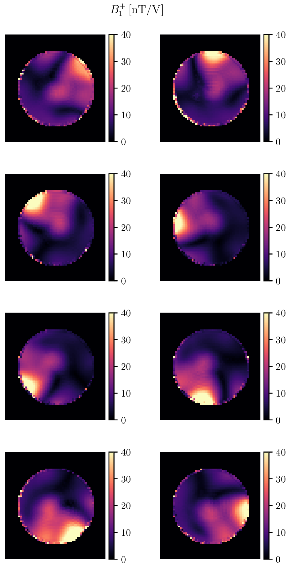
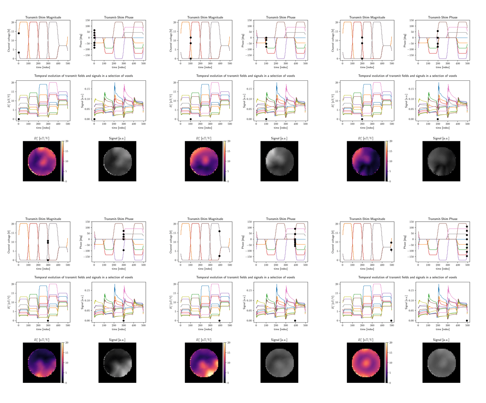
Fig. 2: Top row: realized $$$B_1^+$$$ (left) and simulated signal magnitude (right). This animation illustrates how different parts of the field of view are illuminated at different times, modulating the flip-angles to force magnetisation into a transient state as required for MRF.
The bottom row shows 1D-examples of $$$B_1^+$$$ (left) and signal (right) curves over time from voxels across the phantom, showing how different the realized flip-angle evolutions can be. Note that achieved $$$B_1^+$$$ is quite low due to SAR constraints.
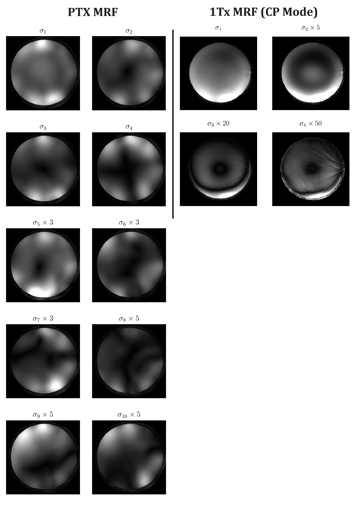
Fig. 3: Left: singular components obtained from temporal low-rank reconstruction of our PTX MRF scan. Different spatially non-uniform modes are imprinted on each component, and interestingly the magnitude is distributed more evenly among components than usual for MRF with static transmit shims.
Right: First four singular components of a static transmit-shim MRF scan of the same phantom. Note that there is substantial scaling applied to the higher components, meaning that the energy content in these is low.
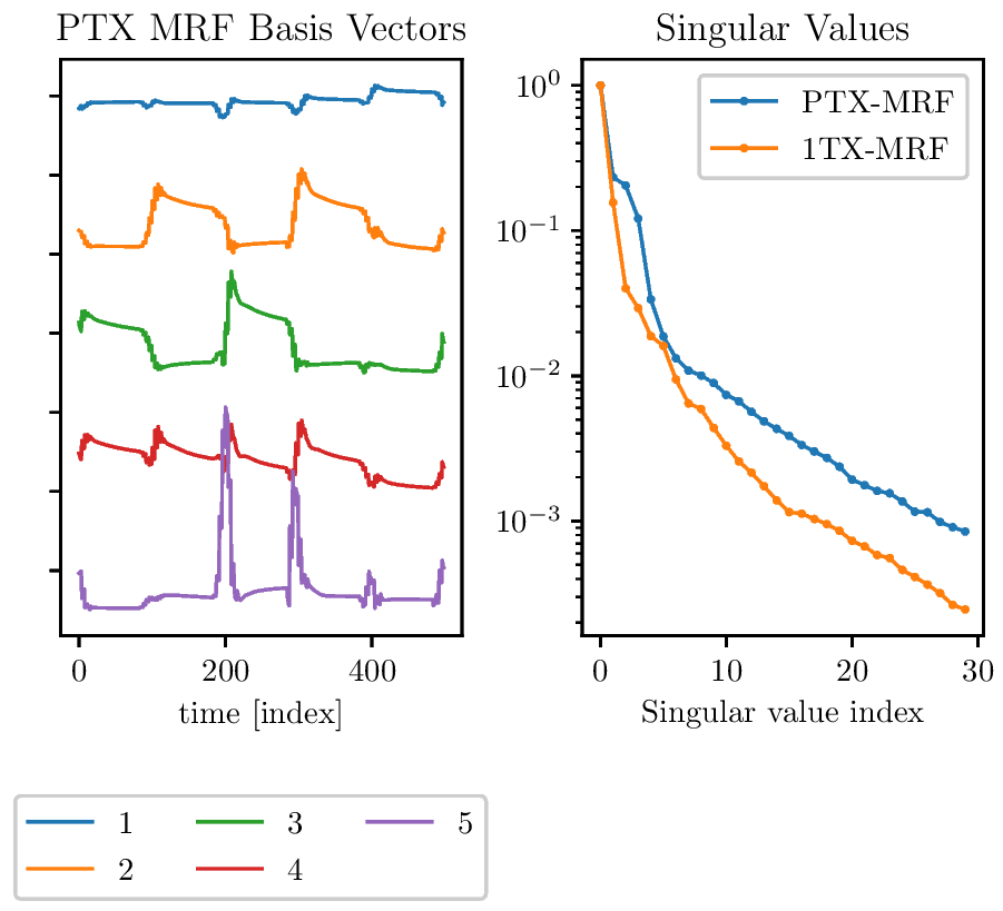
Fig. 4: Left: temporal low-rank basis vectors of PTX-MRF. Curves are shifted to improve visibility, the most significant component's vector is on top. Apart from the first vector of PTX-MRF being nearly flat, no special features stick out. Note that curves appear rugged because the transmit-shim can only be updated every four $T_R$ due to limitations of the real-time SAR-model predictions.
Right: singular values corresponding to the basis on the right in blue, and those of 1Tx MRF on the right. Specifically for the first components the drop-off is slower for PTX-MRF.