0948
Open-Source, Cross-Platform Workflow for MRI Data Acquisition and Image Reconstruction Based on the Pulseq Framework1Division of Medical Physics, Department of Diagnostic and Interventional Radiology, University Medical Center Freiburg, Faculty of Medicine, University of Freiburg, Freiburg, Germany, 2Department of Radiology and Nuclear Medicine, St. Olav's University Hospital, Trondheim, Norway
Synopsis
Keywords: Image Reconstruction, Image Reconstruction, open source vendor-independent sequences
Motivation: To enhance efficiency, transparency, and reproducibility of data acquisition and reconstruction in large-scale MRI studies.
Goal(s): To establish an open-source, cross-platform, easy-to-learn data acquisition and reconstruction workflow.
Approach: The Pulseq framework is extended to integrate Siemens’ “Image Calculation Environment” (ICE) and Gadgetron. To validate the workflow, MPRAGE and EPI sequences were developed using the extended Pulseq and executed on three Siemens scanners with comparison to the corresponding product sequences.
Results: The preliminary results show that Gadgetron had comparable reconstruction performance to ICE, and Pulseq sequences generally produced image quality comparable to product sequences. Online Gadgetron and ICE produce images within seconds/minutes after measurements.
Impact: An open-source, cross-platform MRI data acquisition and reconstruction workflow is established by extending the Pulseq framework to link to reconstruction tools. The preliminary results indicate that this workflow has the potential to enable efficient, transparent, reproducible data acquisition and reconstruction.
Introduction
There is currently a worldwide effort to discover potential research and promote clinical use of magnetic resonance imaging (MRI). However, a significant practical barrier is the effort required to harmonize sequences and reconstruction algorithms on vendor-specific development platforms to ensure that data are acquired and reconstructed in a consistent and reproducible manner across different platforms1. In order to directly compare results between platforms or to pool data from multiple platforms for increasing statistical power, open-source science and software tools are desirable for efficiently harmonizing data acquisition and image reconstruction in multi-site MRI studies.To alleviate this challenge, we develop an open-source, cross-platform, easy-to-learn workflow based on the Pulseq2 framework to enable efficient, transparent, reproducible data acquisition. In this project, we extend Pulseq to integrate with Siemens’ “Image Calculation Environment” (ICE) platform and Gadgetron3,4 to establish a complete data acquisition and reconstruction workflow. ICE is integrated into the Siemens magnetic resonance (MR) system, while Gadgetron can be used to reconstruct data from various vendors by employing vendor-independent ISMRMRD5 data format. Two example sequences, Magnetization Prepared RApid Gradient Echo (MPRAGE)6 and Echo-Planar Imaging (EPI)7, were developed based on the extended Pulseq and executed on three Siemens scanners to validate the workflow.
Methods
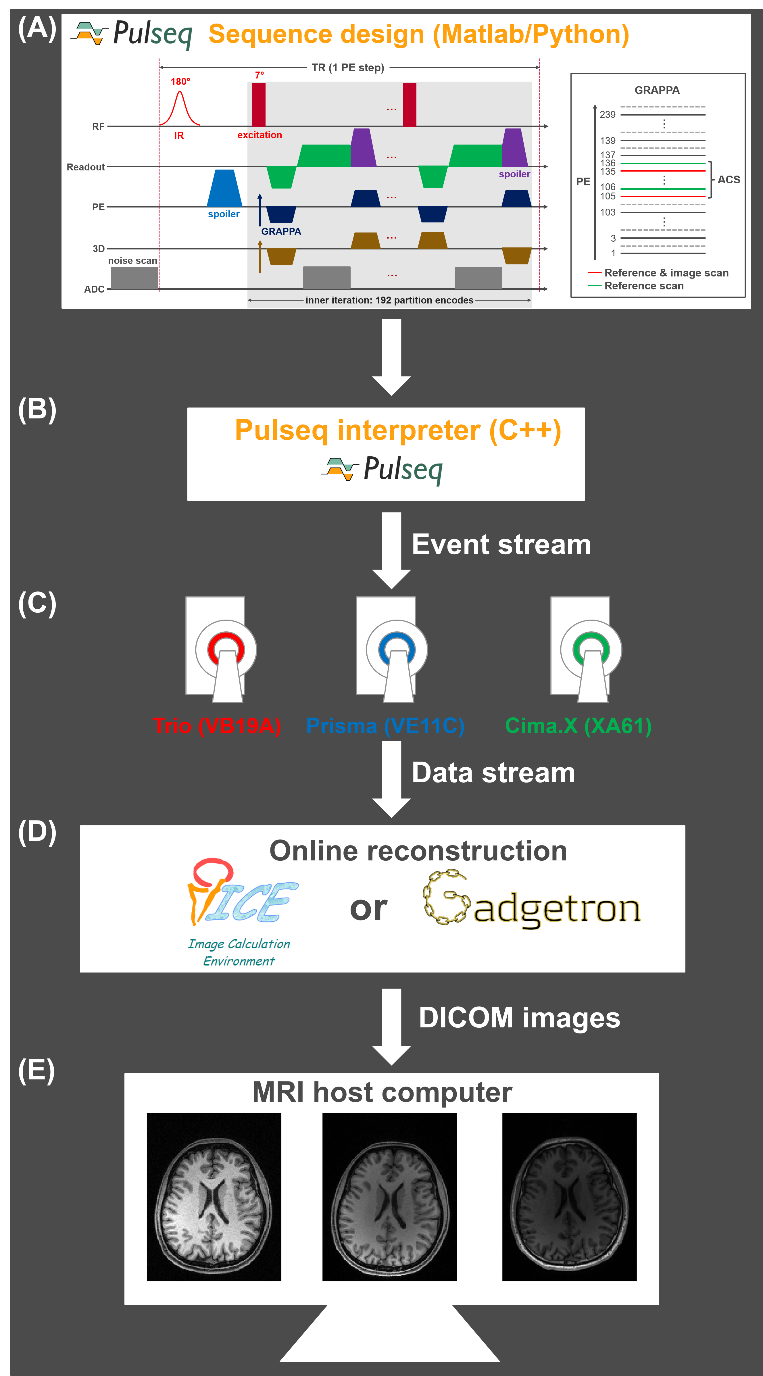
WorkflowThe whole workflow is shown in Figure 1. First, sequences are designed in Pulseq using high-level languages (Matlab or Python) and are exported to a vendor-independent file format (.seq), which can be executed on Siemens, General Electric, and Philips whole-body MR scanners. For interfacing with ICE/Gadgetron, every analog-to-digital converter (ADC) event in the Pulseq sequence must be labeled, and additional necessary information is given using the sequence definitions option (“seq.setDefinition”). Then, the Pulseq interpreter loads the .seq file and produces an event stream to control pulse sequence execution and data acquisition. The acquired data are streamed to ICE/Gadgetron line-by-line and are instantly reconstructed during the measurement. Finally, ICE/Gadgetron sends DICOM images to the host computer within seconds/minutes after the measurement. If Gadgetron is not installed on the scanner, raw data can be exported for offline Gadgetron reconstruction.
Pulse Sequences
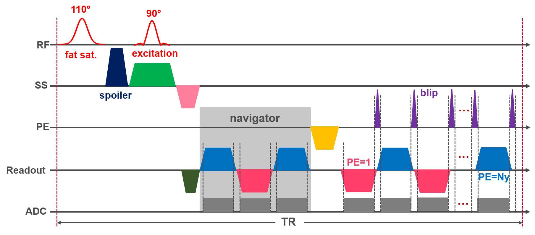
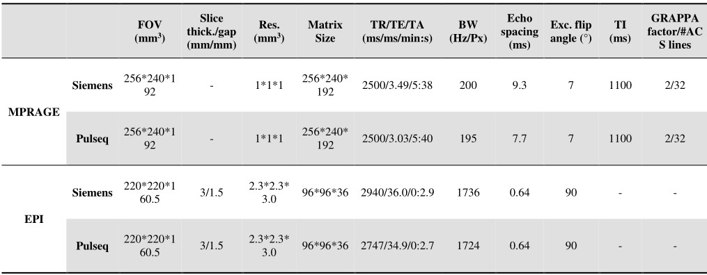
A 3D MPRAGE sequence with a noise scan and two-fold GeneRalized Autocalibrating Partial Parallel Acquisition (GRAPPA)8 acceleration was implemented using Pulseq (Figure 1A). A 2D multi-slice EPI sequence with fat saturation was also implemented in Pulseq (Figure 2). In particular, this EPI sequence relies on ramp sampling to accelerate data acquisition, and similarly to the product EPI, uses a three-echo navigator for Nyquist ghost correction. In addition, the vendor MPRAGE and EPI protocols were configured to closely match the Pulseq MPRAGE and EPI sequences, respectively. The parameters of these four sequences are listed in Table 1 (with a minor modification for EPI on Trio to avoid mechanical resonances of the gradient coil). The Pulseq sequence source code is readily available online9.
Measurement
A water-filled phantom doped with a NiCl/NaCl mixture and the brain of a healthy male volunteer were scanned with the aforementioned four sequences on three 3T Siemens scanners: Trio (VB19A), Prisma (VE11C), and Cima.X (XA61) (SIEMENS Healthineers, Erlangen, Germany) equipped with 20-channel, 20-channel, and 12-channel receive-only radiofrequency coils, respectively. In our lab, Gadgetron is only installed on the Prisma scanner, enabling online Gadgetron reconstruction for Pulseq there. Other data were reconstructed offline with Gadgetron.
Results and Discussion
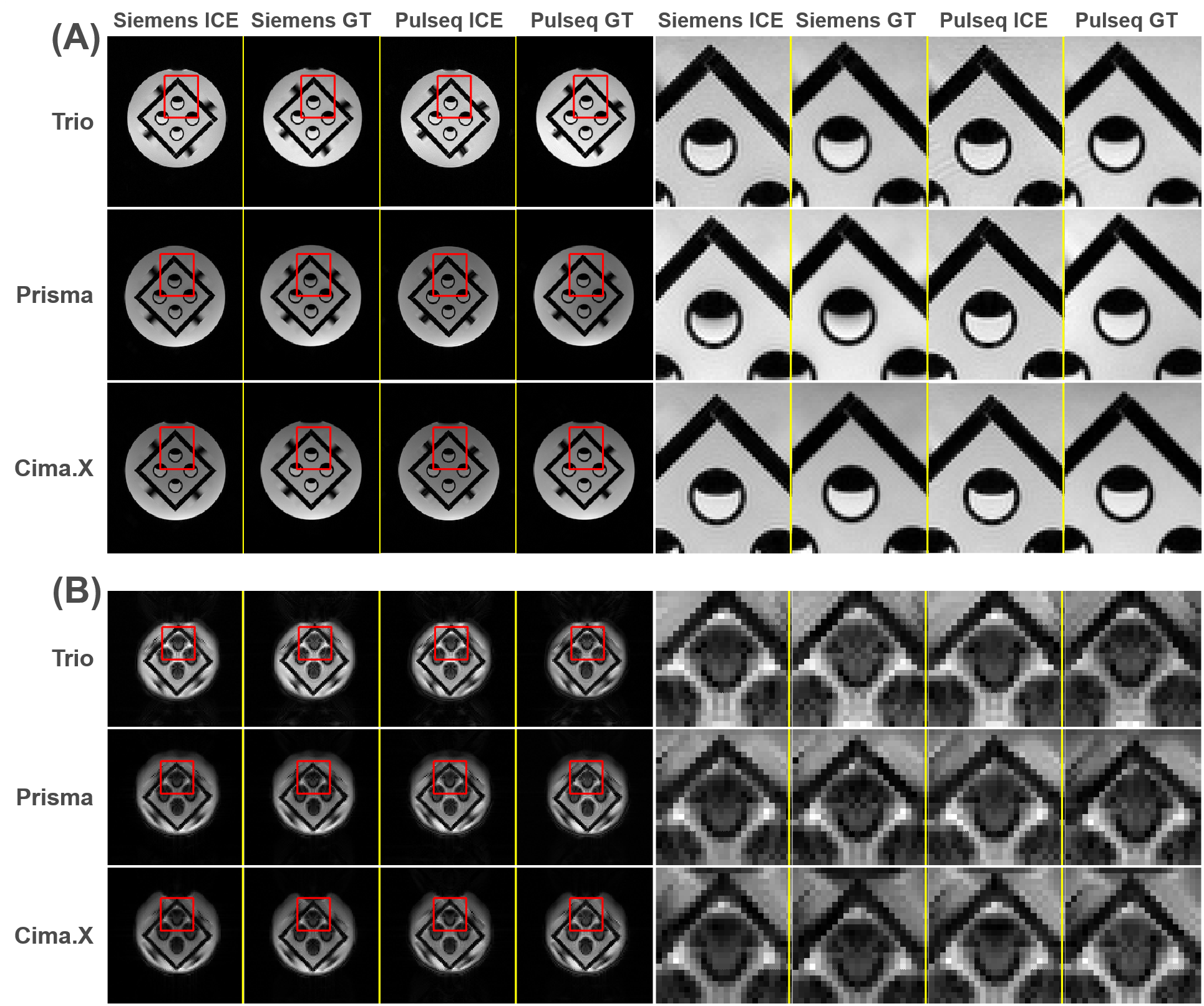
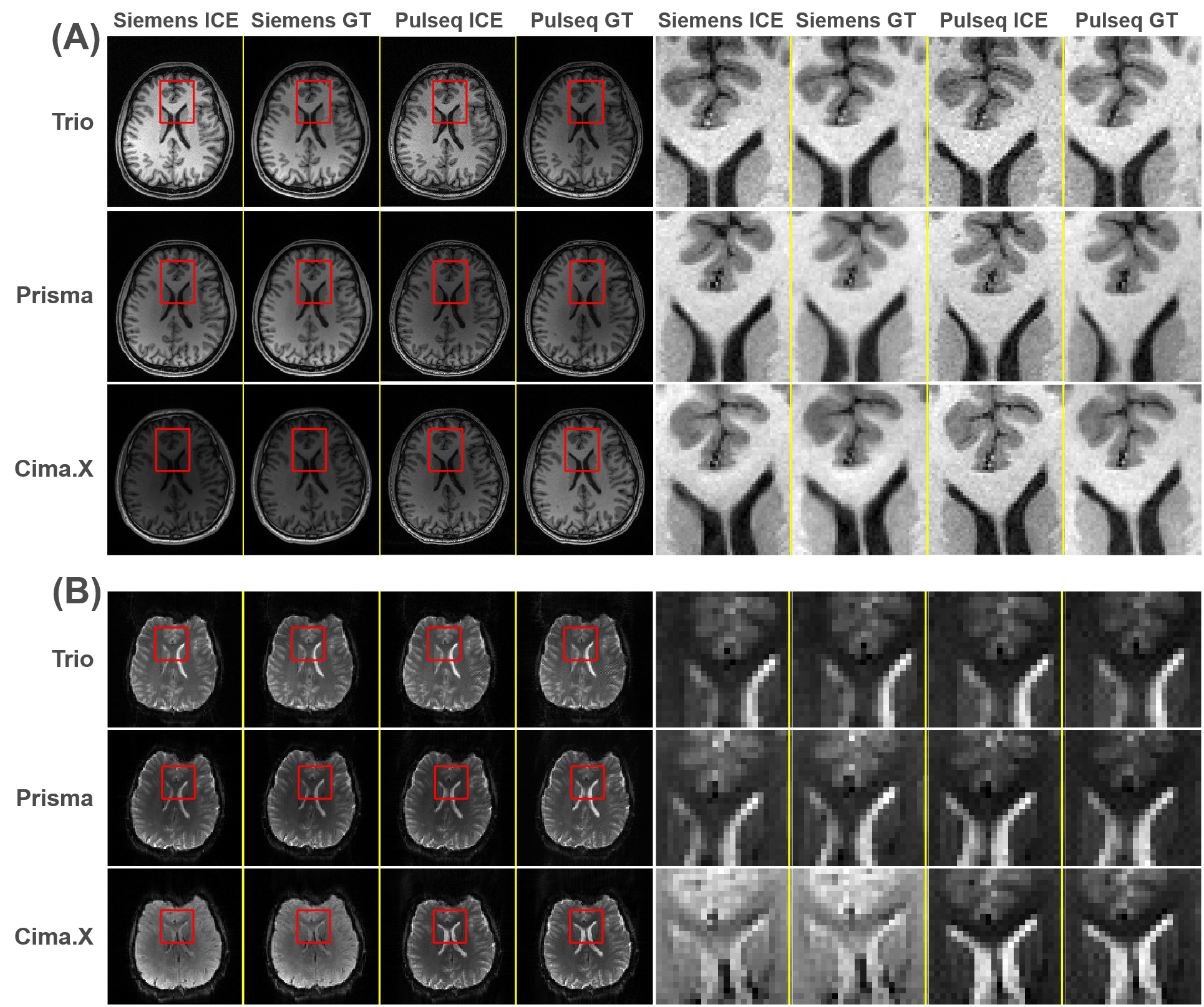
The phantom results are shown in Figure 3. For both MPRAGE and EPI, Gadgetron has comparable reconstruction performance to ICE, and the images acquired with Pulseq sequences align with corresponding product sequences. The images from Prisma and Cima.X show slightly lower noise levels and better structure delineation than those from the older Trio scanner equipped with the head coil with fewer channels.The in vivo results (Figure 4) agree well with the phantom results (Figure 3), except that in Cima.X, the image contrast of the product EPI sequence is slightly different from that of the Pulseq EPI sequence. One possible explanation is that the protocol is different from the source code of the EPI vendor sequence, which is not yet available for Cima.X, leading to a different steady state (although only a single volume is intended to be acquired with this EPI protocol). Noteworthy, however, is the consistency of the Pulseq sequence performance across the three very different systems.
Conclusion
We successfully establish an efficient, open-source, cross-platform MRI data acquisition and image reconstruction workflow based on Pulseq and validate it for MPRAGE and EPI protocols. The preliminary results indicate that this workflow has excellent potential to enhance efficiency, transparency, and reproducibility for data acquisition and reconstruction in large-scale MRI studies.Acknowledgements
This work is supported by research grants NIH R01 EB032378 and NIH U24 NS120056.References
1. Van Horn JD, Toga AW. Multisite neuroimaging trials. Curr Opin Neurol. 2009;22(4):370-378.
2. Layton KJ, Kroboth S, Jia F, et al. Pulseq: A rapid and hardware-independent pulse sequence prototyping framework. Magn Reson Med. 2017;77(4):1544-1552.
3. Hansen MS, Sørensen TS. Gadgetron: An open source framework for medical image reconstruction. Magn Reson Med. 2013;69(6):1768-1776.
4. Xue H, Inati S, Sørensen TS, Kellman P, Hansen MS. Distributed MRI reconstruction using Gadgetron-based cloud computing. Magn Reson Med. 2015;73(3):1015-1025.
5. Inati SJ, Naegele JD, Zwart NR, et al. ISMRM Raw data format: A proposed standard for MRI raw datasets. Magn Reson Med. 2017;77(1):411-421.
6. Mugler JP. Rapid Three-dimential T1-weighted MR Imaging with the MP-RAGE sequence. J Magn Reson Imaging. 1991;1(561-567).
7. Stehling MK, Turner R, Mansfield P. Echo-planar imaging: Magnetic resonance imaging in a fraction of a second. Science (80- ). 1991;254(5028):43-50.
8. Griswold MA, Jakob PM, Heidemann RM, et al. Generalized Autocalibrating Partially Parallel Acquisitions (GRAPPA). Magn Reson Med. 2002;47(6):1202-1210.
9. GitHub. Accessed November 8 2023. https://github.com/pulseq/pulseq/tree/master/matlab/demoSeq.
Figures