0944
Spiral HASTE using variable-flip-angle single-shot spiral-ring TSE for rapid T2-weighted abdominal imaging1Radiation Oncology, City of Hope National Medical Center, Duarte, CA, United States, 2Radiological Sciences, University of California Los Angeles, Los Angeles, CA, United States, 3Radiology and Biomedical Imaging, University of California San Francisco, San Francisco, CA, United States, 4Radiology & Medical Imaging, University of Virginia, Charlottesville, VA, United States, 5Biomedical Engineering, University of Virginia, Charlottesville, VA, United States
Synopsis
Keywords: Data Acquisition, Data Acquisition, Spiral TSE, HASTE, Abdominal imaging
Motivation: Conventional Cartesian HASTE may be susceptible to image blurring, resolution loss, and artifacts in abdominal imaging.
Goal(s): To develop a 2D T2-weighted abdominal spiral HASTE technique to achieve better image sharpness, reduced scan time, and reduced power deposition compared to Cartesian HASTE.
Approach: A variable-flip-angle refocusing RF series was designed and employed to maintain high signal-intensity at middle- and late-echoes with a smooth signal-evolution compared to a constant-flip-angle scheme. Variable-density spiral-ring trajectories combined with L1-ESPIRiT reconstruction were performed to accelerate the data acquisition.
Results: Whole-abdomen spiral HASTE images were acquired with 1.5×1.5×6 mm3 spatial-resolution and 25 slices in a 7-second acquisition at 3T.
Impact: This work highlights the utilization of spiral-ring TSE acquisition along with variable-flip-angle RF pulses and L1-ESPIRiT reconstruction for accelerated single-shot abdominal imaging, providing images with improved image sharpness, shorter scan time, and reduced power deposition over conventional Cartesian HASTE.
Introduction:
Cartesian HASTE, a single-shot technique, uses a very long echo train (e.g., 128) to acquire all k-space data following a single RF excitation.1,2 Partial-Fourier (PF) sampling3 and parallel imaging (e.g., GRAPPA4) are normally introduced to reduce image blurring and artifacts from T2 decay. However, 2D-Cartesian sampling remains time-consuming because of its relatively inefficient k-space coverage when high-spatial-resolution and large-FOV are needed. This becomes more severe in abdominal imaging where different kinds of motion (respiratory, peristatic) exist, and affects the generation of high-quality images.Spiral acquisitions cover k-space more efficiently than Cartesian sampling and are attractive in fast/dynamic imaging, where very short overall scan times are preferred.5-7 Researchers have developed sampling strategies including spiral TSE for fast T2-weighted imaging. Hennig et al.5, for example, has proposed single-shot spiral TSE with annular-ring segments, enabling highly efficient acquisition of high-resolution brain-images in less than 200 ms per slice.
In this work, we developed and optimized a 2D single-shot TSE sequence using annular spiral-ring trajectories for abdominal imaging at 3T. We use a variable-flip-angle approach to flatten the signal evolution as well as maintain a relatively high signal intensity at later echoes, and compressed sensing to further accelerate the data acquisition. Additionally, to achieve images with different effective TEs, we utilize different echo-ordering schemes and compare the performance of spiral-rings with different shifts along the echo-train.
Methods:
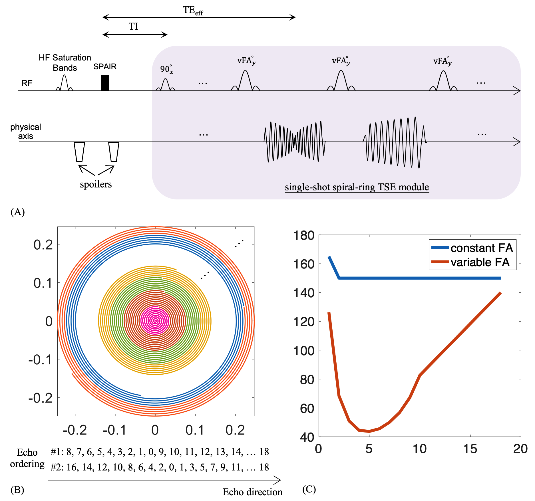
A schematic of the proposed spiral HASTE sequence is shown in Fig.1A. The preparation module consisted of saturation bands along the head-foot direction to reduce flow artifacts from the abdominal aorta and vena cava, and a spectral attenuated inversion recovery (SPAIR) pulse to suppress fat, each followed by large-gradients applied immediately to spoil unwanted transverse-magnetization. A single-shot TSE module with linear-variable-density spiral-ring readouts was used for data acquisition. The central spiral-in-out ring was placed at the echo with the effective TE (TEeff), while other echoes were filled with outer spiral-out rings. Specifically, two echo-ordering schemes are shown in Fig.1B.The variable-flip-angle RF series shown in Fig.1C (red line) was calculated for liver specifically at 3T (T1: 800 ms, T2: 35 ms)8 using the EPG algorithm9,10, with a maximum 140o flip angle for SAR reduction. The contrast-equivalent TE (TEequiv) was calculated using the acquired signal and the transverse coherence component of the signal where relaxation effects were ignored.10 A constant-flip-angle scheme (blue line) was implemented for comparison.
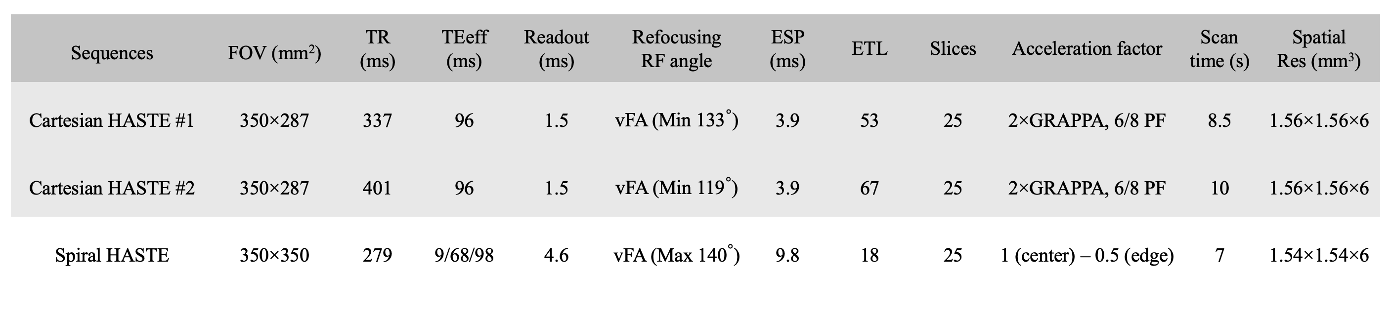
Experiments were performed on a 3T scanner (MAGNETOM Vida, Siemens Healthcare, Erlangen, Germany) using an 18-channel Ultraflex large coil array. For image reconstruction, L1-ESPIRiT11 was performed on all undersampled datasets. For healthy volunteer studies, images were acquired using spiral HASTE (7s/25slices) and Cartesian HASTE (two versions: 8.5s/ and 10s/25slices) as a reference. Other sequence parameters are given in Table-1.
Results and Discussion:
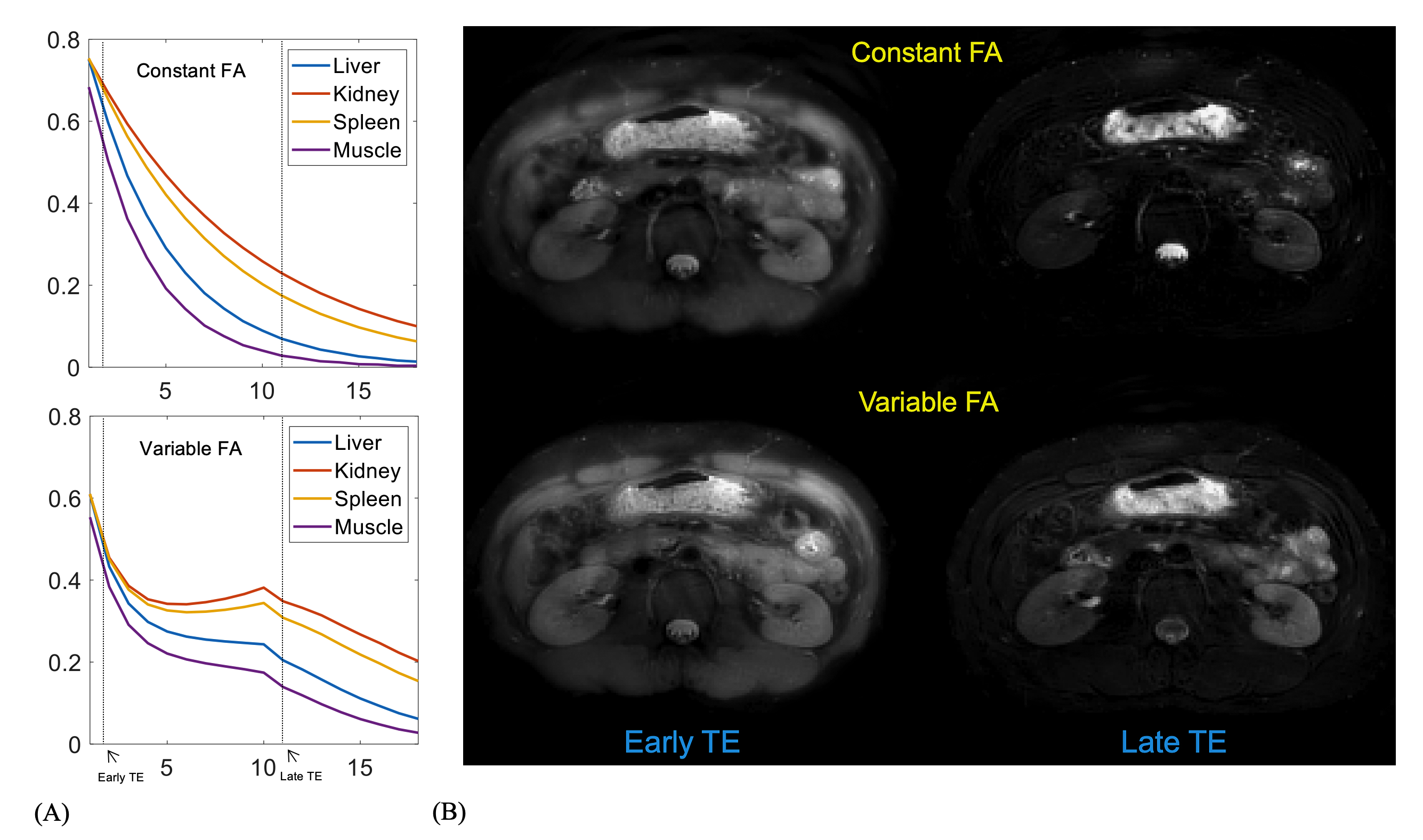
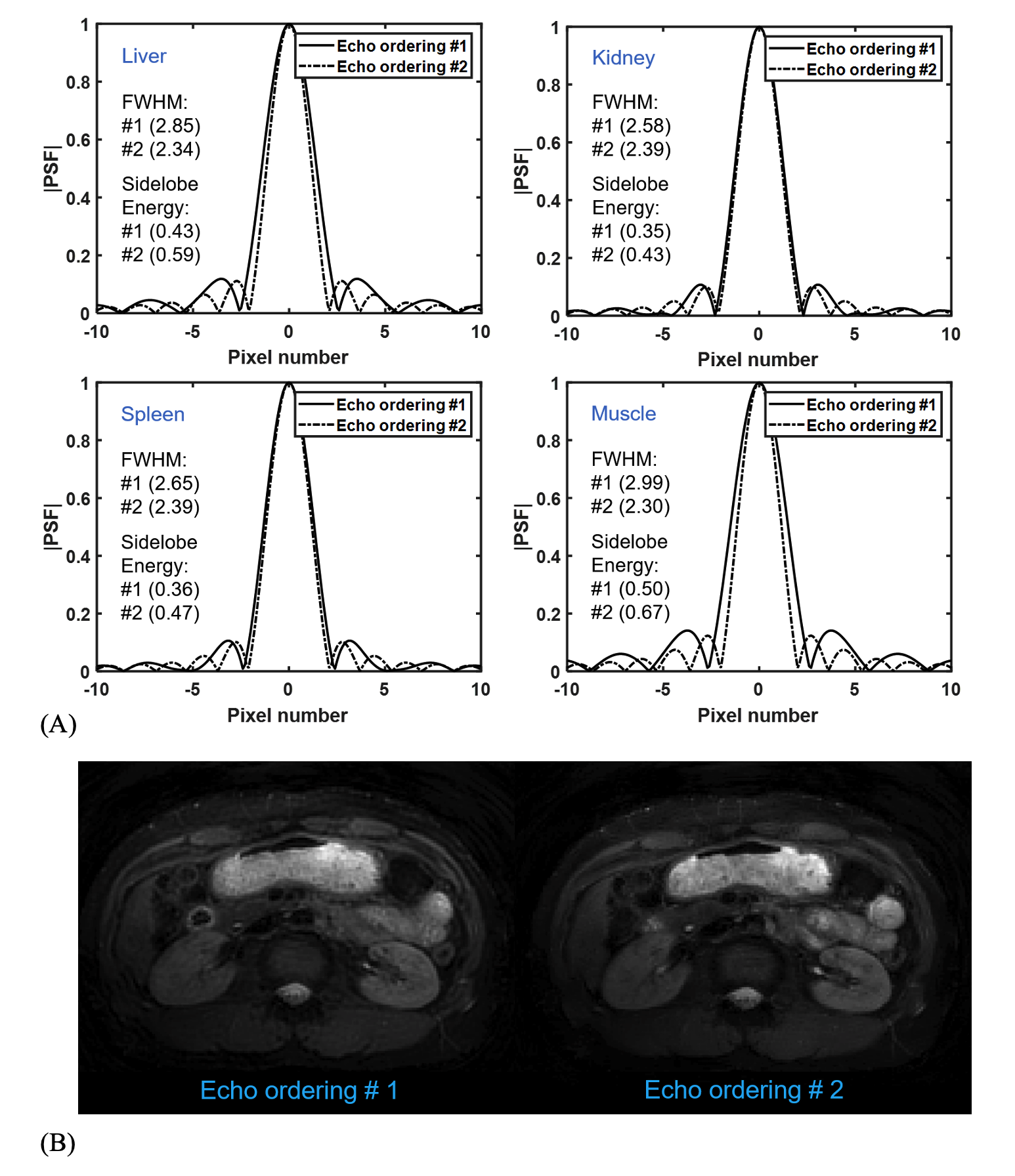
Fig.2A shows the signal evolutions for liver, kidney, spleen, and muscle, using the constant-flip-angle (top) and variable-flip-angle (bottom) schemes. Note that all signals from the variable-flip-angle approach are much higher than those for the constant-flip-angle counterpart at middle and late echoes, and the signal pathways at the bottom are relatively flattened with a smooth plateau. The performance of using the variable-flip-angle approach can be seen in Fig.2B, where images from constant-flip-angle (top) show strong blurring at early-TE and contrast loss at late-TE because of substantial T2-decay signal variation, while images from variable-flip-angle (bottom) depict reasonable image contrast and sharpness.Fig.3 illustrates the comparison of spiral HASTE with two proposed echo-ordering schemes. Simulation results of center lines of 2D PSFs are shown in Fig.3A for each echo-ordering and each tissue. Note that the PSF was generated based on the designed undersampled k-space and was then normalized to [0, 1] by dividing by its own peak. The echo-ordering #2 produces a smoother frequency response, yielding a narrower main lobe of the PSF for all tissues compared to #1, especially in tissues with short T2 values (liver and muscle). However, the sidelobe energy is increased in echo-ordering #2 relative to that for #1; this can be mitigated by using L1-ESPIRiT. Fig.3B shows one example of in-vivo abdomen-images, demonstrating the performance of improved image sharpness when using echo-ordering #2 rather than #1.
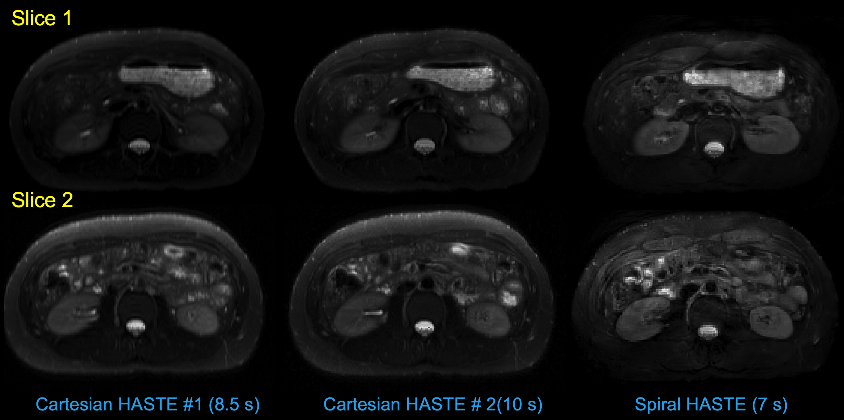
Fig.4 shows abdomen-images of spiral HASTE using L1-ESPIRiT (right) with a 7-s scan time, compared to Cartesian HASTE images using GRAPPA and PF with 8.5-s scan time (left) and 10-s scan time (middle). Images from Cartesian HASTE show obvious blurring along the phase-encoding direction, especially when using shorter scan time, while spiral HASTE yields the best image sharpness and shortest scan time, as well as a 20% reduced power deposition (1.51 versus 1.94 W/kg).
Acknowledgements
No acknowledgement found.References
[1] Semelka RC, Kelekis NL, Thomasson D, Brown MA, Laub GA. HASTE MR imaging: description of technique and preliminary results in the abdomen. J Magn Reson Imaging 1996; 6:698-699.[2] Patel MR. Klufas RA, Alberico RA, Edelman RR. Half-Fourier acquisition single-shot turbo spin-echo (HASTE) MR: Comparison with fast spin-echo MR in diseases of the brain. AJNR Am J Neuroradiol 1997; 18:1635-1640.
[3] McGibney G, Smith MR, Nichols ST, Crawley A. Quantitative evaluation of several partial Fourier reconstruction algorithms used in MRI. Magn Reson Med. 1993;30:51-59.
[4] Griswold MA, Jakob PM, Heidemann RM, Nittka M, Jellus V, Wang J, Kiefer B, Haase A. Generalized auto-calibrating partially parallel acquisitions (GRAPPA). Magn Reson Med. 2002;47:1202–1210.
[5] Hennig J, Barghoorn A,Zhang S, Zaitsev M. Single shot spiral TSE with annulated segmentation. Magn Reson Med. 2022;88:651-662.
[6] Wang Z, Allen SP, Feng X, Mugler JP, Meyer CH. SPRING-RIO TSE: 2D T2-Weighted Turbo Spin-Echo Brain Imaging using SPiral RINGs with Retraced In/Out Trajectories. Magn Reson Med. 2022;88:601-616.
[7] Wang Z, Cao X, Qing K, Mugler JP, Meyer CH. Distortion-free diffusion imaging using single-shot diffusion-prepared turbo-spin-echo sequence with spiral-ring readouts and magnitude stabilizers. In Proceedings of the 32nd Annual Meeting of ISMRM, Toronto, CA, 2023.p.2018.
[8] de Bazelaire CM, Duhamel GD, Rofsky NM, Alsop DC. MR imaging relaxation times of abdominal and pelvic tissues measured in vivo at 3.0 T: preliminary results. Radiology. 2004;230(3):652-659.
[9] Hennig J, Weigel M, Scheffler K. Calculation of flip angles for echo trains with predefined amplitudes with the extended phase graph (EPG)-algorithm: principles and applications to hyperecho and TRAPS sequences. Magn Reson Med 2004; 51: 68– 80.
[10] Busse RF, Hariharan H, Vu A, Brittain JH. Fast spin echo sequences with very long echo trains: design of variable refocusing flip angle schedules and generation of clinical T2 contrast. Magn Reson Med. 2006;55(5):1030-1037.
[11] Uecker M, Lai P, Murphy MJ, Virtue P, Elad M, Pauly JM, Vasanawala SS, Lustig M. ESPIRiT–an eigenvalue approach to auto-calibrating parallel MRI: where SENSE meets GRAPPA. Magn Reson Med. 2014;71:990–1001.
Figures