0943
Echo-planar Imaging-based Rapid Simultaneous MR Angiography and Venography with Dedicated Flow-related Ghosting Suppression1Institute of Biophysics, Chinese Academy of Sciences, Beijing, China, 2The Innovation Center of Excellence on Brain Science, Chinese Academy of Sciences, Beijing, China, 3University of Chinese Academy of Sciences, Beijing, China, 4Siemens Shenzhen Magnetic Resonance Ltd, Shenzhen, China, 5Institute of Artificial Intelligence, Hefei Comprehensive National Science Center, Hefei, China
Synopsis
Keywords: Data Acquisition, Vessels, Ghosting suppression, Sequence design, Low-rank reconstruction
Motivation: Ghost artifact of large vessels impede the use of echo-planar imaging(EPI) for accelerated MR Angiography(MRA) or MR Venography(MRV) acquisitions.
Goal(s): To analyze the physical principles of flow-related ghost, and develop targeted technical approaches to suppress the artifact, enabling EPI-based rapid and high-fidelity simultaneous MRAV.
Approach: By employing techniques including point-spread function modeling, alternating flow-compensation scheme, flow-related phase-based reconstruction, and automatic detection and correction algorithms, rapid in-vivo vascular imaging were achieved.
Results: DEPSAV-II achieved cerebral MRAV with suppressed ghost artifacts and comparable vascular depiction with GRE-based methods in 6 minutes, thereby improving the accessibility of comprehensive vascular examination in routine clinical practices.
Impact: Developed a suite
of techniques to overcome flow-related ghosting of large vessels which tackling
EPI-based rapid arteriovenous-imaging methods.
DEPSAV-II’s simultaneous MRAV
and significant time-reduction may benefit clinical studies and practices like
small vessel diseases requiring comprehensive arterial and venous examination.
Introduction
Conventional time-of-flight (TOF) MR angiography(MRA) and susceptibility-weighted MR venography(MRV) rely on gradient recalled echo (GRE) acquisition, which are inherently time-consuming and require separate scan for arteries and veins, limiting their clinical utility1,2. Over the decades, endeavors have been made to incorporate high-speed echo-planar imaging(EPI), especially multi-shot EPI, into MRA&MRV acquisitions3–5. Among these practices, we introduced a preliminary technique in last year's abstract using the flow-compensated multi-shot dual-contrast 3D-EPI for simultaneous MRAV(DEPSAV) to further boost scan efficiency6. Collectively, these EPI-based innovations have shown a substantial reduction in time required for MRA and MRV.However, a major challenge in these methods is the unresolved issue of “ghost” artifacts due to the flow-related phase variation, shown as conspicuous false “vessels” along the phase-encoding direction near large vessels7,8.
In this study, we first elucidated the physical principles behind the emergence of this artifact. Building on the theory, we introduced sequence modifications and reconstruction algorithms to achieve automatic detection and correction of large vessel artifacts. The suite of techniques was termed as DEPSAV-II and its feasibility for fast cerebral vascular imaging was evaluated.
Methods
Theory: flow-related ghost artifactEPI sequence inherently exhibits flow-sensitivity, which is manifested in the flow-related phase modulation along the readout direction. The remaining first-order moment of readout gradients introduces a periodic phase shift between odd and even lines(Figure.1(A)). This periodic modulation across the whole k-space, resembling typical odd-even line variation in EPI, causes point-spread function as:
$$\begin{aligned}G_\varphi[n_y]&=\sin\left.\left(\frac{\Delta\varphi}2\right)\frac{N_y}{n_i}(-1)^{n_y}e^{-j\pi(2n_i-1)n_y/N_y}\right.\\&\frac{\sin^2\left(\pi n_in_y/N_y\right)}{\sin\left(\pi n_y/N_y\right)}\sum_{m=-n_i}^{n_i-1}\delta[n_y-\frac{N_ym}{2n_i}]\quad(eqa1)\end{aligned}$$, indicating artifacts akin to Nyquist ghosts9(Figure.1(B)).
Alternating flow-compensation(FC) scheme
In DEPSAV with center-out trajectory, the data acquired with odd-echo is flow-compensated, nulling the flow-related phase, while even-echo data has constant phase of $$$\Phi_{e\nu en}(TE)\propto\frac{1}{3}Gr2+\frac{1}{2}Gh2+hr$$$. Interestingly, if FC for even-echo data, then the phase of odd-echo is $$$\Phi_{odd}(TE)\propto-(\frac{1}{3}Gr2+\frac{1}{2}Gh2+hr)$$$.
As shown in Figure.2(A), DEPSAV-II employed an alternating FC gradient scheme, i.e., applying gradients to achieve FC of odd-echoes for shots in upper half of k-space, and FC of even-echoes with reversed polarity for lower half of k-space acquisition.
Data reorganization and reconstruction
Based on flow-related phase, all the k-space data can be reorganized into two “under-sampled” k-space (Figure.2(B)). Theoretically, within the same group, phase modulations are approximately consistent, thereby suppressing the ghost artifacts. In contrast, two groups possess distinct FC properties, leading to specific signal variation patterns of vessel/artifact.
Specifically, with the low-rank regularized reconstruction framework of DEPSAV6, a regular reconstruction is performed for the upper and lower k-space jointly, correcting global artifacts resulting from field-inhomogeneity and inter-shot phase-variations. This yields a “hybrid-phase image”. Subsequently, the two reorganized data groups are reconstructed into the “flow-compensated image” and “flow-dephased image", yielding distinct signal characteristics for vessel and artifact components.
Automatic ghost artifact detection
As shown in Figure.2(D,E), hybrid-phase image blends two flow-related phases, resulting in the most pronounced artifacts. Conversely, the flow-phase based reconstructions exhibit relatively lower artifact levels and are characterized by FC states.
Furthermore, utilizing this signal feature allows for the automatic detection of vessels and artifacts shown in Figure.1(D). A dual-channel weighted difference is performed between hybrid-phase image, flow-compensated image and flow-dephased image. Filtering-enhancement and connectivity region extraction are then utilized, enabling the automatic extraction of vessel and artifact regions.
Artifact correction
Point-spread function reveals that the distribution of artifact is dispersed and localized. Per Equation 2, contaminated voxels can be restored through linear correlations10.$$\begin{aligned}\widetilde{a}_{m,artifact}&=\sum^\text{tissue}k(n)\cdot a_n+PSF_{n,vessel}(y_{\widetilde{m}n})a_{n,\nu essel}\text{ (eqa2.1)}\\\\a_m&\approx\sum^\text{neighbor}{ k ^ { - 1 }}(m)\cdot\widetilde{a_n}\text{ (eqa2.2})\end{aligned}$$
Employing the classical region restoration algorithm in computer-vision achieves the restoration of contaminated tissue voxels, thus effectively correcting the ghost artifacts.11
Experiment
High-resolution (0.6×0.6×0.9mm3) images of DEPSAV-II were obtained in-vivo and compared with conventional GRE on a Siemens 3T Prisma system. The key parameters were shown in Table.1.
Results
Figure.1(C) displays representative images of weighted difference, where line profile manifest characteristics consistent with PSF-prediction, confirming the hypothesis that artifacts stem from flow-related phase modulation of EPI readout.Figure.3(B,C) exhibits two representative results of effective detection and correction of large vessel ghosting artifacts.
Figure.4 present in-vivo vascular imaging results for comparison of DEPSAV-II and parallel-imaging accelerated GRE which showed comparable vascular depictions.
Discussion & Conclusion
This study successfully addressed the issue of ghost artifacts of large vessels in EPI-based MRA acquisitions for the first time. The introduced methods, including alternating FC scheme, flow-phase based data-reorganization, and signal feature-based artifact identification, can be applied to other EPI methods and thus enhance utility of EPI-based fast acquisition for MRA and MRV. Notably, DEPSAV-II images achieved similar quality in depicting cerebral arteries and veins with significantly reduced scan time, while mitigating the image blurring and g-factor noise introduced by high-fold under-sampling.Acknowledgements
This work was supported in part by National Natural Science Foundation of China (82271985, 82001804, 81961128030), Youth Innovation Promotion Association CAS (2022093), National Science and Technology Innovation 2030 Major Program (2022ZD0211900, 2022ZD0211901), Ministry of Science and Technology of China grant (2019YFA0707103), and National Nature Science Foundation of China grant (31730039).References
1. Chen Y, Liu S, Buch S, Hu J, Kang Y, Haacke EM. An interleaved sequence for simultaneous magnetic resonance angiography (MRA), susceptibility weighted imaging (SWI) and quantitative susceptibility mapping (QSM). Magn Reson Imaging. 2018;47:1-6. doi:10.1016/j.mri.2017.11.005
2. Du YP, Jin Z. Simultaneous acquisition of MR angiography and venography (MRAV). Magn Reson Med. 2008;59(5):954-958. doi:10.1002/mrm.21581
3. Slavin GS, Riederer SJ, Ehman RL. Two-dimensional multishot echo-planar coronary MR angiography. Magn Reson Med. 1998;40(6):883-889. doi:10.1002/mrm.1910400614
4. Yang GZ, Gatehouse PD, Keegan J, Mohiaddin RH, Firmin DN. Three-dimensional coronary MR angiography using zonal echo planar imaging. Magn Reson Med. 1998;39(5):833-842. doi:10.1002/mrm.1910390521
5. Liu W, Zhou K. 3D Flow Compensated Interleaved EPI with Partial Fourier Acquisition: A Feasibility Study for Fast Intracranial TOF-MRA.Proc of ISMRM 2021
6. Wu Y, Weng DH, et al. Modified Dual-contrast multishot 3D echo-planar imaging for distortion-free and fast acquisition of simultaneous MR angiography and venography (DEPSAV). Proc of ISMRM 2023
7. Wielopolski PA, Simonetti OP, Duerk JC. Echo-Planar Imaging Angiography. In: Schmitt F, Stehling MK, Turner R, eds. Echo-Planar Imaging: Theory, Technique and Application. Springer; 1998:253-309. doi:10.1007/978-3-642-80443-4_8
8. Slavin GS, Riederer SJ. Gradient moment smoothing: A new flow compensation technique for multi-shot echo-planar imaging. Magn Reson Med. 1997;38(3):368-377. doi:10.1002/mrm.1910380304
9. Reeder SB, Atalar E, Bolster BD, McVeigh ER. Quantification and reduction of ghosting artifacts in interleaved echo-planar imaging. Magn Reson Med. 1997;38(3):429-439. doi:https://doi.org/10.1002/mrm.1910380312
10. Patzig F, Mildner T, Schlumm T, Müller R, Möller HE. Deconvolution‐based distortion correction of EPI using analytic single‐voxel point‐spread functions. Magn Reson Med. 2021;85(5):2445-2461. doi:10.1002/mrm.28591
11. He K, Sun J. Image Completion Approaches Using the Statistics of Similar Patches. IEEE Trans Pattern Anal Mach Intell. 2014;36(12):2423-2435. doi:10.1109/TPAMI.2014.2330611
Figures
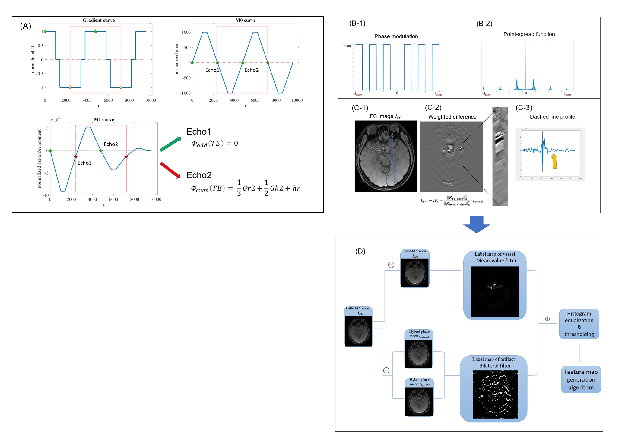
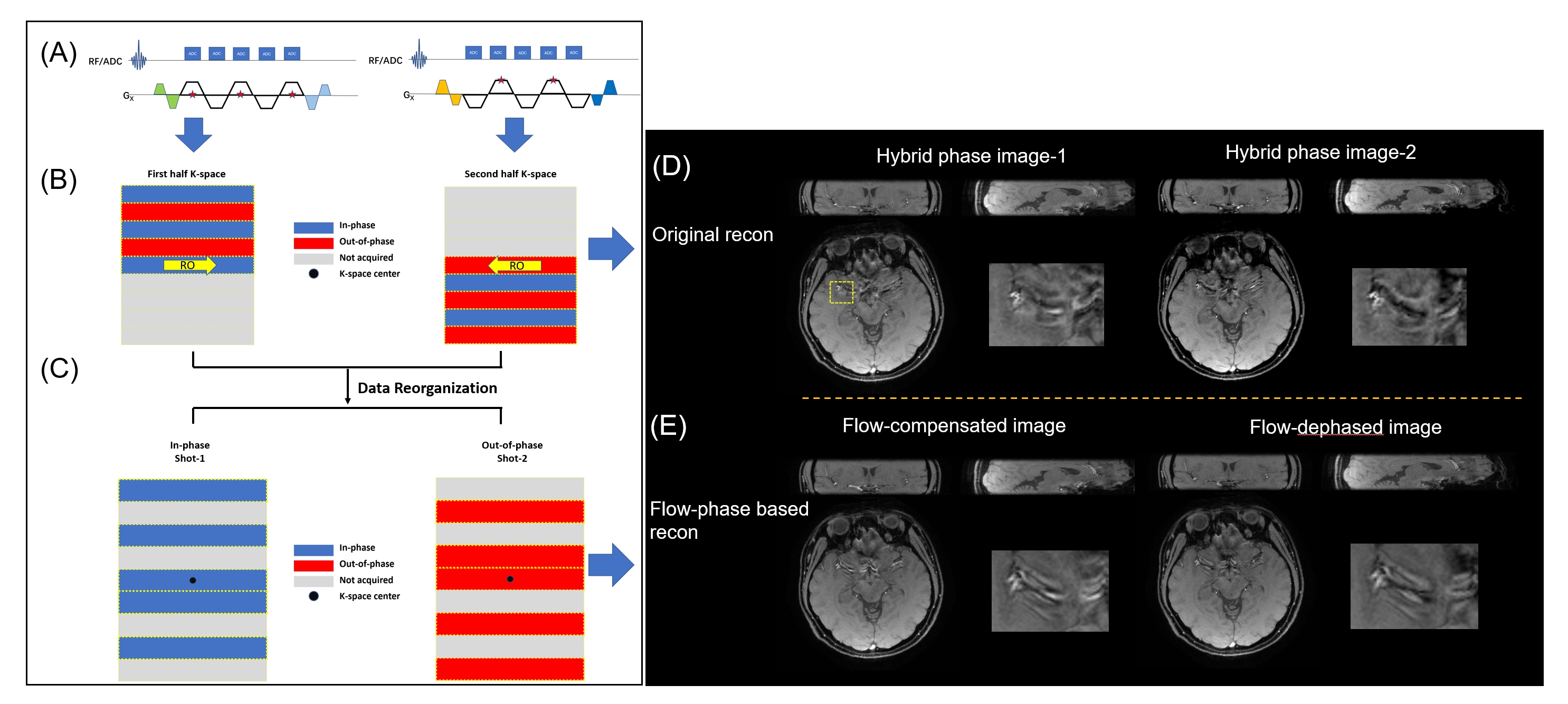
Figure 2. Schematic representation of the alternating flow compensation (FC) scheme used in DEPSAV-II. (B) Corresponding flow-related phase distribution in normal k-space and its reconstruction results(D). (C) Additionally, based on the flow phase state, data can be reorganized into flow-compensated group(blue) and flow-dephased group(red), and the respective reconstruction results (E) showed different FC characteristics.
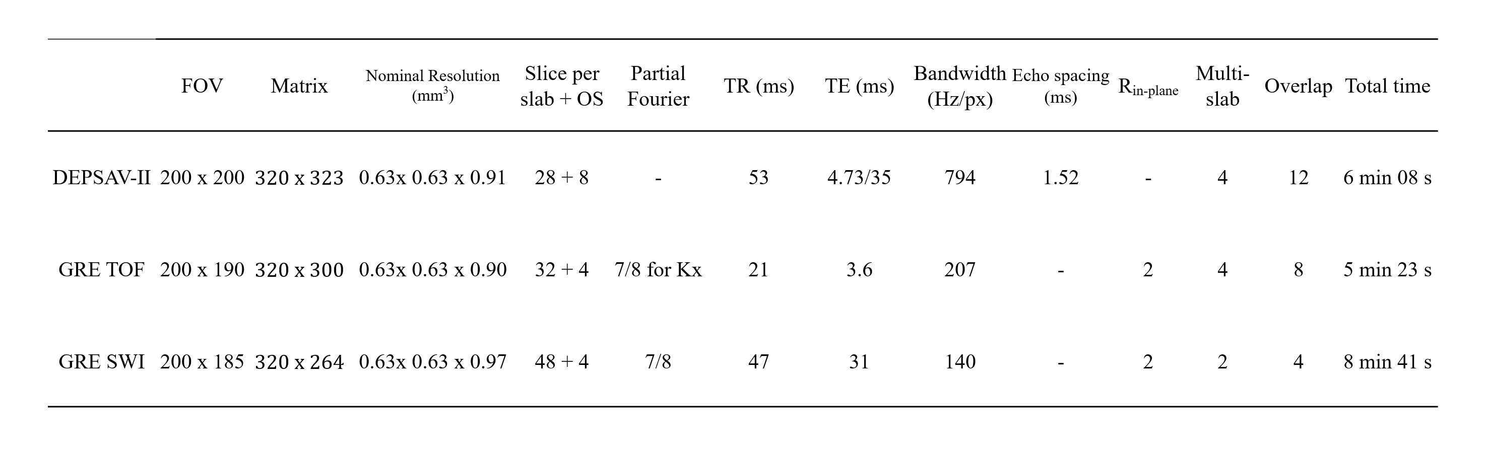
Table 1. The sequence parameters for the comparison experiments of GRE and DEPSAV-II for high-resolution MR Angiography and MR Venography.
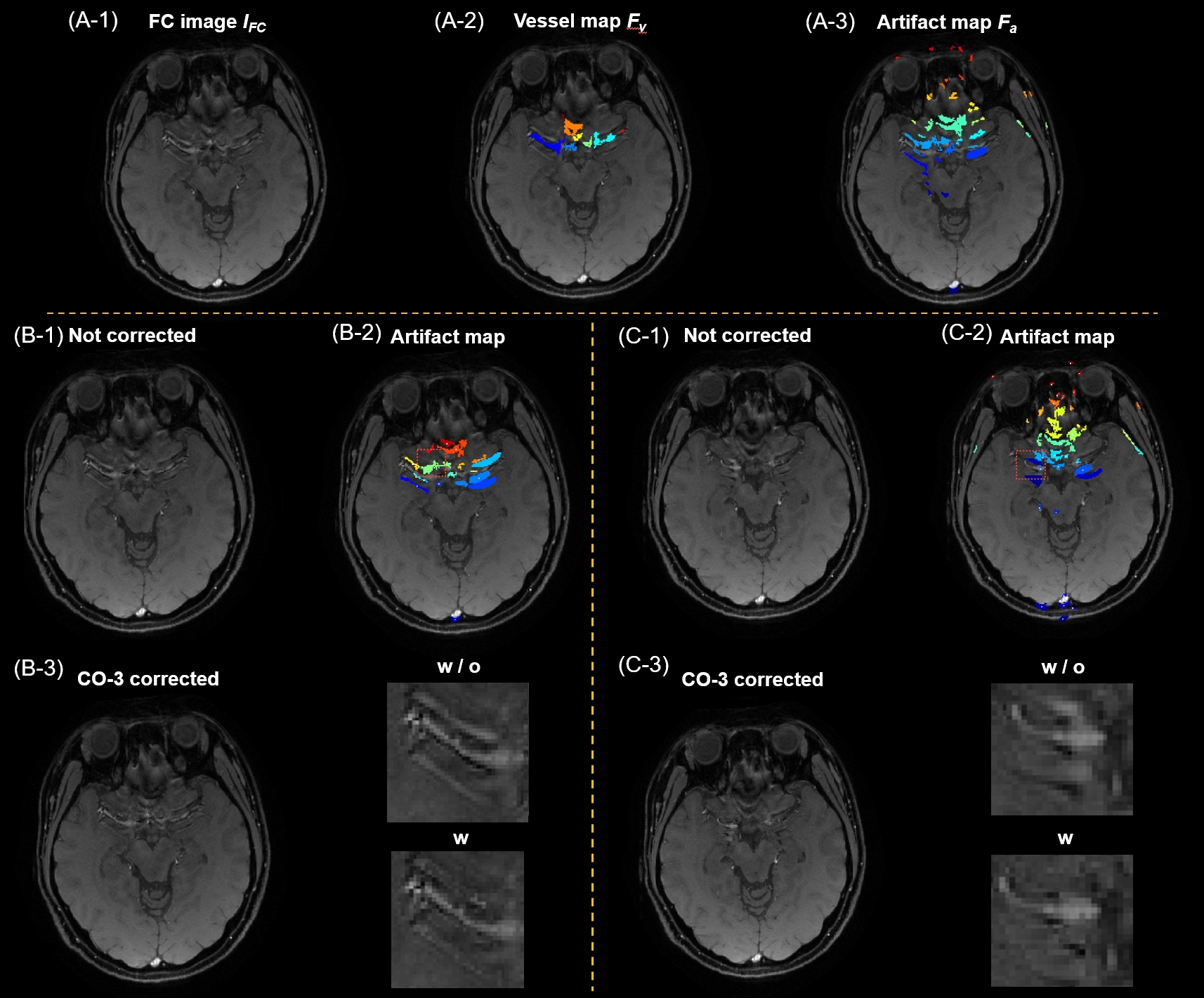
Figure 3. (A) Automatically identified vessel and artifact maps based on signal characteristics in the flow phase-based reconstruction and post-processing. (B) and (C) showed the results of two representative planes with different shape of vessels & artifacts before and after correction, demonstrating that, upon accurate artifact region identification, the image restoration algorithm effectively suppressed large vessel artifacts and produced smooth tissue recovery.
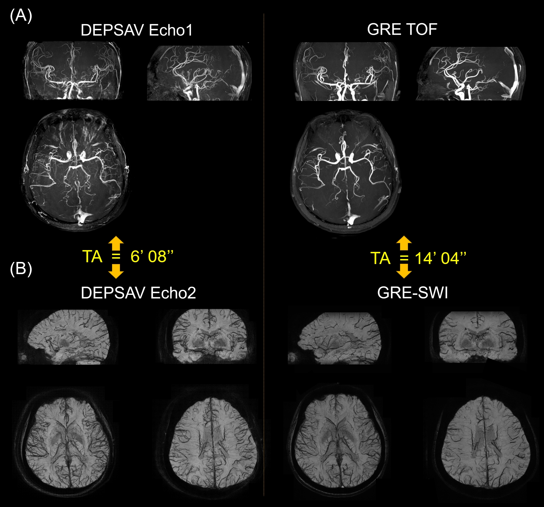
Figure 4. The dual contrast images acquired using DEPSAV-II showing the comparative results of MRA and MRV images using GRE acquisition, with largely reduced scan time.