0941
3D-Yarnball Acquisition and Reconstruction Advancement Yields High Resolution T1-Weighted Whole Brain Images in Just Over 1 Minute1University of Alberta, Edmonton, AB, Canada, 2Western University, London, ON, Canada
Synopsis
Keywords: Data Acquisition, Data Acquisition
Motivation: This study is motivated by the creation of high-resolution T1-weighting whole brain images in considerably less time (1 minute) than currently required for standard MP-RAGE (~4 minutes).
Goal(s): The sampling efficient 3D-Yarnball trajectory offers a potential imaging solution, but trajectory/sequence design and reconstruction aspects remain to be explored.
Approach: Variably under-sampled Yarnball trajectories with 2-10 ms duration were compared in healthy brain, along with different methods of steady-state sequence excitation. Iterative, off-resonance correcting, wavelet-regularized reconstruction was applied to Yarnball for the first time.
Results: Yarnball sequence and reconstruction consideration enabled high-quality 0.77 mm isotropic whole brain images in 1 minute
Impact: The image acquisition, sequence, and reconstruction investigation of this work enabled robust, high-quality 0.77 mm isotropic T1-weighted whole brain images in just over 1 minute. The goal of this work is to facilitate considerably shorter MRI protocols.
Introduction
3D-Yarnball k-space acquisition was originally introduced within the context of spoiled steady-state T1-weighted human brain imaging, and a 10 ms readout duration (TRO) was arbitrarily selected for initial demonstration of sampling efficiency1. However, while long spiraling readouts are efficient, they are prone to off-resonance1,2. Here, the purpose is to compare long TRO with shorter TRO, which necessarily require under-sampling to achieve the same scan duration. Image acquisition with and without fat-saturation is compared, and an iterative, off-resonance correcting reconstruction3 is considered for Yarnball, all for the first time.Methods
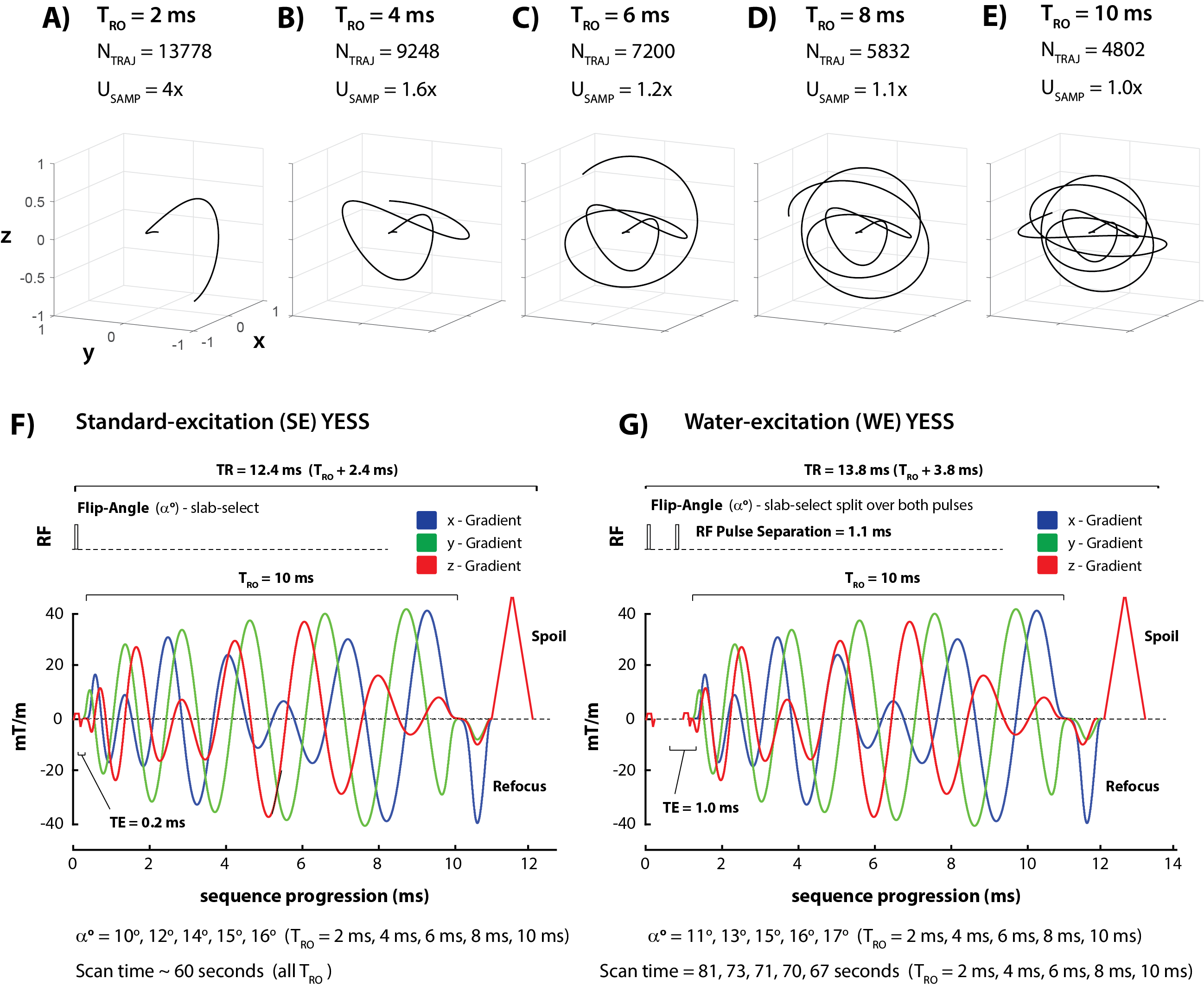
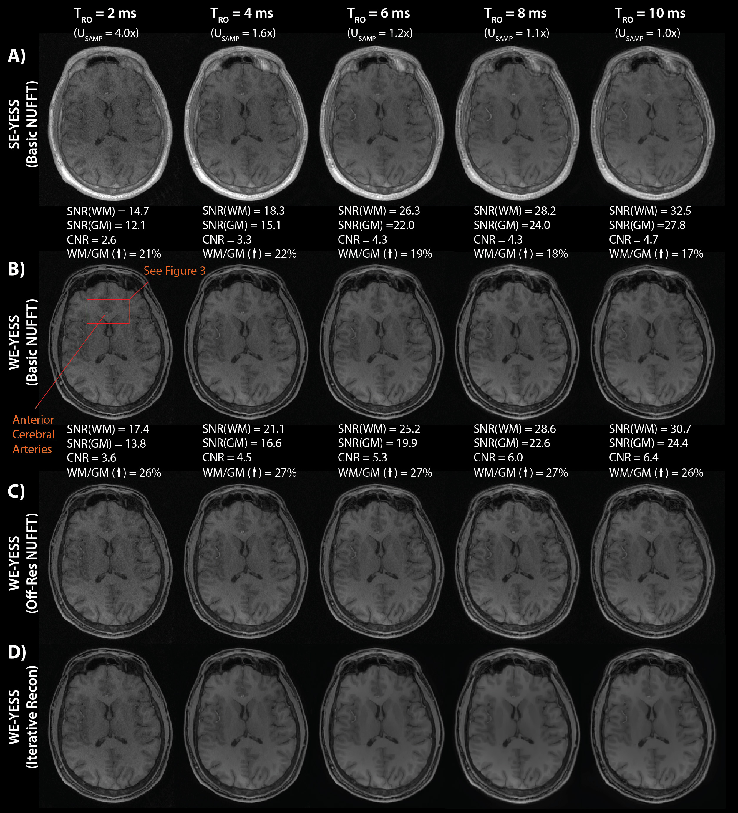
Five different Yarnball readouts (TRO=2,4,6,8,10 ms) were developed for 0.77 mm isotropic voxels and a 240 mm FoV (Figure 1A-E). Each was fully sampled to ~15% of the k-space radius (used for coil profile estimation), and under-sampled beyond that to achieve the requisite number of trajectories (NTRAJ) for a 60 second standard-excitation Yarnball-Encoded Spoiled Steady-state (SE-YESS) scan (Figure 1F). SE-YESS was also compared with water-excitation (WE-YESS) for fat suppression (Figure 1G). Note that TR=TRO+ 2.4 ms(SE-YESS) or 3.8 ms(WE-YESS), accounting for excitation, refocus and spoiling. Flip-angle (αo) was determined for maximum signal difference (and thus CNR) between white matter (WM) and gray matter (GM) using T1(WM)=1400 ms, and T1(WM)=875 ms.Four healthy volunteers (F–24 years/F–45 years/M–27 years/M–47 years) were scanned on a Siemens Prisma 3T with a 20 channel head-neck coil, and for each volunteer, both SE-YESS and WE-YESS images were acquired for all five TRO. An off-resonance map was also acquired from two (interleaved) SE-YESS images with TE=0.2/0.8 ms in a total time of 15 seconds (voxels=2.5 mm isotropic, TRO=1.5 ms, NTRAJ=1891). Basic NUFFT reconstruction was first used to compare SNR and CNR between the different TRO and YESS sequences (using Rayleigh distributed background noise), and to demonstrate under-sampling and off-resonance artifacts/blurring. Next, a time-segmented off-resonance correcting NUFFT4 was considered. Finally, images were created with off-resonance correcting wavelet-regularized (2 levels/dimension, λ=20), BFISTA5 based (NIT = 5) iterative reconstruction (Gitlab/MatMRI3, ~3 minutes using custom NUFFTs on 4 Titan-RTX graphics cards). Given their conspicuity, size and separation, the precallosal segments of the anterior cerebral arteries were used as ‘resolution elements’ to compare both resolution between different TRO, and image reconstruction effectiveness. Resolution, SNR/CNR and off-resonance artifacts are used to suggest the ‘best’ TRO and YESS sequence for T1-weighted brain imaging.
Results
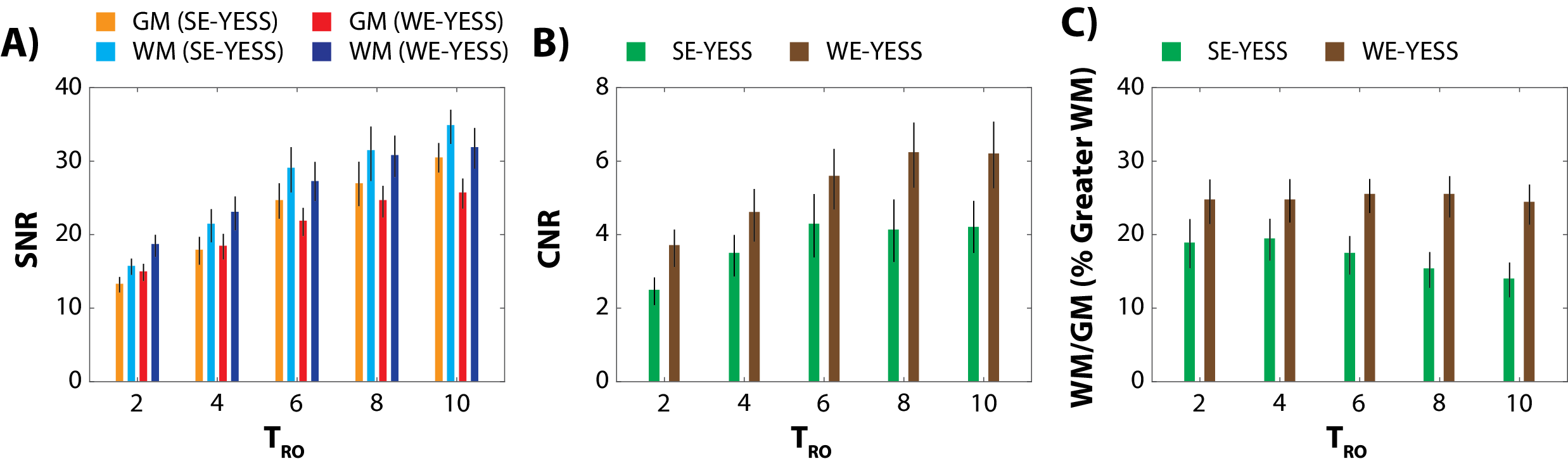
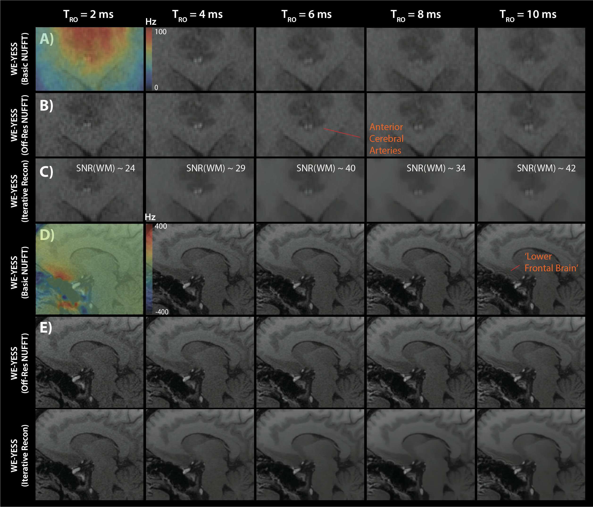
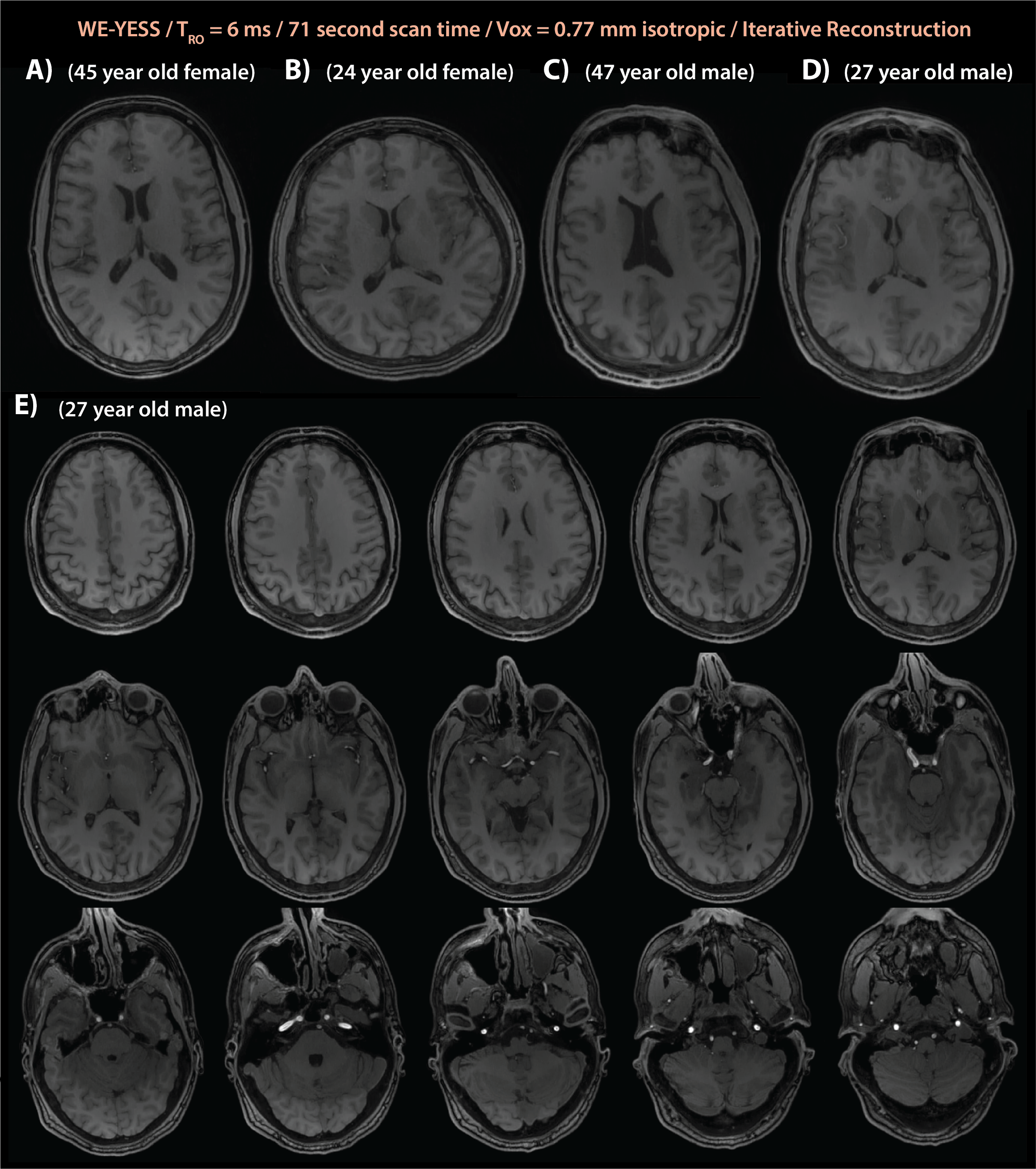
Representative images in Figure 2 demonstrate the absence of coherent aliasing artifact in the 4x under-sampled TRO=2 ms Yarnball images, as well as progressive SNR and CNR increase with TRO for both SE-YESS and WE-YESS, measured from the basic NUFFT reconstruction (Figure 2A-B). These consistent increases were observed over all four volunteers (Figure 3A-C). Interestingly, the CNR of WE-YESS is as much as 50% greater than SE-YESS (Figure 3B), and the WM/GM ratio of 1.25 for WE-YESS is considerably greater than the 1.17 for SE-YESS, which decreases with TRO (Figure 3C). For this reason, further results are presented for WE-YESS only. Off-resonance smearing is effectively demonstrated with the precallosal segments of the anterior cerebral arteries, which are here used as ‘resolution elements’ (Figures 2B/4A). At this location, the tissue is ~100 Hz off resonance (Figure 4A), and the resultant smearing at long TRO=10 ms is clearly visible. However, this smearing is effectively removed with the off-resonance correcting NUFFT (Figures 2C/4B). Iterative image reconstruction visibly reduces image noise while maintaining resolution, as observed through ‘artery resolution element’ distinction. However, the SNR in WM (estimated within uniform WM regions) remains lower for TRO<4 ms (Figures 2C/4B). Large tissue boundary off-resonance (~300 Hz) yields a ringing artifact at long TRO highlighted in the ‘lower frontal brain’ region of Figure 4D. This ringing is not removed with the off-resonance correcting NUFFT (Figure 4E) or iterative reconstruction (Figure 4F). Thus, image noise for short TRO<4 ms, and ringing at highly off-resonant tissue boundaries for long TRO>8 ms, promotes the use of TRO=6 ms for T1-weighted YESS imaging. TRO=6 ms images from all 4 volunteers (Figure 5A-D), and slices showing full brain coverage (Figure 5E), demonstrate excellent image quality.Discussion
Yarnball-encoded steady-state (YESS) readout selection (TRO=6 ms) that considered both SNR/CNR and tissue boundary off-resonance ringing, combined with water-excitation sequence selection for elevated CNR, and iterative, wavelet regularized, off-resonance correcting reconstruction3, produced high-quality T1-weighted brain images with 0.77 mm isotropic voxels in 71 seconds at 3T (or 86 seconds total, when combined with the off-resonance scan) (Figure 5). Future work will compare YESS with (~4 minute) MP-RAGE, demonstrate motion robustness, and facilitate considerably shorter MRI protocols.Acknowledgements
No acknowledgement found.References
Stobbe RW, Beaulieu C. Three-dimensional Yarnball k-space acquisition for accelerated MRI. Magn Reson Med. 2021;85(4):1840-1854.
Haskell MW, Nielsen JF, Noll DC. Off-resonance artifact correction for MRI: A review. Nmr Biomed. 2023;36(5).
Varela-Mattatall G, Dubovan PI, Santini T, Gilbert KM, Menon RS, Baron CA. Single-shot spiral diffusion-weighted imaging at 7T using expanded encoding with compressed sensing. Magn Reson Med. 2023;90(2):615-623.
Fessler JA, Lee S, Olafsson VT, Shi HR, Noll DC. Toeplitz-based iterative image reconstruction for MRI with correction for magnetic field inhomogeneity. Ieee T Signal Proces. 2005;53(9):3393-3402.
Ting ST, Ahmad R, Jin N, et al. Fast Implementation for Compressive Recovery of Highly Accelerated Cardiac Cine MRI Using the Balanced Sparse Model. Magn Reson Med. 2017;77(4):1505-1515.
Figures