0936
A new handle on extracellular diffusion and exchange? Single and double diffusion encoded MRS in humans after MSM ingestion1Danish Research Centre for Magnetic Resonance, Copenhagen University Hospital – Amager and Hvidovre, Hvidovre, Denmark, 2Department of Health Technology, Technical University of Denmark, Lyngby, Denmark, 3Clinical Imaging Sciences Centre, Brighton and Sussex Medical School, University of Sussex, Brighton, United Kingdom, 4CIBM Center for Biomedical Imaging, EPFL CIBM-AIT, EPFL Lausanne, Lausanne, Switzerland, 5Cardiff University Brain Research Imaging Centre (CUBRIC), School of Psychology, Cardiff University, Cardiff, United Kingdom, 6Magnetic Resonance Methodology, Institute of Diagnostic and Interventional Neuroradiology, University of Bern, Bern, Switzerland, 7Translational Imaging Center, sitem-insel, Bern, Switzerland
Synopsis
Keywords: Microstructure, Microstructure
Motivation: The investigation of extracellular spaces with diffusion weighted MRI andMRS is limited by the ubiquitous distribution and fast exchange of water, while endogenous metabolites are mostly intracellular.
Goal(s): Investigate the diffusion characteristics of ingested MSM compared to water and endogenous metabolites.
Approach: Multi b-valued single and double diffusion encoded MRS in humans at 3T.
Results: MSM diffuses anisotropically, but considerably faster than intracellular metabolites, and exhibits a substantially different behavior compared to both water and metabolites.
Impact: MSM provides a novel probe of extracellular spaces similar to water but with differences that could be rotted in different transmembrane exchange properties.
Introduction
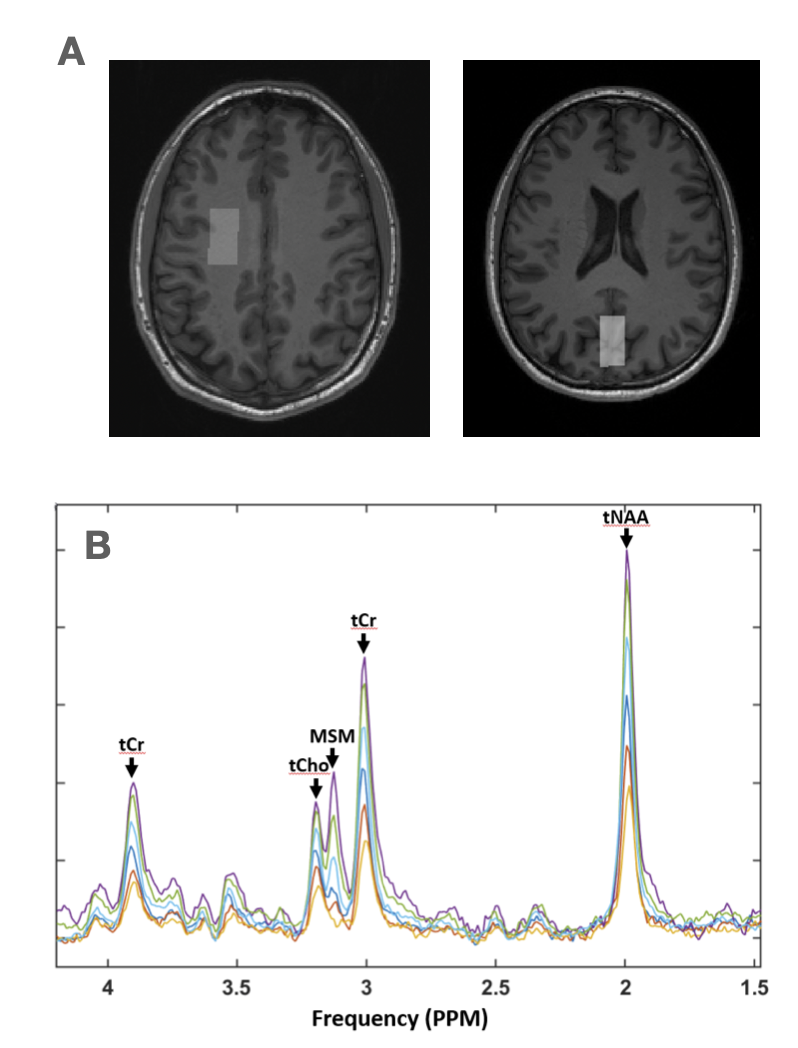
Diffusion weighted magnetic resonance spectroscopy (DWS) offers a unique view into neuronal and glial intracellular morphology through the measurement of endogenous metabolites1. In contrast, a more direct view on the extracellular space is limited through the low abundance of MR-visible molecules such as lactate and potentially fast exchange of omnipresent water. Studies have so far been limited to more invasive experiments in rodents with exogenous administration of e.g. sucrose or tetramethylammonium2,3. In this work we explore methylsulfonylmethane (MSM, (CH3)2SO2) as a potential exogenous diffusion marker suitable for in vivo human use. MSM is a dietary supplement commercialized for use in humans and pets. Interestingly, orally ingested MSM crosses the blood brain barrier and provides a prominent MRS visible singlet at 3.15 ppm with a half-life of 3.5 to 7.5 days4,5. The distribution of MSM in brain tissue is unknown but could offer an access to a complimentary view on microstructure not provided by water or metabolites.Methods
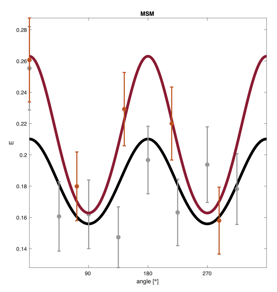
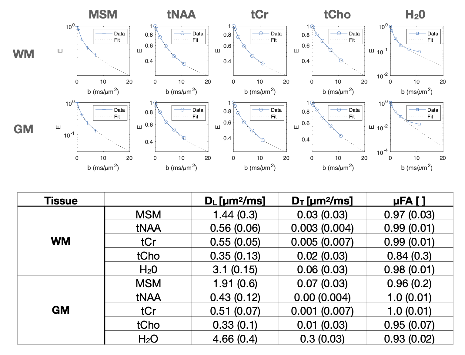
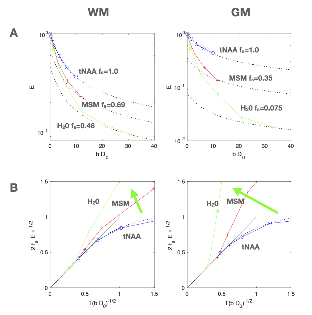
Experiments: 6 healthy male individuals (age 29-61) volunteered for the experiments and ingested 10 g MSM dissolved in water 12-24 hours prior to the scans. Experiments were performed on 3T Siemens Prisma scanners using a sLASER sequence modified with diffusion weighting gradients. Single diffusion encoding was performed with cardiac synchronization and TE/TR= 100 ms/3 cardiac cycles , b = 0 - 11.00 ms/µm2, voxel size 9 cc. Voxel was acquired mainly covering parietal white matter (WM) in 5 scans and occipital gray matter (GM) in 4 scans. MPRAGE images were acquired for positioning. Double diffusion encoding for detection of microscopic anisotropy used metabolite-cycling to preserve water as an internal reference for phase, frequency-drift, and eddy-current correction and motion compensation6,7. Data were acquired twice in one subject in WM (10.6±0.5mL) at a total b=5.200 ms/µm2, with 5 or 8 angles over the full circle in 3 planes (TE/TR: 125/2500 ms; Δ/δ/ε/τ: 32/14/1/39 ms). The experiments were approved by the local ethics committees.Analysis: Data were corrected for phase and frequency drifts, powder averaged over gradient directions in Matlab and quantified for each diffusion condition using LCModel or FiTAID with a MSM singlet added to the basis set8.Single diffusion encoded powder averaged MRS data was fitted to the “Callaghan model” of randomly oriented monodisperse diffusion tensors9,10. tNAA and water data at high b-values and intermediate diffusion times reflect the same “stick” diffusion with a common WM/GM-ratio dependent tortuosity T11. We thus compared tNAA, water and MSM relative to their respective free diffusivities at body temperature set to D0 = 0.9, 3.0 and 1.6 µm2/ms and compared attenuations to the b-1/2 power law scaling expected for “stick” fractions12,13. A maximum stick fraction (fs) was estimated as the extrapolation of the tNAA signal.
Results & Discussion
Clear MSM peaks were observed in all participants (Fig 1) and provided fits with CRLB below 12% and 22% for b = 7 ms/µm2 in white and gray matter respectively. Double diffusion encoded MSM data exhibited a cosinusoidal modulation characteristic of microscopic anisotropy (Fig 2).Metabolite diffusivities and µFA where in line with earlier studies but apparently overestimated for water and MSM (DL exceeding D0) which may be attributed to a violation of a monodisperse scenario reflecting a mixture of both anisotropic intracellular and less anisotropic extracellular signal components (Fig 3). MSM attenuates slower than water but faster than endogenous metabolites in both GM and WM when accounting for their different free diffusivity (Fig 4 A). MSM and water approach a common b-1/2 scaling as tNAA in WM but not in GM (Fig 4 B). An apparently larger “stick” fraction of MSM could be assigned to lower exchange rates from MSM compared to water in dendrites and glial processes.Conclusions
Orally ingested MSM demonstrates a microscopically anisotropic diffusion that is distinctively different from both endogenous metabolites and water. We suggest that MSM is similarly distributed as water but is governed by significantly lower transmembrane permeabilities. A lower exchange rate could facilitate a better understanding of the extracellular space and its interaction with intracellular spaces but further experiments reflecting time dependent diffusion are needed.Acknowledgements
HL is supported by the European Research Council (ERC) (EU Horizon 2020, #804746). AD is supported by a Swiss National Science Foundation Fellowship (SNSF #202962). The authors would like to thank Edward J. Auerbach, Dinesh Deelchand and Małgorzata Marjańska from the Center for Magnetic Resonance Research at the University of Minnesota for assistance in obtaining and implementing the sLASER MRS and DW-MRS sequences.References
[1] M. Palombo, N. Shemesh, I. Ronen, and J. Valette, “Insights into brain microstructure from in vivo DW-MRS,” Neuroimage, vol. 182, no. June 2017, pp. 97–116, 2018, doi: 10.1016/j.neuroimage.2017.11.028.
[2] M. Vincent, M. Gaudin, C. Lucas-Torres, A. Wong, C. Escartin, and J. Valette, “Characterizing extracellular diffusion properties using diffusion-weighted MRS of sucrose injected in mouse brain,” NMR Biomed., vol. 34, no. 4, pp. 1–16, 2021, doi: 10.1002/nbm.4478.
[3] C. D. Kroenke, J. J. H. Ackerman, and J. J. Neil, “Magnetic resonance measurement of tetramethylammonium diffusion in rat brain: Comparison of magnetic resonance and ionophoresis in vivo diffusion measurements,” Magn. Reson. Med., vol. 50, no. 4, pp. 717–726, 2003, doi: 10.1002/mrm.10579.[4] S. E. Rose, J. B. Chalk, G. J. Galloway, and D. M. Doddrell, “Detection of dimethyl sulfone in the human brain by in vivo proton magnetic resonance spectroscopy,” Magn. Reson. Imaging, vol. 18, no. 1, pp. 95–98, 2000, doi: 10.1016/S0730-725X(99)00110-1.
[5] L. G. Kaiser, D. Russell, T. Maschmeyer, R. L. Redfern, and B. A. Inglis, “Methylsulfonylmethane (MSM): A chemical shift reference for 1H MRS of human brain,” Magn. Reson. Med., vol. 83, no. 4, pp. 1157–1167, 2020, doi: 10.1002/mrm.27997.
[6] A. Döring, V. Adalid, C. Boesch, and R. Kreis, “Diffusion-weighted magnetic resonance spectroscopy boosted by simultaneously acquired water reference signals,” Magn. Reson. Med., vol. 80, no. 6, pp. 2326–2338, 2018, doi: 10.1002/mrm.27222.
[7] André Döring, Jessie Mosso, Roland Kreis, Derek K Jones, Chloé Najac, Matt G Hall, Henrik Lundell, Lijing Xin, Itamar Ronen, “Robust double-diffusion-encoded spectroscopy in the human brain using metabolite-cycling", ESMRMB 2023
[8] S. W. Provencher, “Automatic quantitation of localized in vivo 1H spectra with LCModel,” NMR Biomed., 2001, doi: 10.1002/nbm.698.
[9] C. D. Kroenke, J. J. H. Ackerman, and D. A. Yablonskiy, “On the nature of the NAA diffusion attenuated MR signal in the central nervous system,” Magn. Reson. Med., 2004, doi: 10.1002/mrm.20260.
[10] H. Lundell, C. Ingo, T. B. Dyrby, and I. Ronen, “Cytosolic diffusivity and microscopic anisotropy of N-acetyl aspartate in human white matter with diffusion-weighted MRS at 7 T,” NMR Biomed., vol. 34, no. 5, pp. 1–14, 2021, doi: 10.1002/nbm.4304.
[11] H. Lundell, C. Najac, M. Bulk, H. E. Kan, A. G. Webb, and I. Ronen, “Compartmental diffusion and microstructural properties of human brain gray and white matter studied with double diffusion encoding magnetic resonance spectroscopy of metabolites and water,” Neuroimage, vol. 234, no. March, p. 117981, 2021, doi: 10.1016/j.neuroimage.2021.117981.
[12] J. Veraart, E. Fieremans, and D. S. Novikov, “On the scaling behavior of water diffusion in human brain white matter,” Neuroimage, vol. 185, no. October 2018, pp. 379–387, 2019, doi: 10.1016/j.neuroimage.2018.09.075.
[13] J. L. Olesen, L. Østergaard, N. Shemesh, and S. N. Jespersen, “Diffusion time dependence, power-law scaling, and exchange in gray matter,” Neuroimage, vol. 251, no. August 2021, p. 118976, 2022, doi: 10.1016/j.neuroimage.2022.118976.
Figures