0935
Quantification of exchange in the mouse brain using double diffusion encodings with fixed total diffusion-weighting1Wellcome Centre for Integrative Neuroimaging, FMRIB, Nuffield Department of Clinical Neurosciences, University of Oxford, Oxford, United Kingdom, 2Section on Quantitative Imaging and Tissue Sciences, Eunice Kennedy Shriver National Institute of Child Health and Human Development, NIH, Bethesda, MD, United States, 3National Institute of General Medical Sciences, NIH, Bethesda, MD, United States
Synopsis
Keywords: Diffusion Acquisition, Diffusion/other diffusion imaging techniques, Exchange
Motivation: Exchange is an important effect in diffusion MR of the brain but remains difficult to quantify using conventional methods and signal models due to parameter degeneracy.
Goal(s): To develop and demonstrate robust measurement of exchange in the mouse brain.
Approach: A method based on double diffusion encoding was previously developed to probe exchange isolated from other effects, yielding robust exchange time measurements. We apply this method in vivo for the first time.
Results: We report a fast in vivo exchange time of approximately 38 ms as compared to 146 ms in a fixed sample, obtained by averaging through a slice.
Impact: Cellular water exchange reflects not only structural characteristics but has also been linked to metabolism. Quantifying exchange may yield rich information, yet methods to do so are not mature. Here, we demonstrate a unique, isolated measurement of exchange in vivo.
Introduction
The exchange of water between biological microenvironments, namely the intra- and extracellular space, is increasingly recognized as an important effect in diffusion MR of the brain.1-3 Exchange has also recently been linked to steady-state, metabolic activity4-6, and may thus be a valuable biomarker. Despite growing interest, a standard diffusion MR method to measure exchange has yet to emerge. Various methods have been proposed3,7-9 (e.g., NEXI10), but these generally do not isolate exchange – rather, exchange is modelled in tandem with microstructural parameters, resulting in issues of parameter degeneracy. The difficulty of incorporating exchange into compartment signal models may explain, in part, the wide range of reported exchange times $$$\tau_k$$$ in the literature, which span from $$$\tau_k\approx3-500$$$ ms, even in similar tissue.1,3 These disparate reports indicate that the quantification of exchange remains an open problem.Previous work based on diffusion exchange spectroscopy (DEXSY)11 – a double diffusion encoding method – showed that with just two acquisitions per mixing time, one can separate exchange from other effects.12-15 Thus, this method overcomes the issue of parameter degeneracy, and $$$\tau_k$$$ can be quantified without simultaneously estimating parameters such as intra-/extracellular diffusivities. Here, we provide the first proof-of-concept demonstration of this method in mouse brain, in vivo and in a fixed sample, paving the way for future developments towards quantitative exchange imaging.
Theory
The method is based on sub-sampling the 3D parameter space of the DEXSY experiment, which consists of two diffusion encodings $$$(b_1,b_2)$$$, separated by a longitudinal storage/mixing time, $$$t_m$$$. As previously shown12,14, holding the sum of $$$b$$$-values constant – $$$b_s=b_1+b_2$$$ – removes the effect of non-exchanging, Gaussian diffusion, leaving non-Gaussian diffusion and exchange.14,15 These effects can be captured in the log-ratio of two acquisitions: (i) with equal diffusion-weighting $$$b_1=b_2=b_s/2$$$, denoted $$$S_{b_1=b_2}$$$, and (ii) with $$$b_1=b_s,\;b_2=0$$$, denoted $$$S_{b_1=b_s}$$$. The evolution of this log-ratio with $$$t_m$$$ separates exchange from non-Gaussian diffusion, as non-Gaussian diffusion does not vary with $$$t_m$$$, but manifests in the intercept. Exchange can then be fit to a first-order model:$$-\ln\left(\frac{S_{b_1=b_2}}{S_{b_1=b_s}}\right)=C_1\exp\left(-\frac{t_m}{\tau_k}\right)+C_0,$$
where $$$C_0$$$ captures non-Gaussian diffusion, exchange during the encoding, and $$$T_2$$$-$$$T_2$$$ exchange, and $$$C_1$$$ is proportional to the experimentally observable exchange. Taking a log-ratio also removes $$$t_m$$$-dependent effects, namely $$$T_1$$$-relaxation.
Methods
A stimulated echo DEXSY sequence was implemented on a horizontal-bore $$$7$$$-$$$T$$$ Bruker BioSpec 70/20 (Ettlingen, Germany), using an $$$86\;\mathrm{mm}$$$ transmit RF coil and a 4-channel CryoProbe for the receive coil (Bruker, Germany). The sequence uses bipolar gradients to achieve double diffusion encoding with a chosen $$$b_s=3500\;\mathrm{s/mm^2}$$$, varying the amplitude to acquire either $$$S_{b_1=b_2}$$$ or $$$S_{b_1=b_s}$$$ (Fig. 1). Gradients were oriented in the slice direction (rostral-caudal). A standard EPI readout was used for imaging $$$(0.4\times0.4\;\mathrm{mm}$$$ in-plane, $$$0.75\;\mathrm{mm}$$$ slice thickness). Additionally, $$$\delta/\Delta=4.5/8\;\mathrm{ms}$$$, and $$$\mathrm{TE}=21.6\;\mathrm{ms}$$$. $$$\mathrm{TR}$$$ was variable with a recovery time of $$$3.6$$$ or $$$7.2\;\mathrm{s}$$$ ($$$\mathrm{NR}=10$$$ or $$$5$$$, using the median) in the fixed sample or in vivo, respectively. $$$18$$$ mixing times were acquired from $$$t_m=2-500\;\mathrm{ms}$$$, for a per-slice, per-repetition scan time of $$$4\;\mathrm{min}$$$.Animal procedures were approved by the local Animal Welfare and Ethical Review Body. A C57BL6J wild-type adult mouse ($$$n=1$$$, male) was scanned.
Results
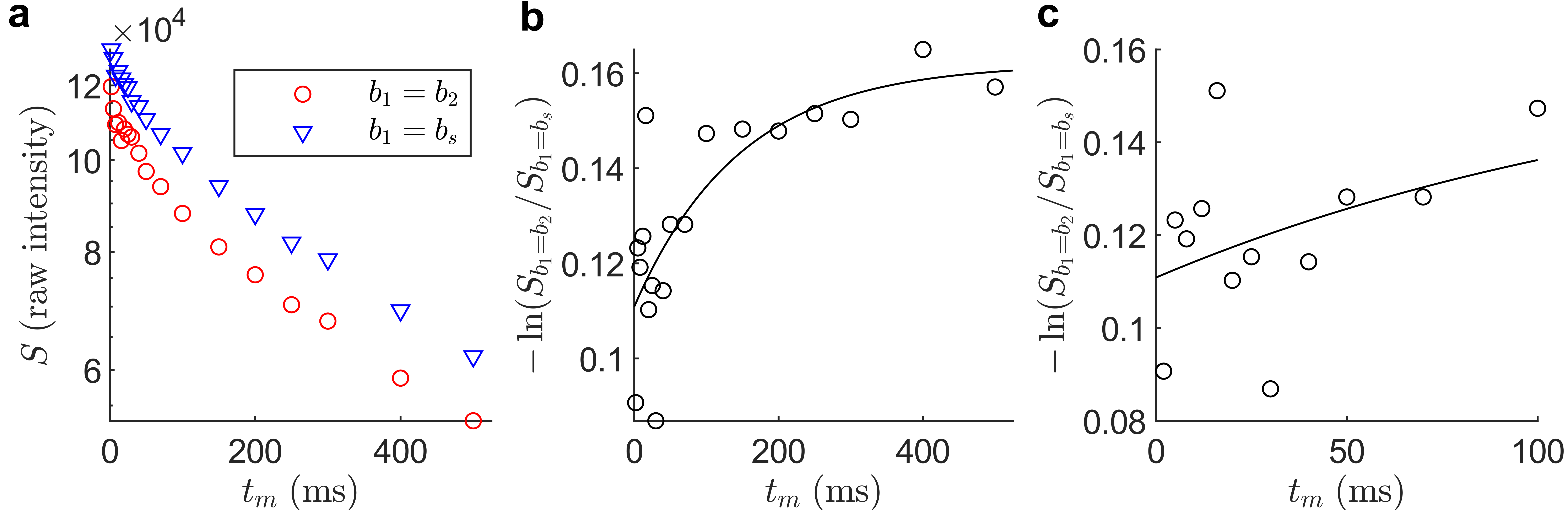
In Fig. 2, the raw in vivo images $$$S_{b_1=b_2}$$$ and $$$S_{b_1=b_s}$$$ at selected $$$t_m$$$ are shown for an exemplar slice. The slice means after masking of the brain were analyzed (rather than individual voxels) due to SNR difficulties arising from the combination of images. In Fig. 3a, the slice means from Fig. 2 are plotted vs. $$$t_m$$$. While both means exhibit decay with $$$t_m$$$ primarily due to $$$T_1$$$-relaxation, $$$S_{b_1=b_2}$$$ exhibits additional decay attributable to exchange. The log-ratio was then fit to Eq. 1, shown in Fig. 3b, yielding $$$\tau_k\approx38\;\mathrm{ms}$$$.Analogous results are shown in Figs. 4 and 5 for a fixed sample, yielding $$$\tau_k\approx146\;\mathrm{ms}$$$.
Discussion and Conclusions
The estimated $$$\tau_k\approx38\;\mathrm{ms}$$$ in vivo lies on the faster end of estimates in the literature1,3 and is of the same order-of-magnitude as previous results $$$\tau_k\approx13\;\mathrm{ms}$$$ obtained in viable, ex vivo neonatal mouse spinal cord6,13,15, which is mostly gray matter. The longer $$$\tau_k$$$ here may result from averaging through a slice containing (less permeable) white matter. The even longer $$$\tau_k\approx146\;\mathrm{ms}$$$ in fixed tissue supports the hypothesis that exchange is linked to activity. Importantly, these results also suggest that exchange cannot be ignored in diffusion MR measurements with typical encoding times of $$$\Delta\sim20-40\mathrm{ms}$$$.The presented results, while preliminary, demonstrate the feasibility of an isolated exchange measurement. No microstructural parameters are estimated in Eq. 1, and the parameters $$$C_0,\;C_1$$$ correspond merely to an intercept and limit in the log-ratio. With further SNR developments, robust in vivo imaging of $$$\tau_k$$$ may be achieved using this method.
Acknowledgements
MT and KLM contributed equally to this work.
TXC and PJB were supported by the IRP of the NICHD.
NHW was funded by the NIGMS PRAT Fellowship Award #FI2GM133445-01.
The Wellcome Centre for Integrative Neuroimaging is supported by core funding from the Wellcome Trust (203139/Z/16/Z), which funds MT. KLM is supported by a Wellcome Trust Senior Research Fellowship (224573/Z/21/Z).
References
1. Nilsson M, van Westen D, Ståhlberg F, Sundgren PC, Lätt J. The role of tissue microstructure and water exchange in biophysical modelling of diffusion in white matter. MAGMA. 2013;26(4):345-70.
2. Jelescu IO, Palombo M, Bagnato F, Schilling KG. Challenges for biophysical modeling of microstructure. Journal of Neuroscience Methods. 2020;344:108861.
3. Olesen JL, Østergaard L, Shemesh N, Jespersen SN. Diffusion time dependence, power-law scaling, and exchange in gray matter. NeuroImage. 2022;251:118976.
4. Springer CS, Baker EM, Li X, et al. Metabolic activity diffusion imaging (MADI): I. Metabolic, cytometric modeling and simulations. NMR in Biomedicine. 2023;36(1): e4781.
5. Springer CS, Baker EM, Li X, et al. Metabolic activity diffusion imaging (MADI): II. Noninvasive, high‐resolution human brain mapping of sodium pump flux and cell metrics. NMR in Biomedicine. 2023;36(1):e4782.
6. Williamson NH, Ravin R, Cai TX, Falgairolle M, O’Donovan MJ, Basser PJ. Water exchange rates measure active transport and homeostasis in neural tissue. PNAS Nexus. 2023;2(3):pgad056.
7. Kärger J. NMR self-diffusion studies in heterogeneous systems. Advances in Colloid and Interface Science. 1985;23:129-48.
8. Åslund I, Nowacka A, Nilsson M, Topgaard D. Filter-exchange PGSE NMR determination of cell membrane permeability. Journal of Magnetic Resonance. 2009;200(2):291-5.
9. Chakwizira A, Westin C, Brabec J, et al. Diffusion MRI with pulsed and free gradient waveforms: Effects of restricted diffusion and exchange. NMR in Biomedicine. 2023;36(1):e4827.
10. Jelescu IO, de Skowronski A, Geffroy F, Palombo M, Novikov DS. Neurite Exchange Imaging (NEXI): A minimal model of diffusion in gray matter with inter-compartment water exchange. NeuroImage. 2022;256:119277.
11. Callaghan PT, Furó I. Diffusion-diffusion correlation and exchange as a signature for local order and dynamics. The Journal of Chemical Physics. 2004;120(8):4032-8.
12. Cai TX, Benjamini D, Komlosh ME, Basser PJ, Williamson NH. Rapid detection of the presence of diffusion exchange. Journal of Magnetic Resonance. 2018;297:17-22.
13. Williamson NH, Ravin R, Benjamini D, et al. Magnetic resonance measurements of cellular and sub-cellular membrane structures in live and fixed neural tissue. eLife. 2019;8:e51101.
14. Williamson NH, Ravin R, Cai TX, et al. Real-time measurement of diffusion exchange rate in biological tissue. Journal of Magnetic Resonance. 2020;317:106782.
15. Cai TX, Williamson NH, Ravin R, Basser PJ. Disentangling the effects of restriction and exchange with diffusion exchange spectroscopy. Frontiers in Physics. 2022;10: 805793.
Figures