0930
3D whole brain mapping of creatine kinase metabolic rate using 31P-MR fingerprinting.1Animal imaging and technology core, CIBM Center for Biomedical Imaging, École polytechnique fédérale de Lausanne (EPFL), Lausanne, Switzerland, 2Laboratory for Functional and Metabolic Imaging, Ecole Polytechnique Fédérale de Lausanne (EPFL), Lausanne, Lausanne, Switzerland, 3Laboratory for Functional and Metabolic Imaging, Ecole Polytechnique Fédérale de Lausanne (EPFL), Lausanne, Switzerland, 4Laboratory for Functional and Metabolic Imaging, École Polytechnique Fédérale de Lausanne (EPFL), Lausanne, Switzerland, 5Department of Radiology, Case Western Reserve University, Cleveland, Cleveland, OH, United States, 6Animal imaging and technology core, CIBM Center for Biomedical Imaging, École Polytechnique Fédérale de Lausanne (EPFL), Lausanne, Switzerland
Synopsis
Keywords: MR Fingerprinting, MR Fingerprinting, 31P, MRF, creatine kinase rate, kCK, MRSI, 7T
Motivation: Using 31P MRS combined with magnetization transfer (MT) experiments including saturation transfer or inversion transfer to assess chemical exchange rate of creatine kinase (kCK) in the human brain are time-consuming and limited to 1D-acquisitions.
Goal(s): Acquiring a 3D whole brain kCK map.
Approach: In this abstract, we introduce an advanced, fast 3D-31P-MRF sequence for the human brain at 7T.
Results: The novel 3D-31P-MRF approach is feasible for whole brain mapping of kCK, enabling the investigation of region-specific energy metabolism under various pathological conditions.
Impact: Using the novel 3D-31P-MR Fingerprinting approach for whole brain mapping of kCK enables us to investigate region-specific energy metabolism under various pathological conditions and may enhance our understanding of the underlying molecular and metabolic processes.
Introduction
The chemical exchange rate of creatine kinase (kCK) in the human brain can be assessed by 31P MRS combined with magnetization transfer (MT) experiments, including saturation transfer or inversion transfer [3-7]. These methods are time-consuming, due to the long relaxation time of PCr, the low signal sensitivity in 31P-MRS and multiple measurements with different saturation or inversion delays. Recently, magnetization transfer (MT) phosphorus Magnetic Resonance Fingerprinting (31P-MRF) has been introduced to measure kCK for the human brain [1]. This framework showed a promising reduction in the scan time due to the intrinsically high SNR efficiency of a balanced steady-state free precession (bSSFP)-like scheme. However, MT-31P-MRF so far is still limited to a 1D acquisition. We introduce the advanced development: a fast 3D-31P-MRF sequence for the human brain at 7T. We demonstrate preliminary results of a 3D whole brain kCK map with 2 different head-coil setups.Methods
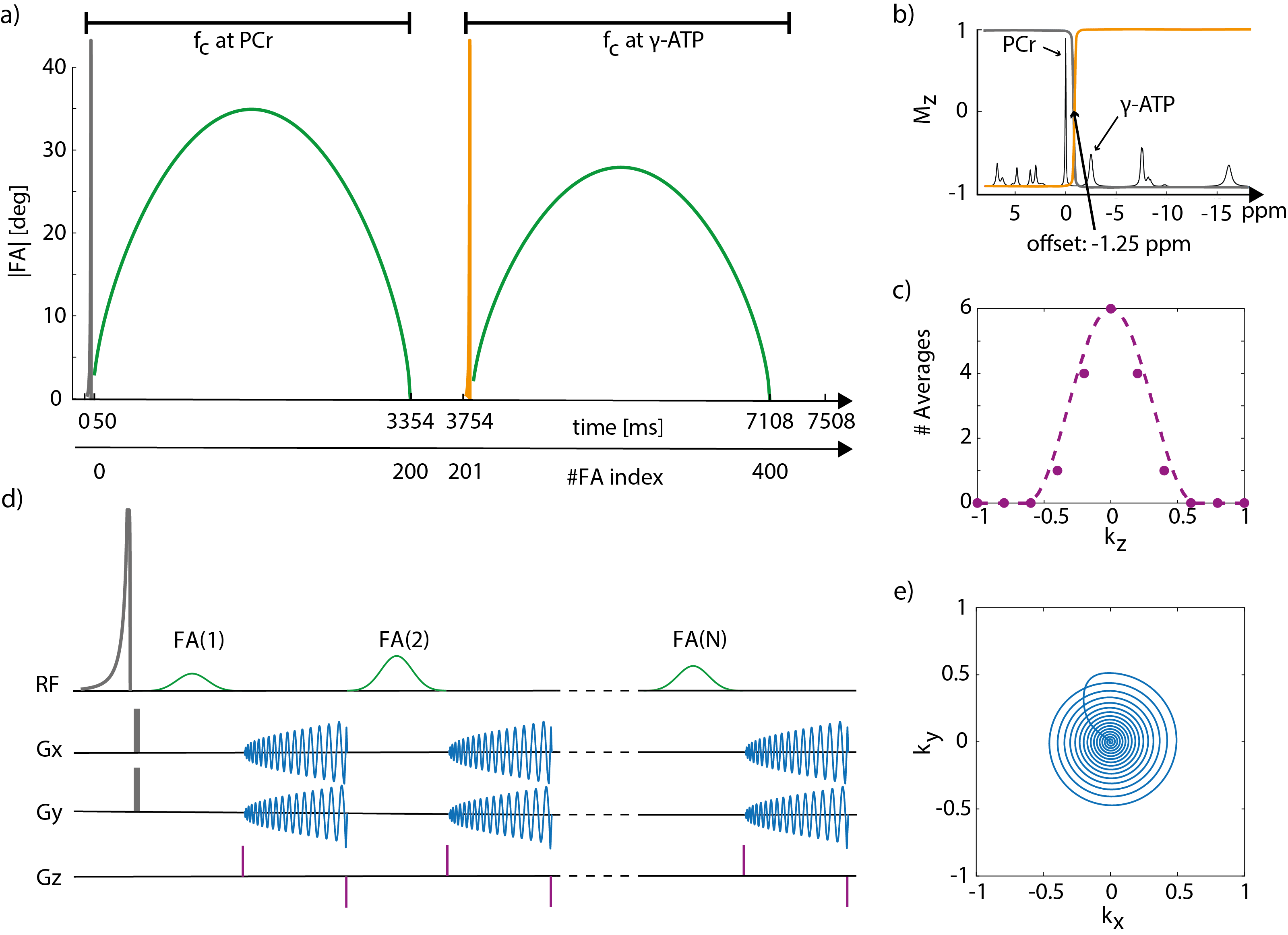
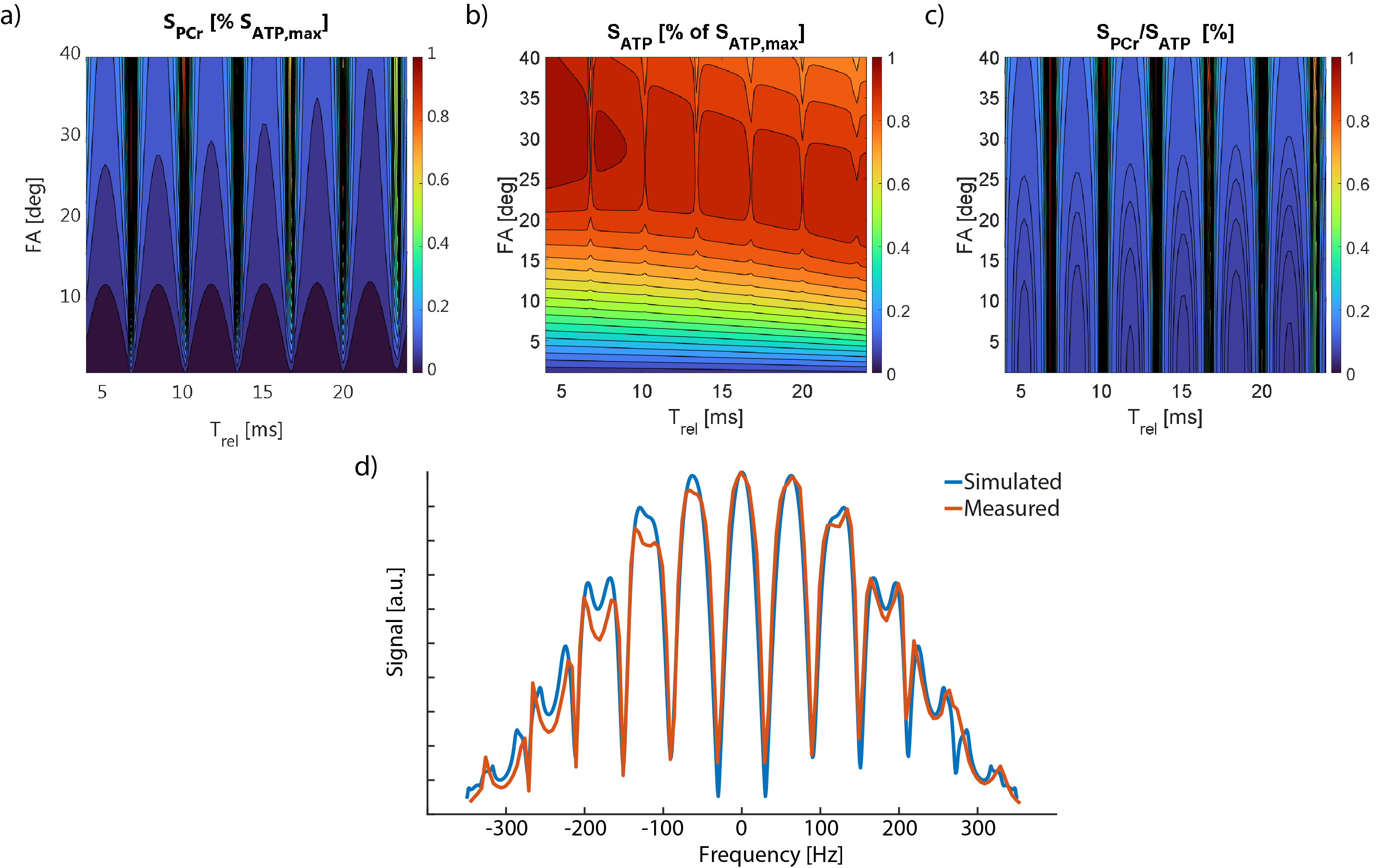
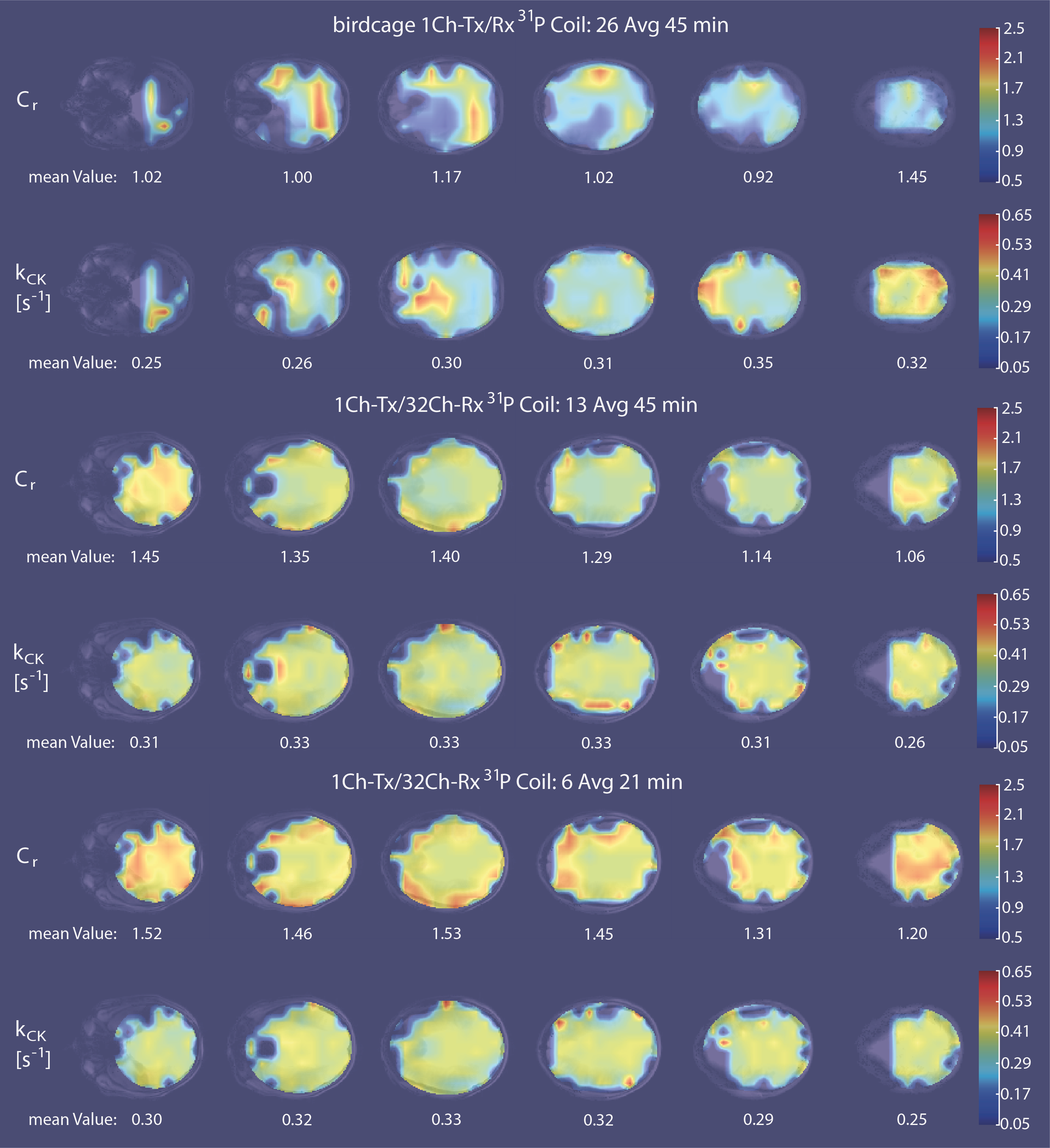
The new 3D-31P -MRF sequence is shown in Figure 1. A 8ms Gaussian pulse is used to selectively excite PCr or g-ATP. The flip angle (FA) pattern consists of 2 sinusoidals (200 FAs/sinusoidal): the first 200 FAs are applied with carrier frequency (fc) on PCr and the second 200 FAs on g-ATP. Both sinusoidals follow an asymmetric inversion pulse (Figure 1b) to introduce an inversion transfer effect. The repetition time (TR) between the RF pulses is optimized to minimize signal leakage from ATP to PCr and vice versa (Figure 2a-c). The time between pulses is used for the encoding gradients in the z-direction and the spiral gradients. A nonlinear spiral trajectory with a maximum |kxy| filling of 0.5 is chosen for denser sampling in the k-space center. In kz, a Hanning weighted stack of spirals is used (Figure 1c). Two healthy participants (1 female; 1 male; age 28 and 29 years), who provided written informed consent, were included. All MR experiments were performed on a 7T/68 cm MR scanner (Siemens Medical Solutions, Erlangen, Germany). One participant was scanned with a single channel 1H/31P birdcage coil. A whole brain map with a matrix size of 12x12x11 (FOV: 230x230x220mm3) was acquired for 45 min (26 averages). The other participant was scanned with a 1Ch-Tx/32Ch-Rx 31P coil (Rapid) and a 1H 1Ch birdcage. A matrix size of 16x16x15 (FOV: 230x230x225mm3) was achieved in the same scan time with 13 averages. For both scans, a B0 map with the same FOV and resolution in z direction was acquired to incorporate the values in the dictionary for the matching process [1]. Dictionaries were simulated using the Bloch-McConnell equations. The dictionary contained signal evolutions for kCK ranging from 0.05 s-1 to 0.65 s-1 in 0.02 s-1 steps, the off resonance B0 from -25Hz to 25Hz in 1Hz steps and Cr (MPCr/MATP) from 0.5 to 2.5 in 0.1 steps. Other parameters were set as assumed values: T1PCr=6s, T1ATP=1 s, CB1=0.7, T2PCr=160 ms and T2ATP=55 ms.Results/Discussion
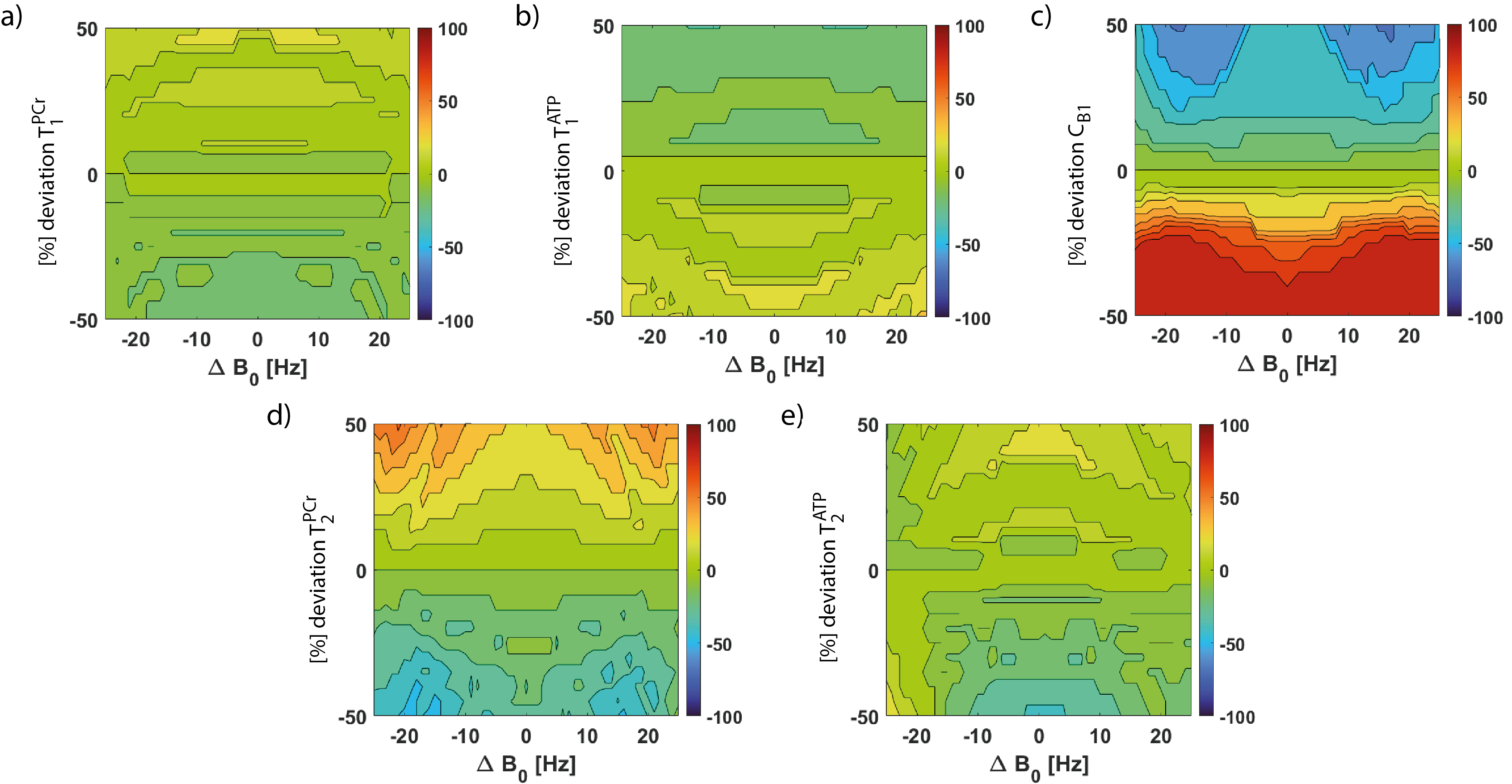
Quantification errors of KCK, due to errors from assumed values used for the dictionary, are investigated in Figure 3. KCK estimation is insensitive to deviations of the assumed values (with a quantification error < 20%), when the errors of T1PCr , T1ATP, T2PCr and T2ATP within a range of |25%| and CB1 within a tight range of |15%|. CB1 values show the need to be either correctly assumed or known prior to the dictionary generation to avoid bias in KCK estimation. The excitation profile was validated in phantom (Figure 2d) and showed a signal leakage of less than 15% beyond 300 Hz. Full brain kCK and concentration ratio Cr maps were achieved in 45 min with a birdcage coil and 21 min with a 1Tx/32Rx coil (Figure 4). We report for the first time the 3D whole brain mapping of kCK in humans with a classical birdcage coil setting and an advanced 32-channel 31P array coil. The kCK values reported here are on average 20% lower than literature values reports (0.37s-1) [1,4,5]. This could be due to a deviation of the assumed CB1. In this preliminary result, we fixed CB1 according to previous in-vivo measurements to 0.7 assuming a homogeneous excitation. To improve estimation accuracy and reproducibility, a subject-specific B1 map should be included in the next step.Conclusion
We conclude that the novel 3D-31P-MRF approach is feasible for whole-brain mapping of kCK, enabling the investigation of region-specific energy metabolism under various pathological conditions.Acknowledgements
This work was supported by the Swiss National Science Foundation (grants n° 320030_189064). We acknowledge the CIBM Center for Biomedical Imaging for providing expertise and resources to conduct this study.References
[1] M. Widmaier, S.-I. Lim, D. Wenz, and L. Xin, “Fast in vivo assay of creatine kinase in human brain by 31P magnetic resonance fingerprinting,” Jun. 2022. https://www.researchsquare.com/article/rs-1708658/v1
[2] Ma D, Gulani V, Seiberlich N, et al. Magnetic resonance fingerprinting. Nature. 2013;495(7440):187-192. doi:10.1038/nature11971
[3] Wang CY, Liu Y, Huang S, Griswold MA, Seiberlich N, Yu X. 31P magnetic resonance fingerprinting for rapid quantification of creatine kinase reaction rate in vivo. NMR in Biomedicine. 2017;30(12):e3786. doi:10.1002/nbm.3786
[4] Ren J, Sherry AD, Malloy CR. Efficient 31P band inversion transfer approach for measuring creatine kinase activity, ATP synthesis, and molecular dynamics in the human brain at 7 T. Magnetic Resonance in Medicine. 2017;78(5):1657-1666. doi:10.1002/mrm.26560
[5] Lei H, Zhu XH, Zhang XL, Ugurbil K, Chen W. In vivo 31P magnetic resonance spectroscopy of human brain at 7 T: An initial experience. Magnetic Resonance in Medicine. 2003;49(2):199-205. doi:10.1002/mrm.10379
Figures