0928
Using NORDIC with 23Na MRI to study dynamic changes in tissue sodium concentration1Sir Peter Mansfield Imaging Centre, University of Nottingham, Nottingham, United Kingdom, 2NIHR Biomedical Research Centre, Nottingham Univ. Hospital NHS Trust and Univ. Nottingham, Nottingham, United Kingdom
Synopsis
Keywords: Non-Proton, Non-Proton
Motivation: To perform 23Na MRI to study dynamic studies of changes in tissue sodium concentration in-vivo.
Goal(s): To demonstrate the application of NOise Reduction with DIstribution Corrected (NORDIC) PCA denoising to 23Na MRI data.
Approach: Dynamic timeseries of 23Na GRE data and NORDIC denoising to validate measurement of a known change in sodium concentration in a phantom, and dynamic changes in calf muscle in response to exercise.
Results: NORDIC PCA denoising allows detection of the temporal change in sodium in a phantom with good spatial resolution. This is applied to study the dynamics of sodium changes in muscle in response to exercise.
Impact: NOise Reduction with DIstribution Corrected (NORDIC) PCA denoising provides the potential to improve low SNR 23Na MRI measures to study dynamic changes in sodium on a spatially resolved level. Here, applied to study 23Na changes in calf muscle on exercise.
Introduction
Sodium (23Na) MRI has intrinsically low signal‐to‐noise‐ratio (SNR), due to the low 23Na ion concentration in vivo (15–30 mmol/L muscle), short relaxation times, and the gyromagnetic ratio being ~ 4 x lower than for 1H [1]. This results in the need for long scan times (~10–20 min) and large voxel sizes (e.g. 4 mm isotropic) compared to 1H-MRI, and means that any study of dynamic changes in 23Na in-vivo is challenging. Recently, the NOise Reduction with DIstribution Corrected (NORDIC) PCA denoising method was published that uses local low rank principal component analysis. NORDIC has been shown to provide impressive improvements in image SNR for neuroimaging data, e.g. for high spatial resolution diffusion tensor imaging (DTI) [2] and functional MRI [3] data. Thus, NORDIC enables higher spatial and temporal resolution data, particularly benefitting data in the thermal noise dominated regime.Here, we assess NORDIC denoising for non-proton 23Na MRI data, with the goal of applying this to study dynamic 23Na changes in-vivo in the muscle on exercise.
Methods
Data has been collected for phantom validation of measuring dynamic 23Na changes, and in-vivo in response to exercise:
Data Acquisition: All MRI data was collected on a 3T Philips Ingenia scanner, using a 23Na birdcage leg coil (PulseTeq Ltd) to acquire 23Na MRI scans, and the Q-body coil to collect 1H measures. 23Na MRI data were acquired using a 3D GRE short TR sequence (3x3x30mm3, 10 slices, TE/TR=1.26/13ms, FA=46o) for a high sampling rate (4.4s per acquisition) For each scan a final noise image was collected with no RF and no gradients. For all acquisitions the ‘SNR Maxima’ and ‘SNR clip factor’ were set to 100 and 0.01 respectively to improve the data’s dynamic range, both magnitude and phase data were saved.
Phantom validation: To assess the accuracy of quantifying the change in sodium concentration dynamically, data was collected of a 120 mm diameter cylindrical phantom, this was filled with an initial concentration of 20mmol/L. A 50ml 400mmol/L NaCl solution was added at a rate of 250ml/hr to the phantom using an MR compatible infusion pump. This increased the sodium concentration in the phantom to 30mmol/L in 12-minutes with a stirrer mounted in the phantom ensuring mixing of the sodium solution through the infusion period (Figure 1). During the infusion 23Na MRI was continuous acquired.
In-vivo data collection: 23Na MRI of the calf muscle was collected in 5 volunteers whilst they performed plantar flexion using an MRI-compatible ergometer (Trispect, Ergospect, Innsbruck Austria), Figure 4. First 1H mDIXON scans were acquired to image the calf. A 43-minute protocol was then performed during which baseline data was collected for 13 minutes, following by repeated plantar flexion exercise at 50% of maximum voluntary contraction for 3.5-minutes, followed by 30-minutes recovery.Data analysis: Data analysis was carried out in MATLAB. Nordic was performed on the MRI data. Linear regression of the reference bottles (10, 20, 30, 40 mmol/L) to convert voxel intensity to 23Na concentration, mmol/L was performed and applied to the phantom/calf (correction performed both using an average image and dynamically). An ROI was placed in the phantom and in the gastrocnemius muscle in the calf and the mean and variance in each region was measured.
Results
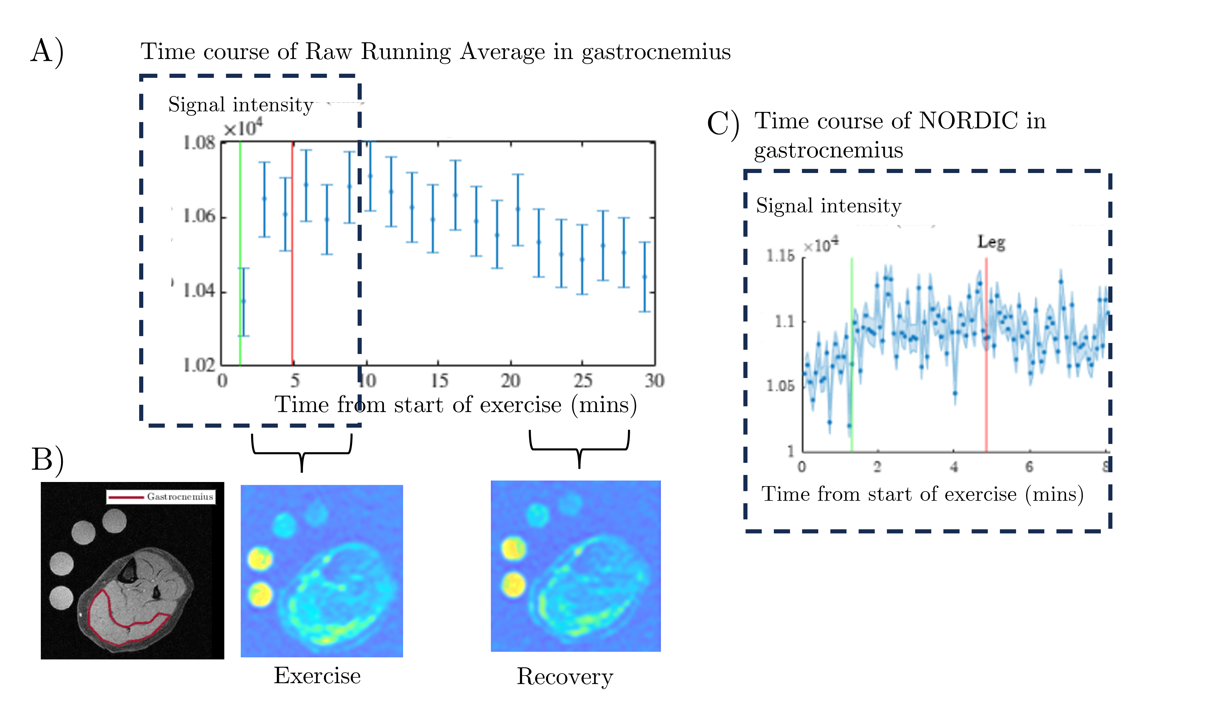
Figure 2 shows the timecourse of the mean signal and spatial variance within the phantom during the infusion. Figure 3 shows the timecourse of the mean sodium concentration measured in the phantom during infusion when performing dynamic-by-dynamic sodium concentration calibration using the reference bottles. Note, the large variance for raw data, whilst applying NORDIC provides high R2 and low root-mean-square-error, particularly using a running average (over 44s). Figure 4 shows In-vivo raw and NORDIC corrected 23Na MRI calf data collected before, during and after exercise. Figure 5 shows the raw moving average time course over the gastrocnemius, 23Na images on exercise (0:88- 5:52mins) and recovery (24:56-30:28 mins) and the high temporal resolution timecourse of the NORDIC data to study dynamically the sodium change in the muscle.Discussion
This work demonstrates the NORDIC denoising gains for 23Na MRI which is in the low SNR regime dominated by thermal noise. We validate on a phantom that it is possible to dynamically quantify sodium concentration by applying NORDIC denoising, and that this can be used to study in-vivo the spatial and temporal changes in muscle sodium on exercise.Conclusion
NORDIC can be used to correct low SNR 23Na MRI data to study spatially localised sodium changes in muscle dynamically. This methodology could in future also be applied to 23Na functional MRI brain studies.Acknowledgements
No acknowledgement found.References
1. Madelin, G., Lee, J. S., Regatte, R. R., & Jerschow, A. (2014). Sodium MRI: Methods and applications. In Progress in Nuclear Magnetic Resonance Spectroscopy (Vol. 79, pp. 14–47). Pergamon.
2. Moeller, S., Pisharady, P. K., Ramanna, S., Lenglet, C., Wu, X., Dowdle, L., Yacoub, E., Uğurbil, K., & Akçakaya, M. (2021). NOise reduction with DIstribution Corrected (NORDIC) PCA in dMRI with complex-valued parameter-free locally low-rank processing. NeuroImage, 226, 117539.
3. Vizioli, L., Moeller, S., Dowdle, L., Akçakaya, M., De Martino, F., Yacoub, E., & Uğurbil, K. (2021). Lowering the thermal noise barrier in functional brain mapping with magnetic resonance imaging. Nature Communications, 12(1), 5181.
Figures
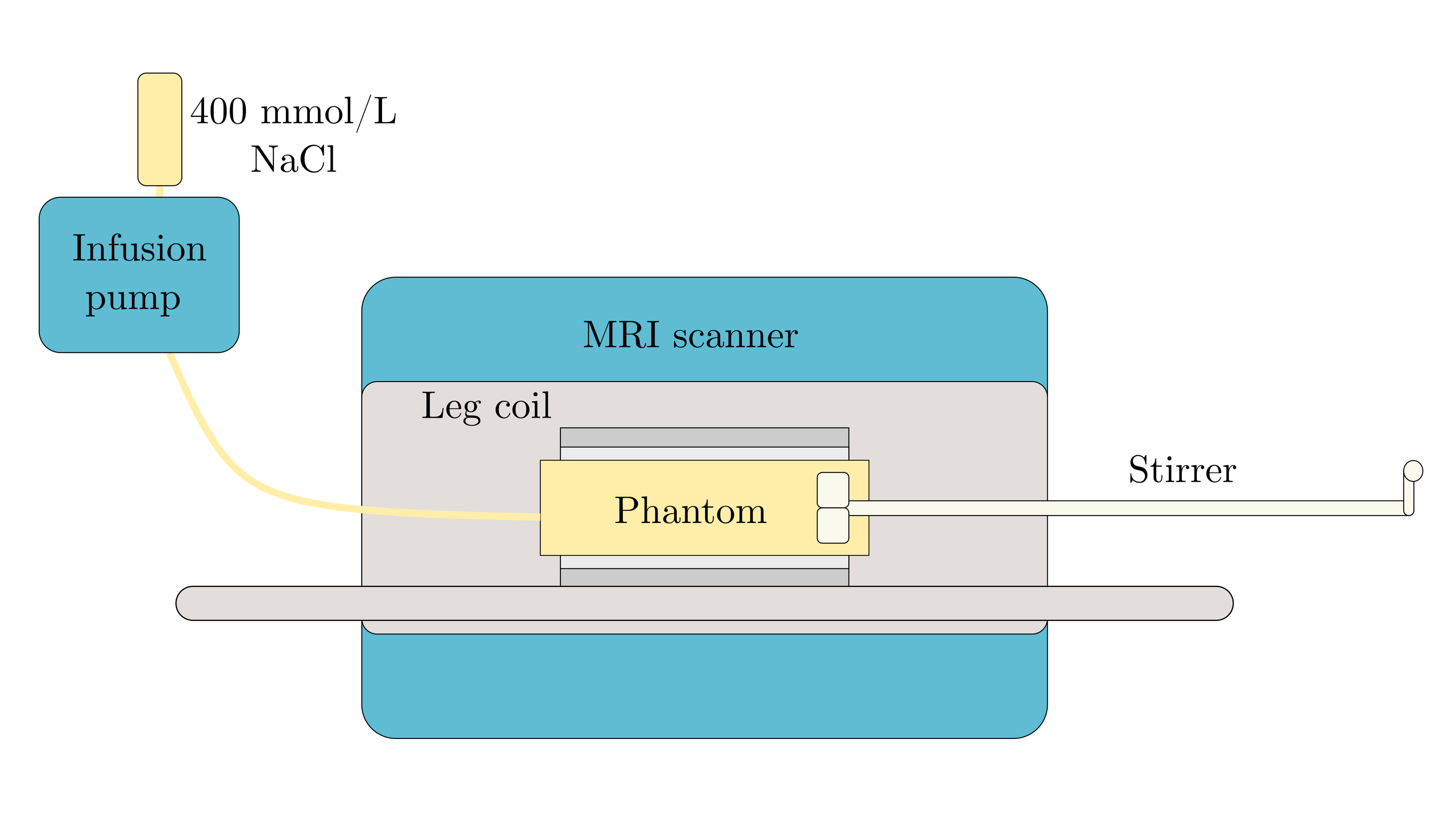
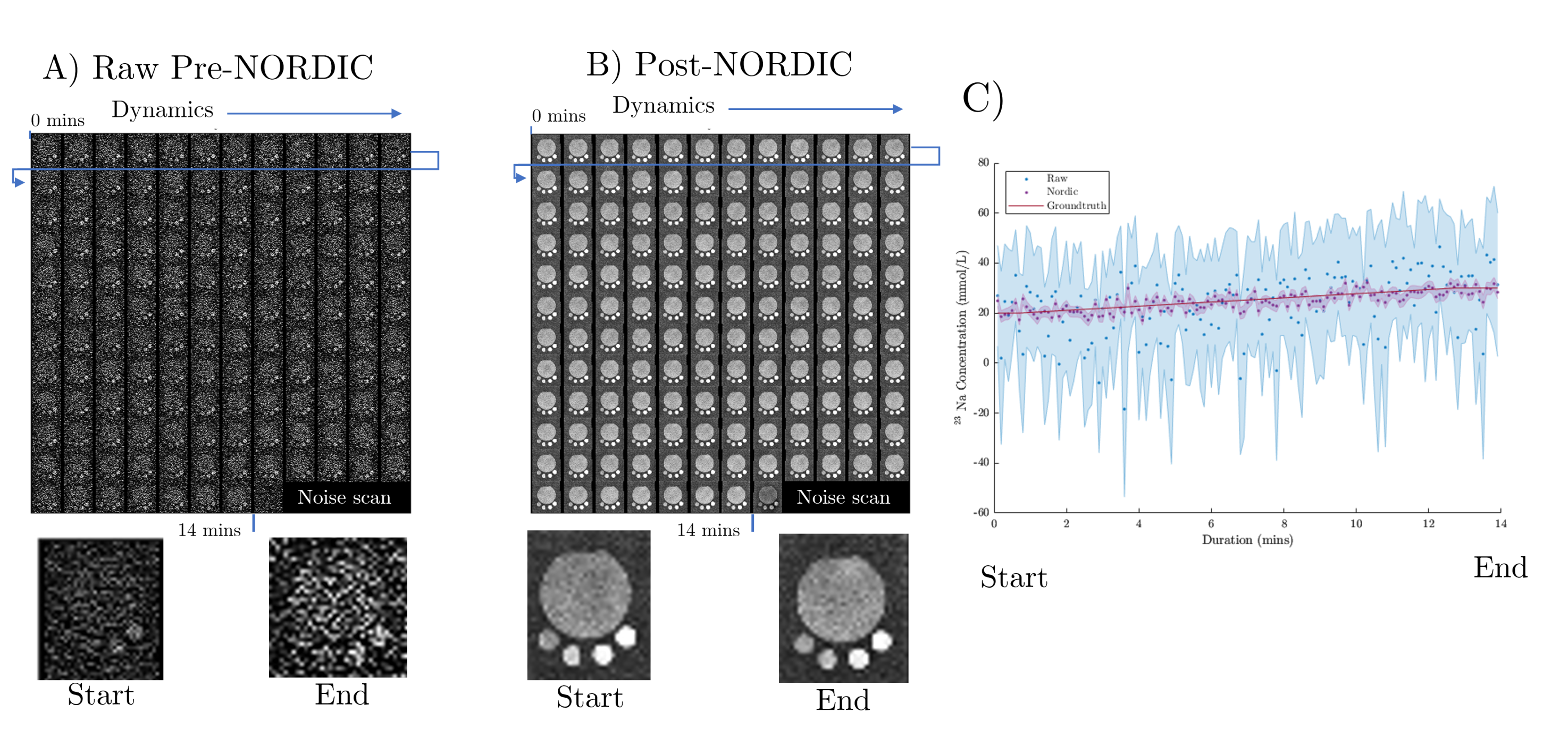
Figure 2: Images time series of the mean sodium concentration and spatial variance from within the phantom during infusion of sodium as the sodium concentration increases from 20 mmol/L to 30 mmol/L over 14 mins. 23Na images collected every 4.4s and shown for A) raw data before NORDIC and B) after NORDIC. Also shown are enlarged images at the start (nominal 20 mmol/L) and end (nominal 30 mmol/L) of infusion. C) Timeseries of the signal from the phantom and variance within the phantom for raw data and data after NORDIC correction using average bottle calibration.
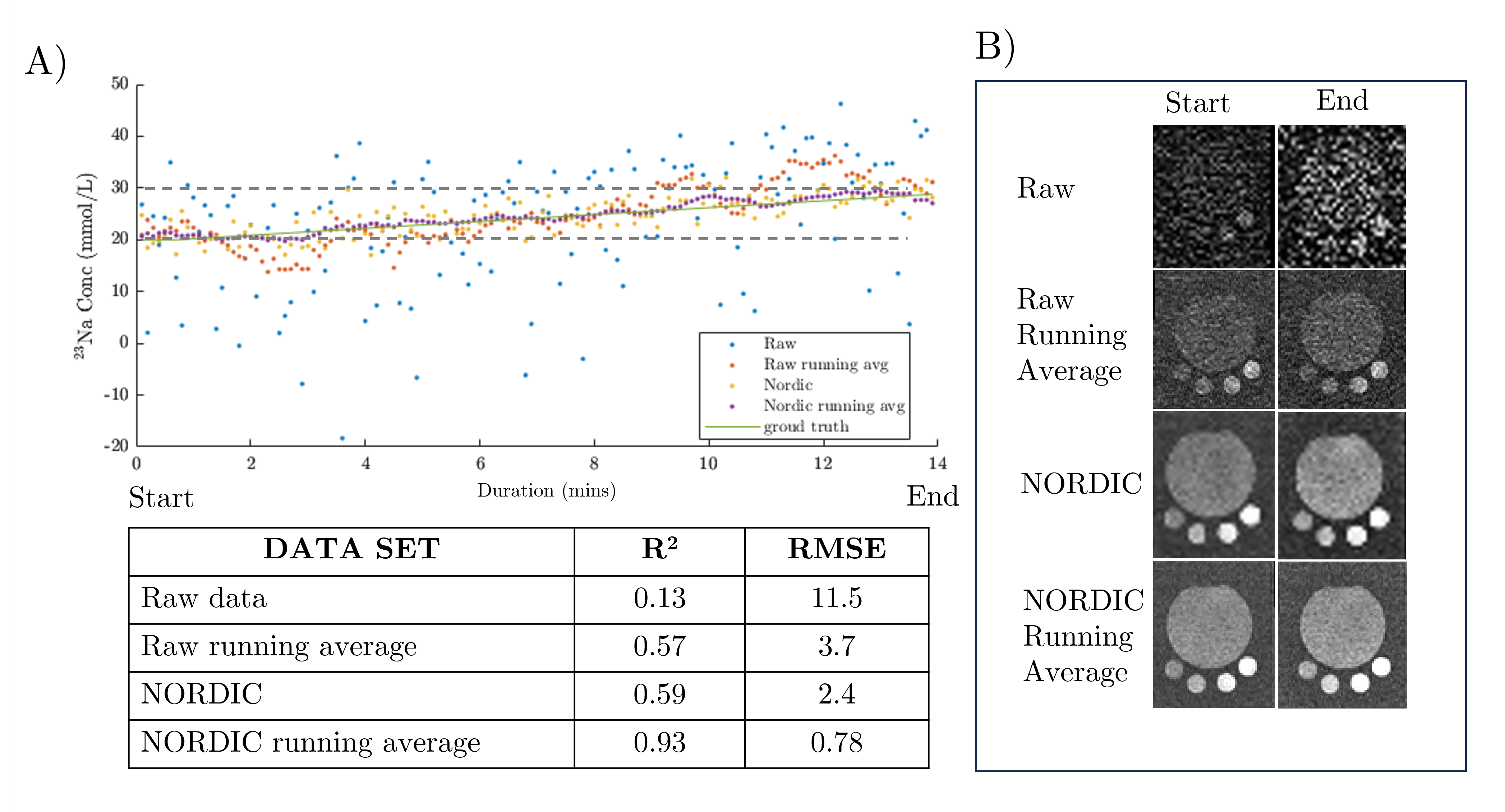
Figure 3: Timecourse of the mean sodium concentration measured in the phantom during the infusion. This is computed dynamic-by-dynamic sodium concentration calibration using the reference bottles is performed and applied to the phantom. Note, for the raw data has large variance, which is reduced with applying a moving average. By using NORDIC dynamic-by-dynamic bottle calibration is possible with, the lowest variance for the moving average with NORDIC. See Table for R2 and root-mean-square-error (RMSE) metrics. B) Example images at the start and end of the infusion.
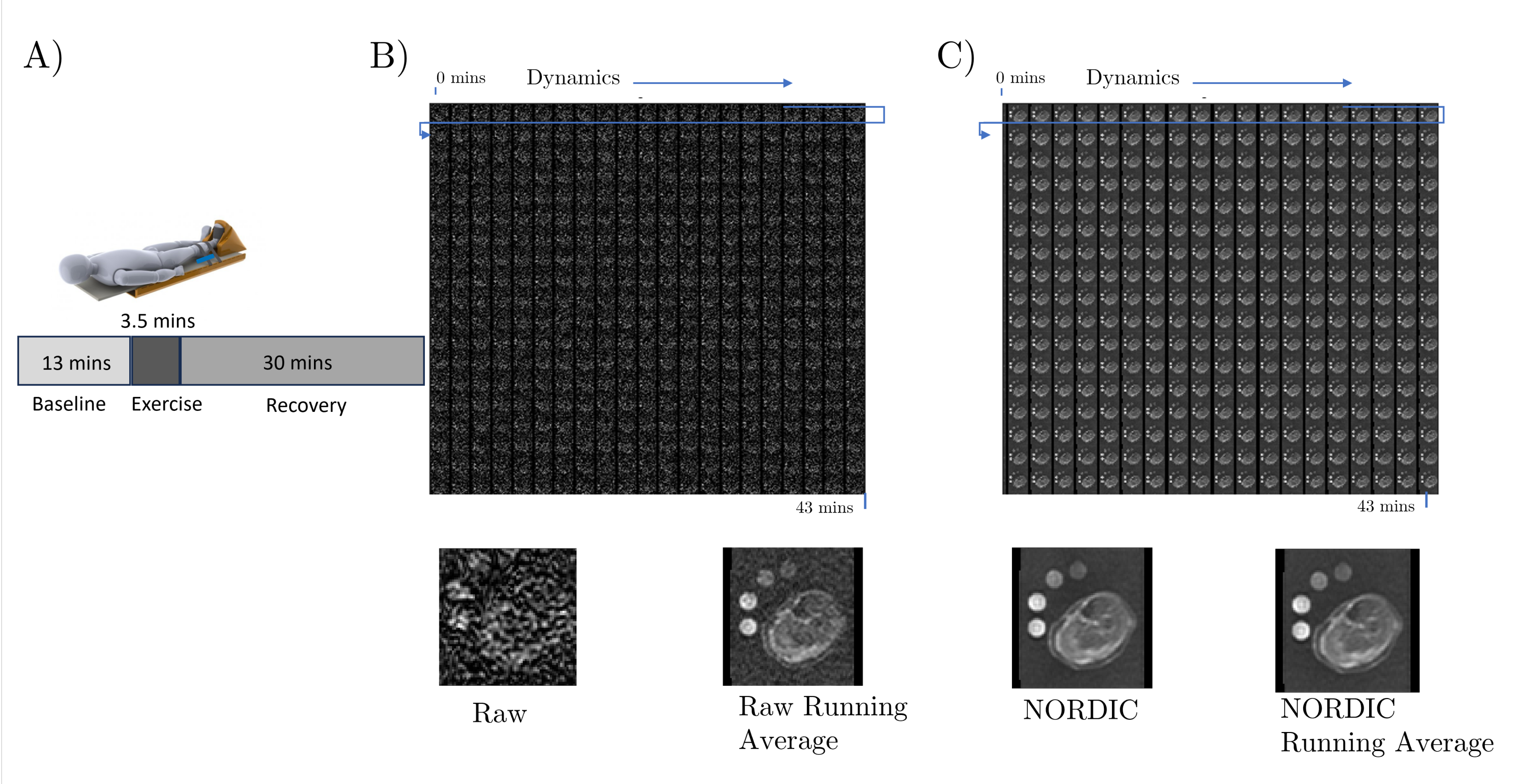
Figure 4: In-vivo muscle collection showing A) the paradigm and Trispect ergometer for plantar flexion exercise to fatigue the gastrocnemius. B) Timeseries of the raw 23MRI calf images collected data before NORDIC and C) after NORDIC.