0923
3D Seiffert spiral k-space trajectories for a functional sodium (23Na) MRI protocol at 3T1NMR Research Unit, Queen Square MS Centre, Department of Neuroinflammation, UCL Queen Square Institute of Neurology, Faculty of Brain Sciences, University College London, London, United Kingdom, 2Department of Medical Physics and Biomedical Engineering, University College London, London, United Kingdom, 3Philips Healthcare, Best, Netherlands, 4Quantitative Imaging Group, Centre for Medical Image Computing (CMIC), Department of Medical Physics & Biomedical Engineering, University College London, London, United Kingdom, 5Department of Brain & Behavioral, University of Pavia, Pavia, Italy, 6Digital Neuroscience Centre, IRCCS Mondino Foundation, Pavia, Italy
Synopsis
Keywords: Non-Proton, Non-Proton
Motivation: With advancing hardware, scan durations of 23Na-MRI have decreased, enabling novel applications that probe dynamic or functional processes; existing sequences, though, may not fully exploit the possible acceleration.
Goal(s): Implement and demonstrate a highly efficient ultrashort echo time non-Cartesian sequence based on 3D Seiffert spirals, for temporally-resolved applications of 23Na-MRI.
Approach: A possible acquisition protocol for functional brain 23Na-MRI at a temporal resolution of 47s was tested on a healthy volunteer at rest, at 3T.
Results: Image quality and SNR are high considering the temporal resolution, with compressed sensing successfully reducing noise. Further work will optimise the sequence analytically, ensuring homogeneous k-space sampling.
Impact: Dynamic 23Na-MRI at 3T is possible at sub-minute temporal resolution with a 3D Seiffert spiral k-space trajectory. Unlike for other sequences, efficient k-space coverage is achieved without downsides of unpleasant acoustic noise, or exciting mechanical resonances of scanner hardware.
Introduction
Sodium (23Na-)MRI is valued for unique insights into physiology1,2 and increasingly also dynamic functioning3–6. Due to rapid transverse relaxation and low MR-sensitivity, ultrashort echo time non-Cartesian sampling is usually employed7,8. We recently reported9 initial simulations of a 3D Seiffert spiral sequence10 for 23Na-MRI, characterised by highly efficient, pseudorandom and isotropic (under)sampling of k-space, suited to compressed sensing (CS) reconstruction and dynamic measurements.This work aims to demonstrate a possible acquisition protocol with 3D Seiffert spirals for in vivo brain functional 23Na-MRI, an application where time-efficient sampling is essential.
Theory
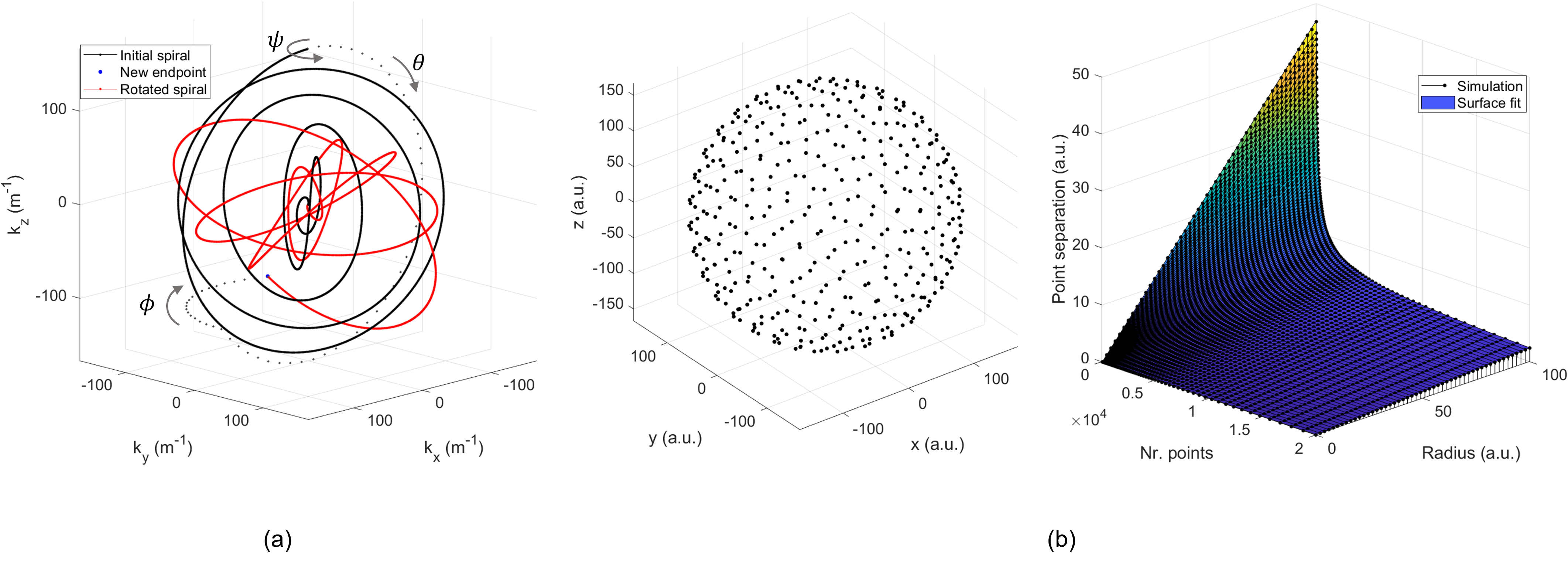
The theoretical formulism of 3D Seiffert spiral trajectories has been described previously9–11. Briefly, a spiral waveform is: (i) defined by the Jacobi elliptic functions; (ii) scaled from $$$k_r=0$$$ to $$$k_r=k_{max}$$$; (iii) rotated to fill k-space.A suitable rotational scheme is essential for complete and homogeneous k-space coverage. With the initial spiral endpoint $$$k_x=k_y=0,k_z=k_{max}$$$, we implemented a three-step $$$zyz$$$ axis rotation with angles $$$\psi,\theta,\phi$$$ (Figure 1); while $$$\theta,\psi$$$ are dictated by the Fibonacci grid endpoint, $$$\psi$$$ is tiny golden angle stepped12 ($$$\psi$$$=32.03°) around the spiral’s axis.
Elsewhere, the number of readouts was calculated by Nyquist testing random k-space points10. This is too computationally intensive for online sequence calculations. Instead, we numerically determined the relationship of the Fibonacci point separation with number of points and sphere radius (Figure 1), deriving an upper limit for the number of readouts at which the Nyquist criterion equals the Fibonacci point separation:$$N_r=\frac{1}{UF}\left( 3.24\frac{k_{max}}{\Delta k}\right)^{2.03},[1]$$ where $$$UF$$$ is an undersampling factor, and substituting for nominal resolution: $$$k_{max}=\frac{1}{2\Delta x}$$$, and FOV: $$$\Delta k=\frac{1}{FOV}$$$. Depending on spiral curvature and homogeneity of coverage, varying degrees of undersampling are possible without discernible artifacts.
Methods
Acquisition: a healthy participant (F, 54y) was scanned at rest on a 3T Philips Ingenia CX with a single-tuned 23Na birdcage head coil (RAPID Biomedical). 3D Seiffert spiral parameters9 were: FOV=240x240x240mm3, matrix=60x60x60, TE/TR=0.22/20ms, FA=45°, TRO=12.9ms, spiral length=20, m=0.025, UF=8, scale=0.75, Gmax=10mTm-1, Smax=100Tm-1s-1, bandwidth=140kHz, NSA=2, Tdyn=47s, dynamics=10. Both gradient spoiling and rewinding with RF phase cycling for balanced steady state free precession (bSSFP) were explored. The point spread function (PSF) was simulated with biexponential T2* attenuation and the sample distribution was visualised by gridding of density compensation factors (DCFs)13.Reconstruction: images were reconstructed offline using a conventional non-uniform FFT14 (NUFFT) and iterative CS (BART15) using total variation (TV) and wavelet regularisation, at strengths 0.0–0.005. SNR in NUFFT images was calculated16 in white matter (WM) and vitreous humour (VH) ROIs.
Results
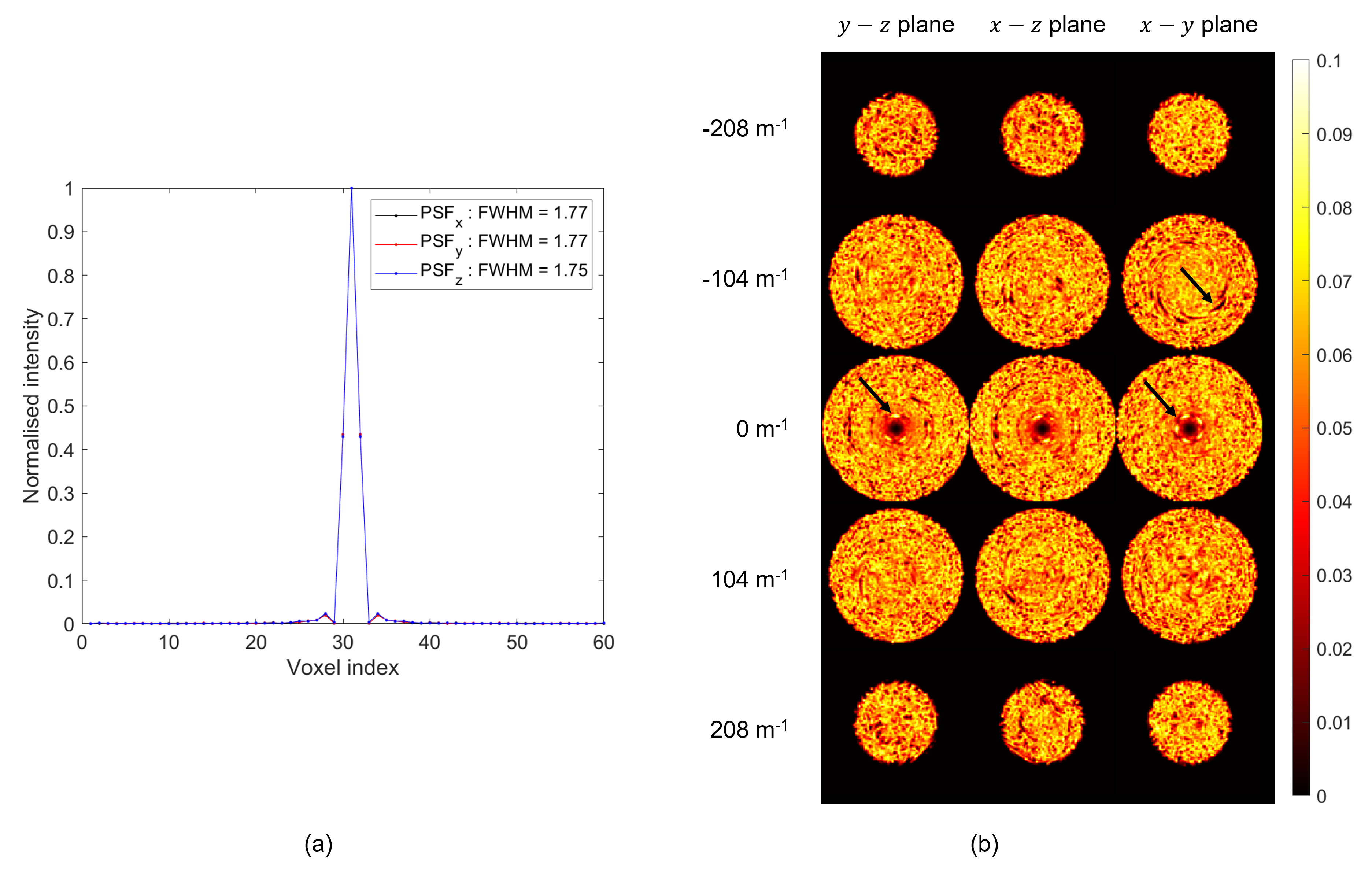
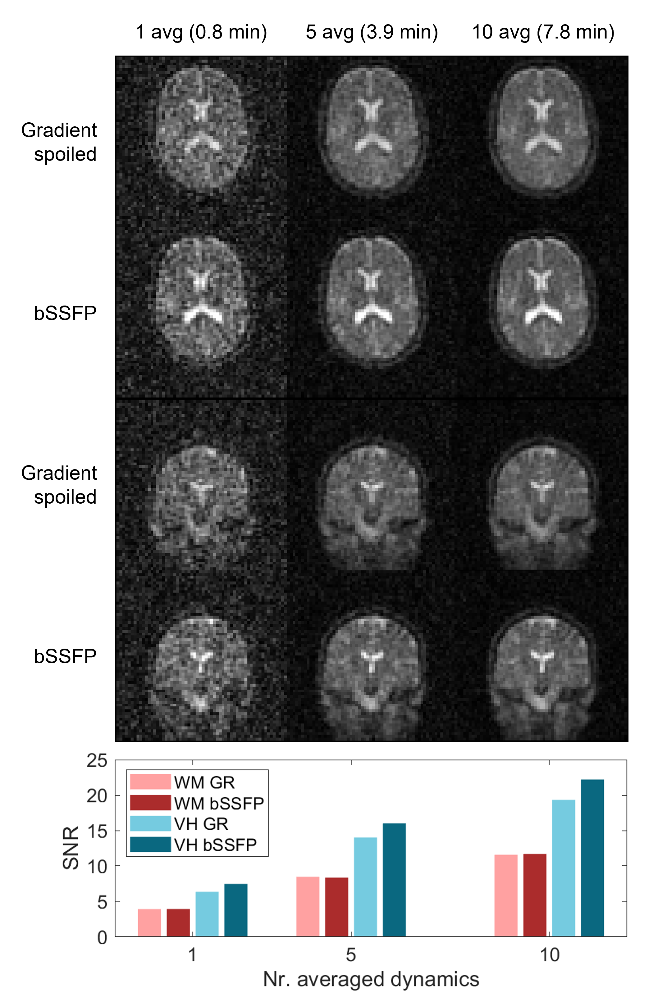
Figure 2 shows PSFs (FWHM=1.75–1.77 voxels) and gridded k-space DCFs. The latter exhibits a sample distribution with fair global homogeneity, but some local inhomogeneities.Figure 3 displays NUFFT reconstructed images with gradient spoiling and bSSFP for one dynamic and averages of five and ten dynamics. A bar chart also shows SNR, which is the same in WM, but higher in VH for bSSFP compared to gradient spoiling. Image quality is good considering the scan time, although single-dynamic images suffer from structured noise.
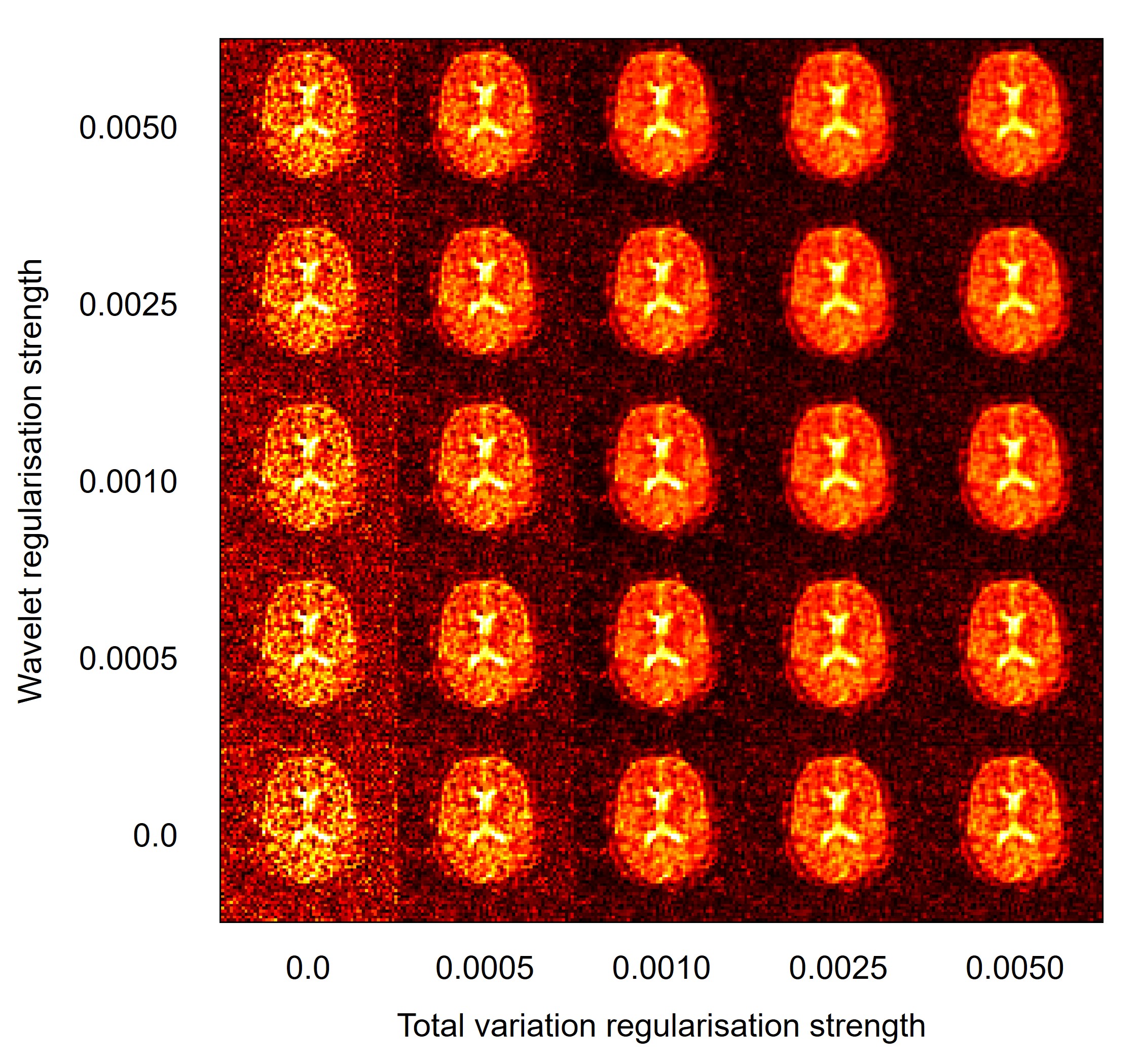
Figure 4 contains CS reconstructed images of one dynamic at various regularisation strengths. TV regularisation is more effective at removing noise, at a cost of blurring.
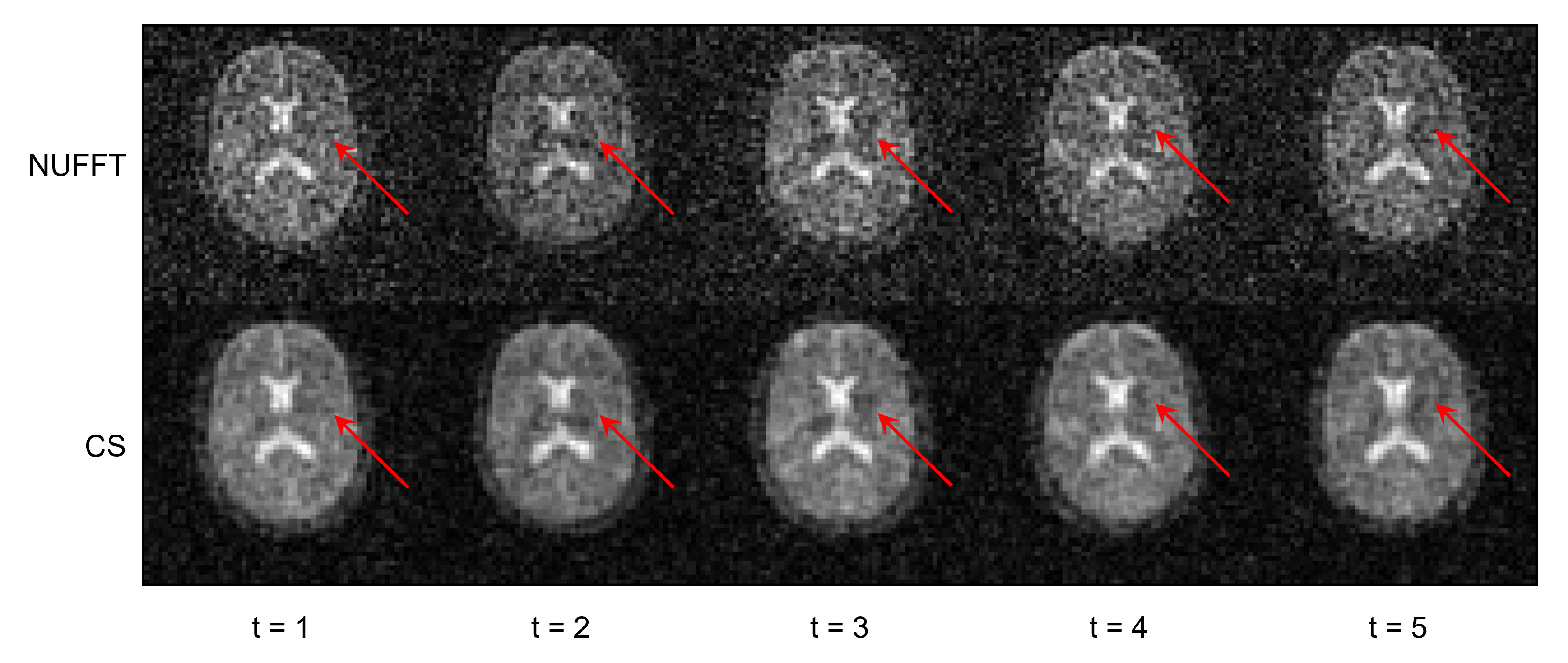
Figure 5 shows five dynamics of a timeseries. Although CS is successful at denoising, coarser noise in low SNR structures like deep grey matter could mimic regionally coherent signal fluctuations.
Discussion and conclusion
We present initial results at 3T of a 23Na-MRI protocol with 3D Seiffert spirals at sub-minute temporal resolution. Image quality is good and SNR compares favourably with literature (e.g. for 1 dynamic, SNRWM≈4, and for a 19min 3D cones16 acquisition, SNRWM≈7, although the comparison is unfair due to different actual resolutions). Images with bSSFP showed higher SNR in fluids with long T2 due to T2/T1 contrast; this could introduce sensitivity to changes in sodium microenvironment, although modelling is needed. Practical benefits of the trajectory include low-pitched acoustic noise, particularly versus 3D cones (avoiding the “sweep” across mechanical resonances17). Gradient waveforms are subject to full safety assessments of the scanner software.Results are encouraging and optimisation will continue, especially to investigate the source of structured noise that could cause misclassification of signal changes as brain activity (Figure 5). Possible culprits are PSF sidelobes and point coherences in k-space (Figure 2). Inhomogeneities in sample distributions can cause noise fluctuations18 (coloured noise19) across k-space and should be avoided20.
Ongoing work aims to demonstrate sensitivity to changes in 23Na concentration accompanying brain function, as well as in phantoms. The sequence will be further optimised analytically, compared to 3D cones, and assessed for quantitative 23Na-MRI. Finally, other applications of dynamic 23Na-MRI and acceleration of conventional 23Na-MRI will be explored.
Acknowledgements
SR: EPSRC-funded UCL Centre for Doctoral Training in Intelligent, Integrated Imaging in Healthcare (i4health) (EP/S021930/1) and the Department of Health’s NIHR-funded Biomedical Research Centre at University College London Hospitals.
CGWK: Horizon2020 (Research and Innovation Action Grants Human Brain Project 945539 (SGA3)), BRC (#BRC704/CAP/CGW), MRC (#MR/S026088/1), Ataxia UK, Rosetrees Trust (#PGL22/100041 and #PGL21/10079). CGWK is a shareholder in Queen Square Analytics Ltd.
References
1. Madelin G, Lee JS, Regatte RR, Jerschow A. Sodium MRI: Methods and applications. Prog Nucl Magn Reson Spectrosc. 2014;79:14-47.
2. Thulborn KR. Quantitative sodium MR imaging: A review of its evolving role in medicine. Neuroimage. 2018;168:250-268.
3. Gandini Wheeler-Kingshott CAM, Riemer F, Palesi F, et al. Challenges and Perspectives of Quantitative Functional Sodium Imaging (fNaI). Front Neurosci. 2018;12.
4. Bydder M, Zaaraoui W, Ridley B, et al. Dynamic 23Na MRI - A non-invasive window on neuroglial-vascular mechanisms underlying brain function. Neuroimage. 2019;184:771-780.
5. Akbari A, Lemoine S, Salerno F, et al. Functional Sodium MRI Helps to Measure Corticomedullary Sodium Content in Normal and Diseased Human Kidneys. Radiology. 2022;303(2):384-389.
6. Scheipers AK, Nagel AM, Ruck L, et al. Quantitative sodium MRI of the human kidneys at 7T – Before, during and after water load via sliding window evaluation. In: Proc. Intl. Soc. Mag. Reson. Med. 31 (2023). ; 2023.
7. Boada FE, Gillen JS, Shen GX, Chang SY, Thulborn KR. Fast three dimensional sodium imaging. Magn Reson Med. 1997;37(5):706-715.
8. Romanzetti S, Mirkes CC, Fiege DP, Celik A, Felder J, Shah NJ. Mapping tissue sodium concentration in the human brain: A comparison of MR sequences at 9.4Tesla. Neuroimage. 2014;96:44-53.
9. Rot S, Clemence M, Solanky BS et al. 3D Seiffert spirals for efficient k-space sampling in 23Na-MRI: initial phantom simulations. In: Proc. Intl. Soc. Mag. Reson. Med. 31. ; 2023.
10. Speidel T, Metze P, Rasche V. Efficient 3D Low-Discrepancy ${k}$ -Space Sampling Using Highly Adaptable Seiffert Spirals. IEEE Trans Med Imaging. 2019;38(8):1833-1840.
11. Erdös P. Spiraling the Earth with C. G. J. Jacobi. Am J Phys. 2000;68(10):888-895.
12. Wundrak S, Paul J, Ulrici J, et al. A Small Surrogate for the Golden Angle in Time-Resolved Radial MRI Based on Generalized Fibonacci Sequences. IEEE Trans Med Imaging. 2015;34(6):1262-1269.
13. Zwart NR, Johnson KO, Pipe JG. Efficient sample density estimation by combining gridding and an optimized kernel. Magn Reson Med. 2012;67(3):701-710.
14. Barnett AH, Magland J, af Klinteberg L. A Parallel Nonuniform Fast Fourier Transform Library Based on an “Exponential of Semicircle" Kernel. SIAM Journal on Scientific Computing. 2019;41(5):C479-C504.
15. Uecker M, Holme C, Blumenthal M, et al. mrirecon/bart: version 0.7.00. 2021.
16. Riemer F, Solanky BS, Stehning C, et al. Sodium (23Na) ultra-short echo time imaging in the human brain using a 3D-Cones trajectory. Magnetic Resonance Materials in Physics, Biology and Medicine. 2014;27(1):35-46.
17. Schulte RF, Vaeggemose M, Hansen ESS et al. Sequence Comparison for Sodium MRI. In: ISMRM Workshop on MR Imaging of X‐Nuclei (23Na & Friends). ; 2023.
18. Liao J, Pauly JM, Brosnan TJ, Pelc NJ. Reduction of motion artifacts in cine MRI using variable‐density spiral trajectories. Magn Reson Med. 1997;37(4):569-575.
19. Eggers H, Kircheis M, Potts D. Non-Cartesian MRI Reconstruction. In: Advances in Magnetic Resonance Technology and Applications; 2022:73-103.
20. Boyer C, Chauffert N, Ciuciu P, et al. On the Generation of Sampling Schemes for Magnetic Resonance Imaging. SIAM J Imaging Sci. 2016;9(4):2039-2072.
Figures