0916
Intravoxel incoherent motion model for prediction of tertiary lymphoid structures in HCC.1Department of radiology, Sun Yat-sen University Cancer Center, Guangzhou, China, 2Shukun Technology Co., Ltd, Beijing, China
Synopsis
Keywords: Liver, Radiomics, Magnetic resonance imaging, IVIM, Hepatocellular carcinoma, tertiary lymphoid structures
Motivation: Intra-tumoral tertiary lymphoid structures (TLSs) are associated with a favorable prognosis for patients with hepatocellular carcinoma (HCC). Intravoxel incoherent motion (IVIM) sequences describe heterogeneity of tumor components.
Goal(s): We aimed to explore the value of intravoxel incoherent motion (IVIM) sequences in predicting TLSs.
Approach: IVIM quantitative parameters and radiomics features were obtained. A fusion model based on the above and clinical characteristics was constructed. Receiver operating characteristic curve analysis was performed to assess the diagnostic performance of different models for TLSs prediction.
Results: Radiomics features based on IVIM-DWI can be better conducive to preoperative prediction of TLSs in patients than IVIM parameter maps.
Impact: TLSs exhibit considerable promise in prognostic prediction and the identification of appropriate candidates for immunotherapy. The fusion model based on the IVIM-DWI showed great performance in predicting TLSs, assisting the selection of clinical immunotherapy patients.
Introduction
Recently, immunotherapy has successfully shown promising outcomes in various tumors[1]. However, the efficacy of immunotherapy varies from person to person. Currently, some studies have report that intra-tumoral tertiary lymphoid structures (TLSs) are associated with a favorable prognosis for patients with hepatocellular carcinoma (HCC)[2]. However, the confirmation of TLSs presence and maturation stages can solely be identified by postoperative pathological and immunohistochemical evaluation[2,3]. It would be beneficial to determine the evolution of TLSs using a noninvasive and easily feasible method.Medical imaging plays a significant role in diagnosing HCC. Multi-b-value DWI with intravoxel incoherent motion (IVIM) can evaluate the true molecular diffusion and the blood microcirculation perfusion calculated with multi-b-values by a bi-exponential model. IVIM is capable of analyzing non-Gaussian diffusion and may be able to more accurately describe heterogeneity of tumor components[4,5].
To our knowledge, there were no studies focused on the relationship between the IVIM parameters and TLSs. Therefore, this study aimed to prospectively investigate the effectiveness of IVIM sequence for preoperatively predicting TLSs in HCC.
Methods
This prospective study enrolled 128 patients with HCC who underwent MRI examination including IVIM sequence with 8 b-values before surgery between March 2019 and January 2021.For each patient, the whole pathological slide images (WSIs) of hematoxylin-eosin (H&E) were reviewed by a pathologist specialized in diseases of liver and lymphopoietic system. The existence of intra-tumoral TLSs was assessed morphologically on WSIs [6,7]. We classified tumors as TLS+ (at least 1 intra-tumoral TLS) or TLS- (without any TLSs).
IVIM quantitative parameters and radiomics features were obtained based on the whole tumor volume of the lesion delineated. Radiological qualitative or semi-quantitative features were also assessed by two radiologists.
The radiomics score was constructed with the least absolute shrinkage and selection operator (LASSO). Uni- and multivariable logistic regression analyses were performed to identify the independent predictors for theTLSs +/-. Moreover, the optimal AUC radiomics model, IVIM parameters and clinical chracteristics were combined to build the fusion model. The performance of different models for TLSs prediction in HCC was assessed using receiver operating characteristic (ROC) curve analysis and compared using the DeLong method. The Kaplan-Meier (KM) curves was used to analyze early recurrence free survival (RFS) based on TLS+ and TLS- cohorts.
Results
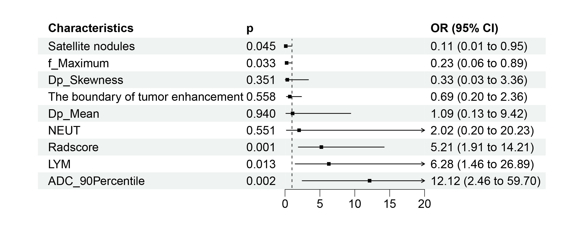
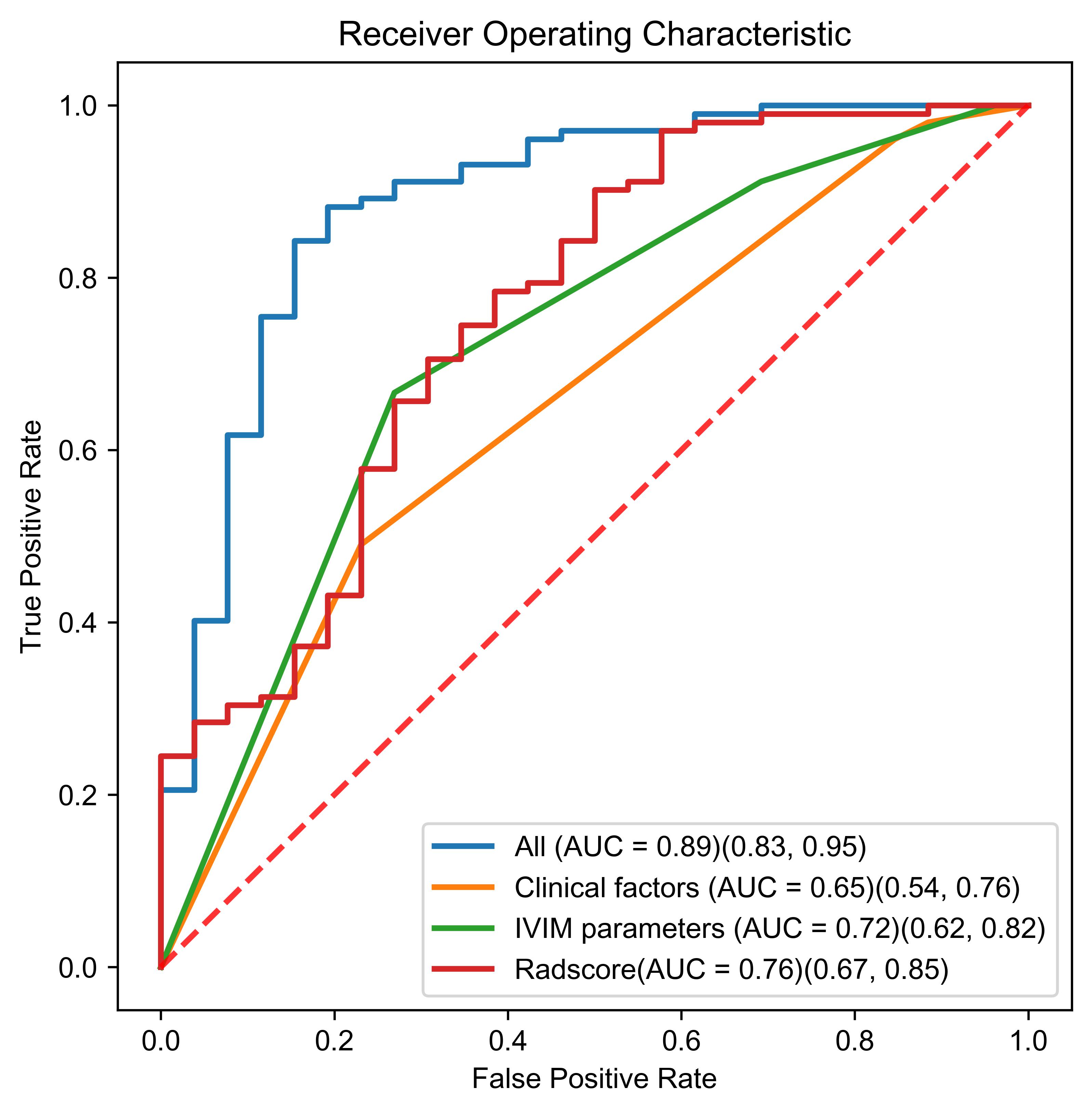
One hundred and two of the 128 HCCs were TLS + HCCs. Multivariable analyses identified Apparent Diffusion Coefficient (ADC), the perfusion fraction (f), satellite nodules, lymphocyte count (LYM), and radiomics score were independent predicted factors in Fig.1.The Radiomics model from IVIM-DWI (AUC = 0.76 (0.67, 0.85)) performed better than IVIM parameter maps (ADC and f, AUC = 0.72 (0.62, 0.82)) and clinical characteristics (satellite nodules and LYM, AUC = 0.65 (0.54, 0.76)). Furthermore, the performance of the fusion model combined with the radiomics score of IVIM-DWI, clinical characteristics, and IVIM parameters was significantly improved (AUC = 0.89(0.83, 0.95), accuracy = 86.72%, sensitivity = 88.24%, specificity = 80.77%) in Fig.2.
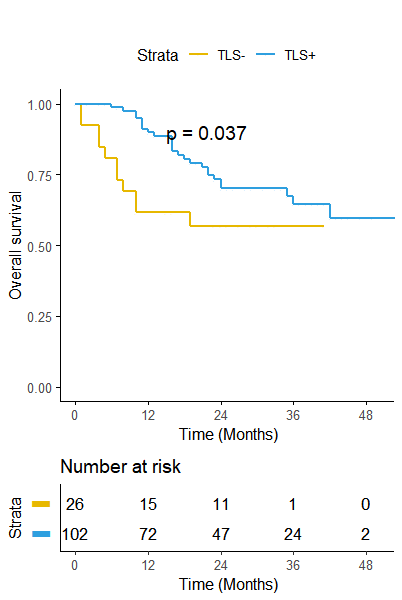
Patients with intra-tumor TLSs display a lower risk of RFS than the patients without intra-tumor TLSs (P = 0.037, Fig.3).
Conclusion
The fusion model based on the IVIM-DWI showed great performance in predicting TLSs. IVIM sequence may be served as a vital supplementary tool for no-invasive prediction of TLSs in HCC, which may assist the selection of clinical immunotherapy patients.Acknowledgements
We thank all patients who participated in this study.References
1.Fridman WH, Zitvogel L, Sautès-Fridman C, Kroemer G (2017) The immune contexture in cancer prognosis and treatment. Nat Rev Clin Oncol 14:717–734.
2.Calderaro J, Petitprez F, Becht E, et al (2019) Intra-tumoral tertiary lymphoid structures are associated with a low risk of early recurrence of hepatocellular carcinoma. Journal of Hepatology 70:58–65.
3.Posch F, Silina K, Leibl S, et al (2018) Maturation of tertiary lymphoid structures and recurrence of stage II and III colorectal cancer. Oncoimmunology 7:e1378844.
4.Meeus EM, Zarinabad N, Manias KA, et al (2018) Diffusion-weighted MRI and intravoxel incoherent motion model for diagnosis of pediatric solid abdominal tumors. J Magn Reson Imaging 47:1475–1486.
5.Comparison of the Diagnostic Value of Mono-exponential, Bi-exponential, and Stretched Exponential Signal Models in Diffusion-weighted MR Imaging for Differentiating Benign and Malignant Hepatic Lesions. Magn Reson Med Sci 20:69–75.
6. Craig AJ, von Felden J, Garcia-Lezana T, et al (2020) Tumour evolution in hepatocellular carcinoma. Nat Rev Gastroenterol Hepatol 17:139–152.
7. Finkin S, Yuan D, Stein I, et al (2015) Ectopic lymphoid structures function as microniches for tumor progenitor cells in hepatocellular carcinoma. Nat Immunol 16:1235–1244.
Figures