0915
Enhancing Hepatocellular Carcinoma (HCC) Diagnosis Through TWIST MRI Sequence1Radiology, Mayo Clinic, Jacksonville, FL, United States
Synopsis
Keywords: Liver, Tumor, Hepatocellular carcinoma
Motivation: Our report targets precise timing in hepatocellular carcinoma (HCC) diagnosis via MRI. Timely recognition of the late hepatic arterial phase is crucial to avoid unnecessary invasive biopsies and minimize patient risks.
Goal(s): Our report evaluates the TWIST technique's effectiveness in HCC diagnosis, aiming to reduce the need for percutaneous biopsies.
Approach: We integrated TWIST into our liver MRI protocols, capturing images at preset intervals, enhancing temporal resolution for HCC diagnosis.
Results: Our report successfully employs the TWIST technique to diagnose HCC by capturing arterial hyperenhancement. This innovation improves diagnostic accuracy and diminishes the necessity for invasive procedures, benefiting patients.
Impact: The implementation of the TWIST technique promises safer and more accurate hepatocellular carcinoma (HCC) diagnosis. This technique minimizes invasive biopsies, reshaping clinical practice and offering a new perspective on non-invasive diagnostic techniques.
Introduction
Hepatocellular carcinoma (HCC) is a common liver malignancy, and its diagnosis can commonly be made based on imaging. According to the Liver Imaging Reporting and Data System (LI-RADS) criteria, establishing an imaging diagnosis relies on demonstrating non-rim arterial hyperenhancement in a lesion through contrast-enhanced cross-sectional imaging, such as magnetic resonance imaging (MRI)1. A key challenge in MRI for HCC diagnosis is the precise timing of acquisition to catch the late hepatic arterial phase, which is when HCC most commonly demonstrates hyperenhancement. Mistiming can lead to insufficient diagnostic criteria, resulting in unnecessary biopsies, with associated risks of bleeding and injury to adjacent structures.Methods
In this report, we introduced the TWIST technique (time-resolved angiography with interleaved stochastic trajectories) into our institutional liver MRI protocols for HCC screening and diagnosis. The TWIST technique, well-established in MR angiography, enables the acquisition of a series of images illustrating the passage of contrast through hepatic arterial vasculature and hepatic parenchyma at predefined intervals, e.g., 7, 14, and 21 seconds2. By obtaining a series of images, we aimed to address the challenge of missing arterial hyperenhancement due to either excessively early or delayed scanning. It's important to note that the TWIST technique enhances temporal resolution while sacrificing spatial resolution.Results
We present two cases of patients referred to our institution for percutaneous biopsies due to suspicious hepatic lesions that did not initially meet the imaging criteria for HCC because of the absence of arterial hyperenhancement. These lesions were in anatomically challenging areas—the caudate lobe (Figure 1) and the hepatic dome (Figure 2), increasing the risk of complications. Through the implementation of the TWIST technique, we successfully diagnosed HCC by demonstrating arterial hyperenhancement without exposing the patients to unnecessary percutaneous biopsies. Our report illustrates the feasibility of incorporating the TWIST technique into liver MRI for HCC diagnosis. The series of images acquired at various time intervals provide a dynamic representation of arterial hyperenhancement, reducing the chances of mistimed scans and improving diagnostic accuracy, ultimately minimizing the need for unnecessary biopsies.Discussion
The integration of the TWIST technique offers an improved approach to address the challenges of timing arterial hyperenhancement detection during liver MRI for HCC diagnosis. The heightened temporal resolution enhances diagnostic accuracy and may reduce the risks and discomfort associated with unnecessary invasive procedures. While the spatial resolution is compromised, the clinical benefits derived from enhanced diagnostic precision outweigh this limitation.Conclusion
By introducing the TWIST MRI sequence into MRI liver protocols for HCC diagnosis, this report offers a pathway to more accurate and safer screening and diagnostic protocols. This technique has the potential to reduce the number of unnecessary biopsies, thereby sparing patients from the risks and discomfort associated with these procedures.Acknowledgements
No acknowledgement found.References
1. Santillan C, Chernyak V, Sirlin C. LI-RADS categories: concepts, definitions, and criteria. Abdom Radiol (NY). 2018 Jan;43(1):101-110. doi: 10.1007/s00261-017-1334-x. PMID: 29038857.
2. Maj E, Cieszanowski A, Rowiński O, Wojtaszek M, Szostek M, Tworus R. Time-resolved contrast-enhanced MR angiography: Value of hemodynamic information in the assessment of vascular diseases. Pol J Radiol. 2010 Jan;75(1):52-60. PMID: 22802762; PMCID: PMC3389854.
Figures
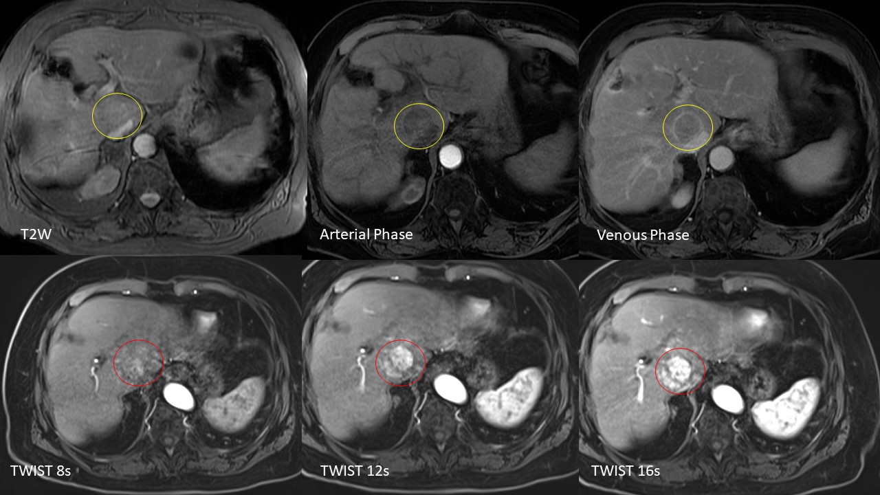
Bottom row: Repeat MRI with TWIST sequences at different time intervals showing clear arterial hyperenhancement of the suspicious lesion in the caudate lobe (red circle), meeting imaging-diagnosis of HCC.
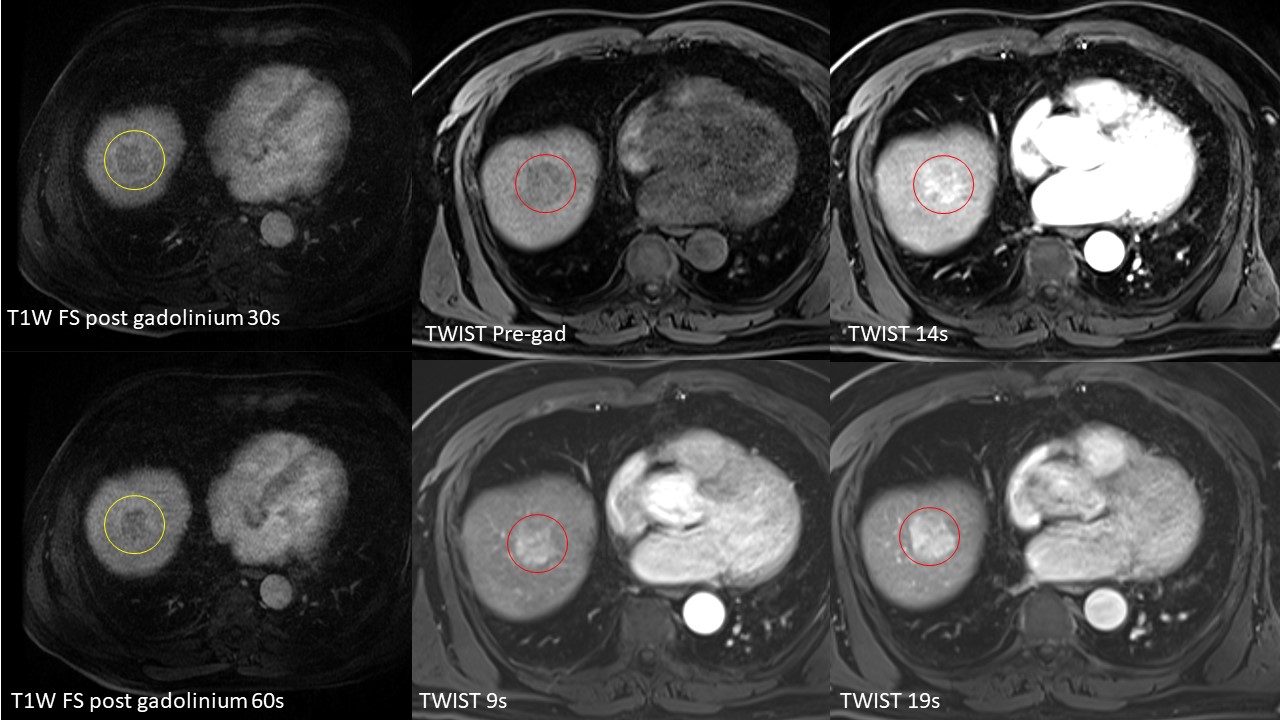
Middle column and right column: Repeat MRI with TWIST sequences shows clear arterial hyperenhancement of the the suspicious lesion in the hepatic dome (red circle), meeting imaging-diagnosis of HCC.