0912
Safe Diffusion Lobes for Diminished Gradient Vibration1Electrical Engineering, Stanford University, Stanford, CA, United States, 2Center for Cognitive and Neurobiological Imaging, Stanford University, Stanford, CA, United States
Synopsis
Keywords: Pulse Sequence Design, Pulse Sequence Design
Motivation: Gradient coil vibrations are a source of loud acoustics, persistent field distortions, potential system damage, and in some cases signal dropout in diffusion MRI. Vibrations are particularly strong at mechanical resonant frequencies.
Goal(s): To design “safe” time-optimal diffusion gradients which avoid mechanical resonant frequencies.
Approach: A convex optimization problem is formed which seeks to maximize b-value for a given duration while constraining the discrete cosine transform of the diffusion lobe. A bisection search is applied to duration to find the shortest feasible solution with the target b-value.
Results: Safe diffusion lobes greatly diminished coil vibration and minimized oscillations in the gradient fields.
Impact: Safe diffusion lobes were shown to avoid specified gradient mechanical resonant frequencies reducing vibrations and persistent k-space oscillations. These waveforms have clinical and research potential to improve auditory comfort, prevent DWI signal dropout and image artifacts, and prevent system damage.
INTRODUCTION
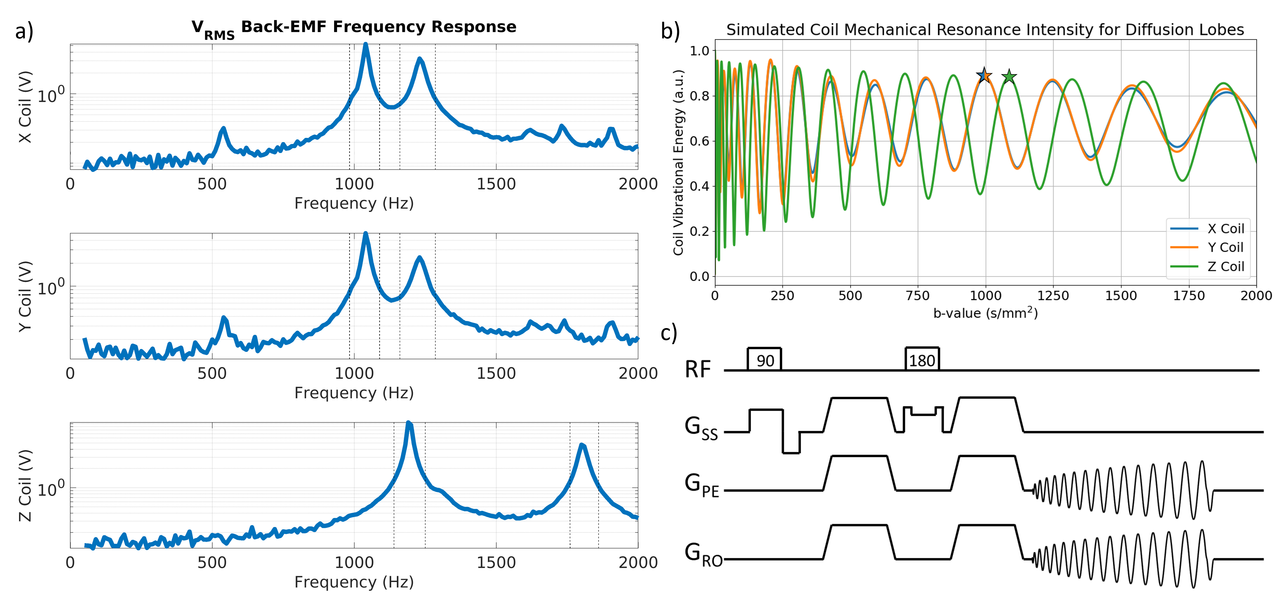
Diffusion weighted imaging (DWI) is a prevalent strategy for applications from acute brain ischemia, to tumor detection and monitoring, to tractography1. A spin-echo DWI sequence consists of large diffusion encoding gradients around the refocusing pulse followed by image readout (Figure 1c). The powerful diffusion gradient lobes can lead to mechanical vibrations in the coils2,3 caused by the large Lorentz forces acting on them. This vibration is exacerbated when gradient switching occurs at coil mechanical resonant frequencies. Stronger vibrations are more likely to cause signal dropout, trajectory errors, loud acoustic waves, and damage to the gradient system2–5. The design of time-optimal diffusion lobes has been shown to be convex and has even been extended to minimize induced eddy currents6,7. Here, a related approach is developed to design time-optimal diffusion lobes with a constrained frequency spectrum to avoid mechanical resonance frequencies.METHODS
The design of safe time-optimal diffusion gradients is adapted from the CODE method6. Here maximizing the objective is equivalent to maximizing the b-value of the finite duration waveform but with a convex form. The allowed duration of the lobe (N) is iterated on using bisection method until the shortest (i.e. time-optimal) feasible solution with b-value greater than equal to the target value is found.$$\begin{aligned}\underset{{\bf{g}}\in{\Bbb{R}}^{N/2}}{maximize}\quad&\sum_{j=0}^M\sum_{i=0}^j{\bf{G}}_i\qquad\qquad\qquad\qquad\qquad\>\mathrm{b-value\>objective}\\subject\>to\quad&\Vert{\bf{g}}\Vert_{2}\leq\mathrm{100mT/m}\qquad\qquad\qquad\quad\>\mathrm{maximum\>amplitude}\\&\Vert{\bf{g}}_{i+1}-{\bf{g}}_{i}\Vert_{2}\leq\mathit{dt}\cdot\mathrm{200T/m}\cdot\mathrm{s}\qquad\mathrm{maximum\>slewrate}\\&{\bf{g}}_{0}=\mathrm{0mT/m}\qquad\qquad\qquad\qquad\quad\>\mathrm{initial\>boundary\>condition}\\&\Vert{}D_{cos}\tilde{\bf{g}}\Vert_{2}\leq\epsilon_{\mathrm{DCT}}\qquad\qquad\qquad\quad\>\>\>\mathrm{DCT\>constraint}\\&|H_{hp}{\bf{g}}|\leq\epsilon_{hp}\qquad\qquad\qquad\qquad\quad\>\>\mathrm{bandlimiting\>constraint}\end{aligned}$$Our optimization problem includes several extensions. First, the solution is constrained to designing a single gradient lobe, then repeated on either side of a spin-echo pulse of duration T180 (Figure 1c). Furthermore, the solution is constrained to be symmetric about the lobe midpoint. Waveform symmetry is enforced by only solving for only N/2 variables and specifying only one boundary condition. This greatly simplifies computational complexity of the problem. The frequency constraint can then be written using the discrete cosine transform ($$$D_{cos}$$$) applied to the full lobe $$$\tilde{\bf{g}}=[{\bf{g}}\>\mathrm{flip}({\bf{g}})]^T$$$. The objective then acts on a concatenation of the full lobe pairs with some intermittent zero padding during the spin-echo pulse $$${\bf{G}}=[\tilde{\bf{g}}\>{\bf{0}}(\mathrm{T_{180}})\>\tilde{\bf{g}}]^T$$$. The solution is also bandlimited to 5kHz by constraining a high-pass filtered signal.
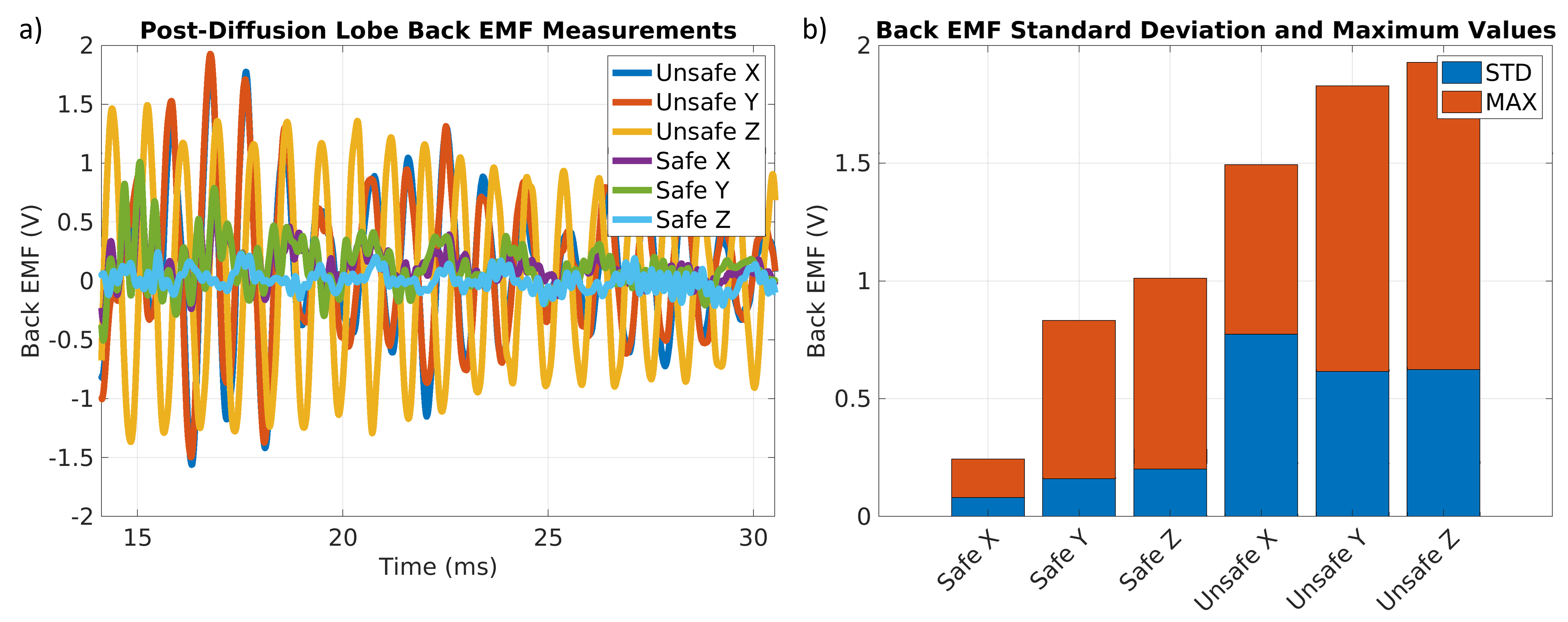
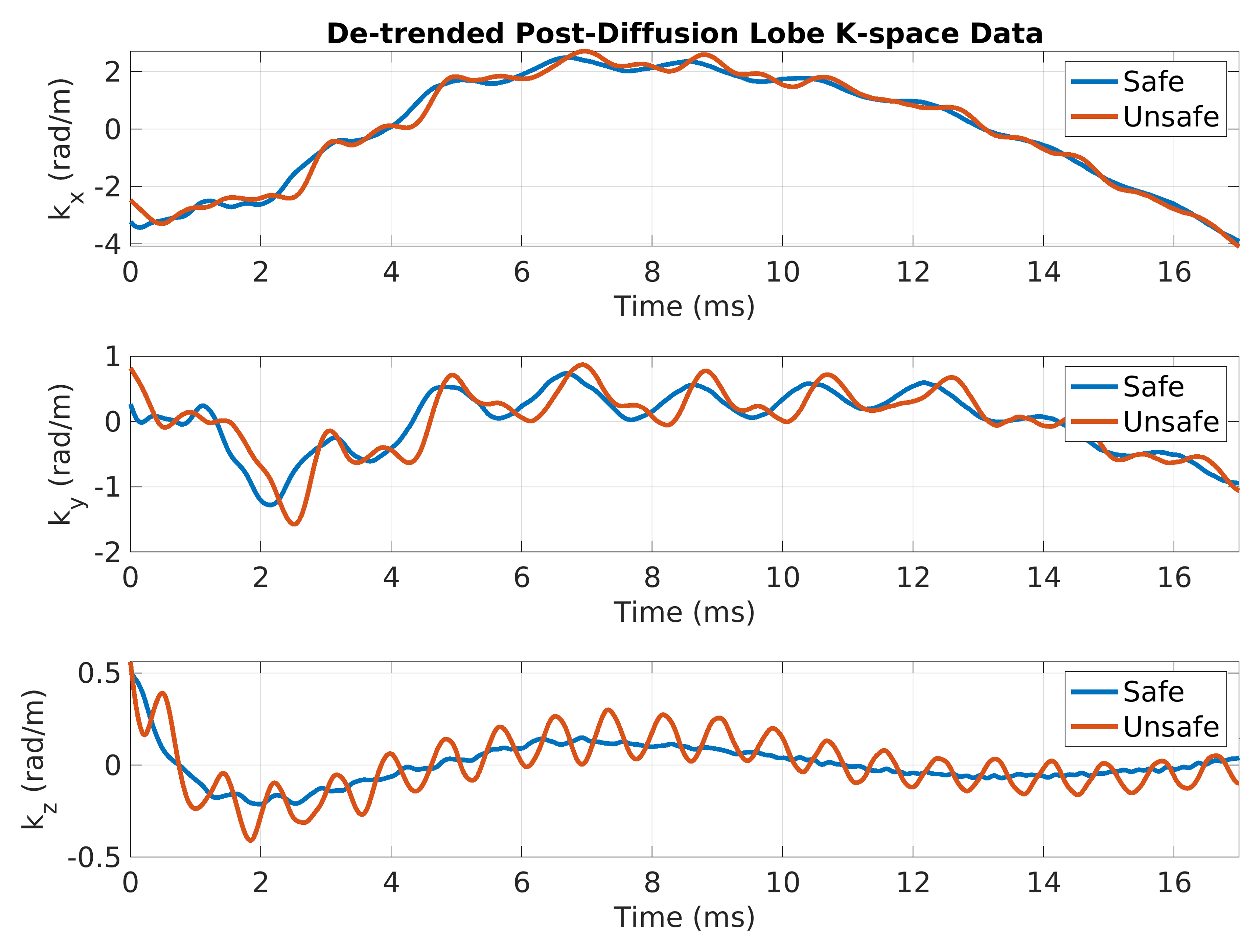
Vibration of the coil following a waveform induces a back-EMF that the current-controlled gradient power amplifier (GPA) bucks in order to maintain zero current. The bridge voltage of the GPA (or back-EMF) then acts as an indicator of vibration magnitude. The EMF is measured for 20ms following each waveform in a set of sinusoids from 50-2000Hz (10Hz spacing), and pairs of safe and unsafe diffusion lobes chosen to maximally excite each gradient axis with b-values close to 1000s/mm2. Relative coil response to lobes is estimated by generating trapezoidal lobes of varying b-value and taking the sum of their power spectra weighted by the back-EMF frequency response. Chosen test lobes include 988s/mm2, on the X and Y coils, and 1078s/mm2 on the Z coil. Additionally, measurements of k-space trajectory over the 20ms period are taken using a field camera (Dynamic Field Camera, Skope, Switzerland).
RESULTS
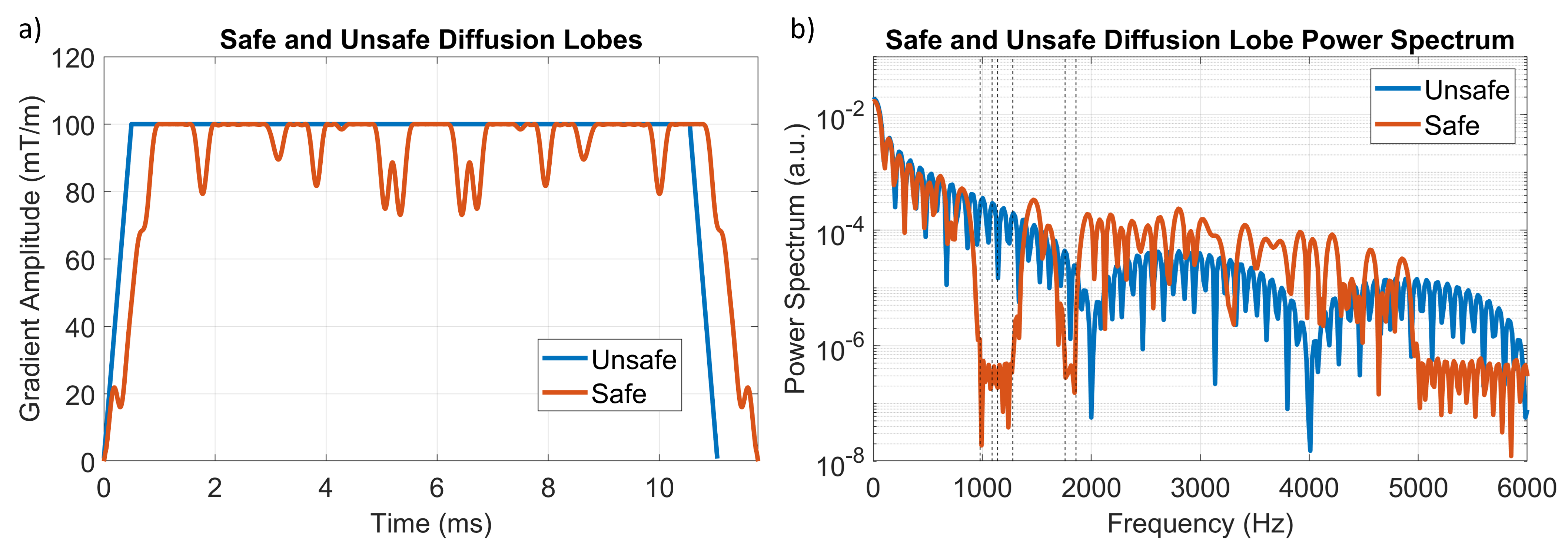
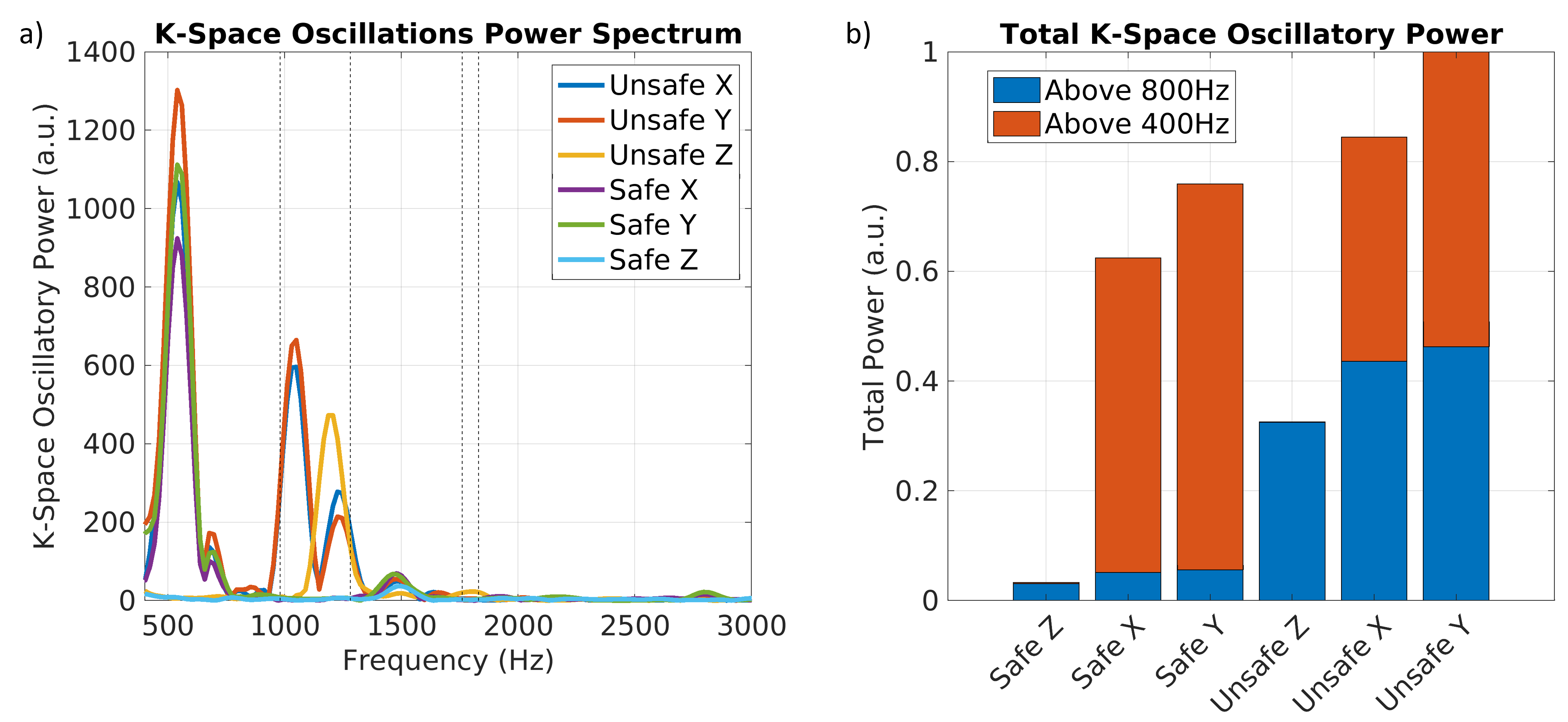
An example safe and unsafe diffusion lobe pair are shown in Figure 2 along with their power spectrums. Back-EMF measurements following worst-case diffusion lobes at approximately 1000s/mm2 are given in Figure 3. De-trended k-space measurements following the diffusion lobes are seen in Figure 4. The power spectra of these measurements are analyzed in Figure 5.DISCUSSION
Back-EMF frequency response measurements showed two primary mechanical resonance peaks at 1040Hz and 1230Hz in X and Y and two at 1190Hz and 1800Hz in Z. These bands were constrained in safe diffusion designs leading to a great reduction in the post-diffusion back-EMF (Figure 3) indicating significant reduction in vibration. We can see from the Skope camera measurements (Figure 4) that there are also oscillations in the magnetic field. The kz measurements show that these oscillations were nearly eliminated using the safe diffusion lobe, but some oscillation remains in kx and ky. We can see from the power spectrum (Figure 5) that oscillations at the target frequencies were nulled on all axes, but a large amount of energy is deposited at 520Hz in kx and ky. The small peak at this frequency in the back-EMF response was ignored, but clearly can be significant for waveforms with high energy at 520Hz. This can easily be addressed by including the band at 520Hz in the frequency constraint.CONCLUSION
A novel design method for time-optimal frequency constrained “safe” diffusion lobes has been outlined. This method has been shown to avoid gradient mechanical resonance frequencies, reducing vibration of the coils and oscillations in the magnetic field. These methods can be extended to the design of other high-power gradient lobes in sequences such as gradient spoilers and crushers to similarly avoid mechanical resonances.Acknowledgements
This work was supported by the National Institutes of Health under grants R01EB009690 and U01EB029427.
We acknowledge the support of the Natural Sciences and Engineering Research Council of Canada (NSERC) through the Postgraduate Scholarship - Doctoral Program (PGSD)
References
1. Chilla GS, Tan CH, Xu C, Poh CL. Diffusion weighted magnetic resonance imaging and its recent trend-a survey. Quant Imaging Med Surg. 2015;5(3):407-422. doi:10.3978/j.issn.2223-4292.2015.03.01
2. Berl MM, Walker L, Modi P, et al. Investigation of vibration-induced artifact in clinical diffusion-weighted imaging of pediatric subjects. Hum Brain Mapp. 2015;36(12):4745-4757. doi:10.1002/hbm.22846
3. Gallichan D, Scholz J, Bartsch A, Behrens TE, Robson MD, Miller KL. Addressing a systematic vibration artifact in diffusion-weighted MRI. Hum Brain Mapp. 2010;31(2):193-202. doi:10.1002/hbm.20856
4. McNulty JP, McNulty S. Acoustic noise in magnetic resonance imaging: An ongoing issue. Radiography. 2009;15(4):320-326. doi:10.1016/j.radi.2009.01.001
5. Winkler SA, Schmitt F, Landes H, et al. Gradient and shim technologies for ultra high field MRI. Neuroimage. 2018;168:59-70. doi:10.1016/j.neuroimage.2016.11.033
6. Aliotta E, Wu HH, Ennis DB. Convex optimized diffusion encoding (CODE) gradient waveforms for minimum echo time and bulk motion–compensated diffusion-weighted MRI. Magn Reson Med. 2017;77(2):717-729. doi:10.1002/mrm.26166
7. Aliotta E, Moulin K, Ennis DB. Eddy current–nulled convex optimized diffusion encoding (EN-CODE) for distortion-free diffusion tensor imaging with short echo times. Magn Reson Med. 2018;79(2):663-672. doi:10.1002/mrm.26709
Figures