0908
Concept 0.13 T bedside MRI for early brain imaging in the neonatal intensive care unit.1A.A. Martinos Center for Biomedical Imaging, Charlestown, MA, United States, 2Radiology, Massachusetts General Hospital, Boston, MA, United States, 3Harvard Medical School, Boston, MA, United States, 4Pediatrics-Neonatology, Massachusetts General Hospital, Boston, MA, United States, 5Harvard-MIT Division of Health Sciences and Technology, Cambridge, MA, United States
Synopsis
Keywords: Magnets (B0), Magnets (B0)
Motivation: Despite the high diagnostic value of MRI, safety concerns and logistical burdens often prohibit the transport of neonatal intensive care unit (NICU) patients to standard MRI scanners.
Goal(s): In response, we aim to design a specialized NICU bedside MRI scanner that prioritizes minimal disruption to care and provides a higher field strength (and signal-to-noise) than currently available portable scanners.
Approach: Using realistic finite element modeling and genetic algorithm optimization, we demonstrate a 131mT Halbach magnet design with a peak-to-peak homogeneity of 421ppm over a 14cm diameter spherical volume.
Results: We present the computer-aided-design prototype of the full portable NICU MRI system.
Impact: The bedside MRI scanner capable of diffusion contrast neuroimaging of neonates could bring a new early evaluation tool for brain conditions such as hypoxic ischemic encephalopathy (HIE).
Introduction
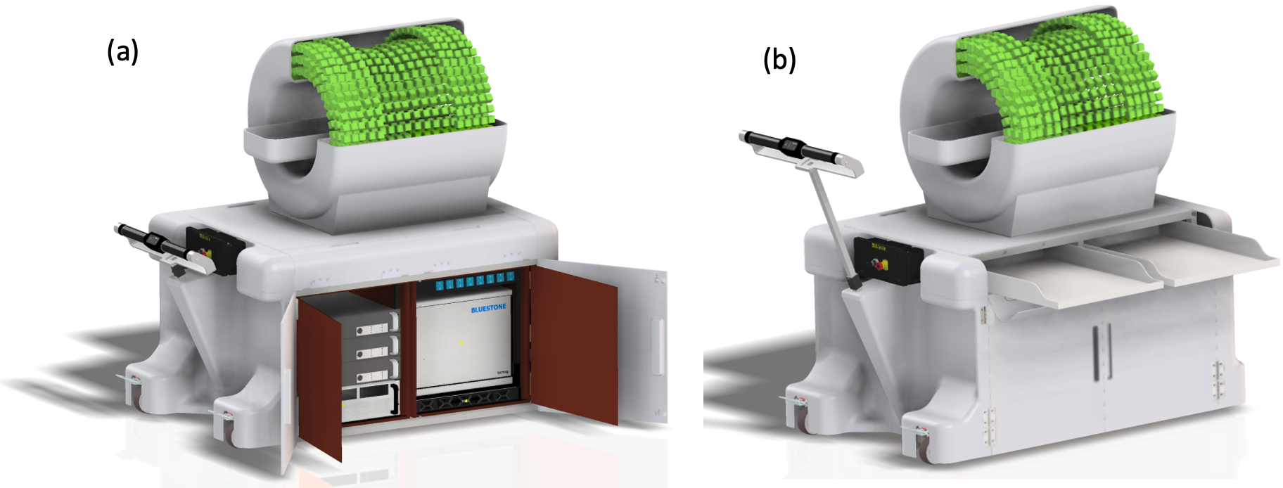
The neonatal brain is highly susceptible to various insults like Hypoxic-Ischemic Encephalopathy (HIE), particularly in preterm births. HIE results from a lack of blood flow and oxygen to the brain at the time of birth and stands out as a primary contributor to neurological problems in children (common cause of cerebral palsy)1,2,6 and deaths among neonates (~23% of newborns)3-5. Effective clinical management of these patients is crucial, as it can greatly reduce disabilities later in life.Diffusion MRI offers distinct advantages over other imaging modalities such as Ultrasound for diagnostic brain imaging in neonates9. Previous efforts have led to specific neonatal scanners located within the NICU, such as the superconducting and permanent magnets designs >1T18-19. These scanners, however, are fixed in one location necessitating patient transport within the NICU, causing disruption to patient care. In contrast, the 64mT Hyperfine Swoop scanner has recently demonstrated bedside neonatal imaging; however, a higher field strength scanner could facilitate more robust diffusion imaging20. Figure1 illustrates our proposed bedside brain MRI scanner for the NICU. A key element of our design is a 0.13T Halbach magnet, delivering twice the field strength of current mobile MRI magnets with similar size and weight10-12.
Methods
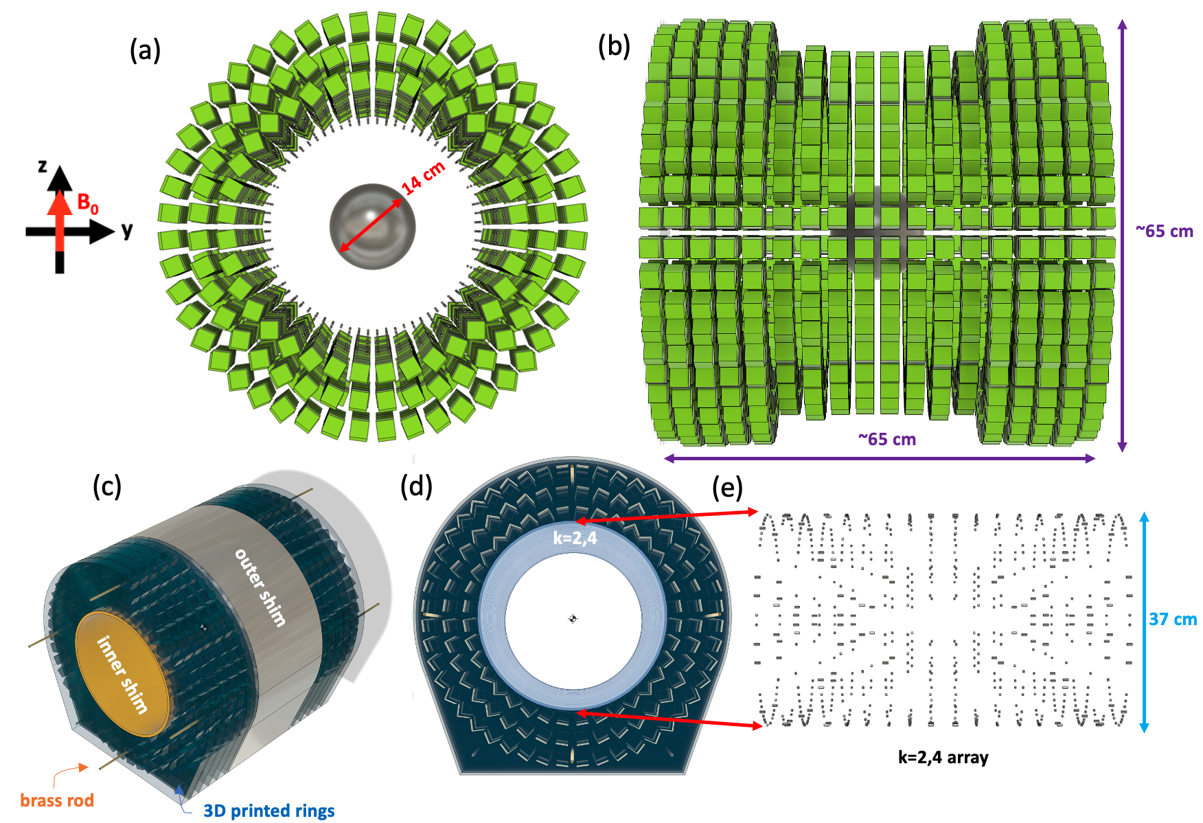
The main Halbach magnet is composed of 1848 NdFeB 1-inch blocks grade N40UH. We selected N40UH due to its high intrinsic coercivity (≥1990kA/m) compared to other NdFeB materials, which minimizes demagnetization effects and interactions between blocks13,14. The dipolar Halbach configuration comprises a total of 18 rings; the central rings consist of 2 layers while the end rings consists of 3 layers to boost the field at the ends and improve the initial homogeneity along the x-axis (Fig.2a,b). A finite-element method (FEM) based magnetostatics simulation is employed using Opera3D software14 (Dassault Systemes, France), utilizing the B(H) curve provided by the manufacturer13,14. A genetic algorithm (GA) optimization is integrated with the FEM simulation14. This integration enables the identification of optimal ring diameters and spacings that yield the highest level of homogeneity over a 14cm diameter-spherical volume (DSV), which is the intended volume for neonatal cranial imaging ranging 0-3 months old16. To further improve homogeneity, we integrated an array of 3mm N40UH cubes (Fig.2d,e). The inner array was optimized by varying the number of stacked blocks in predetermined locations and their orientations using Halbach rings of the k=2 and k=4 configurations. Nevertheless, constructed designs almost always have a higher inhomogeneity than expected due to fabrication and positioning errors15,17. To address these issues, we will implement a two-step shimming process, which consists of distributing 3mm cubes on an inner sleeve insert and outer surface attachments (Fig.2c).Results
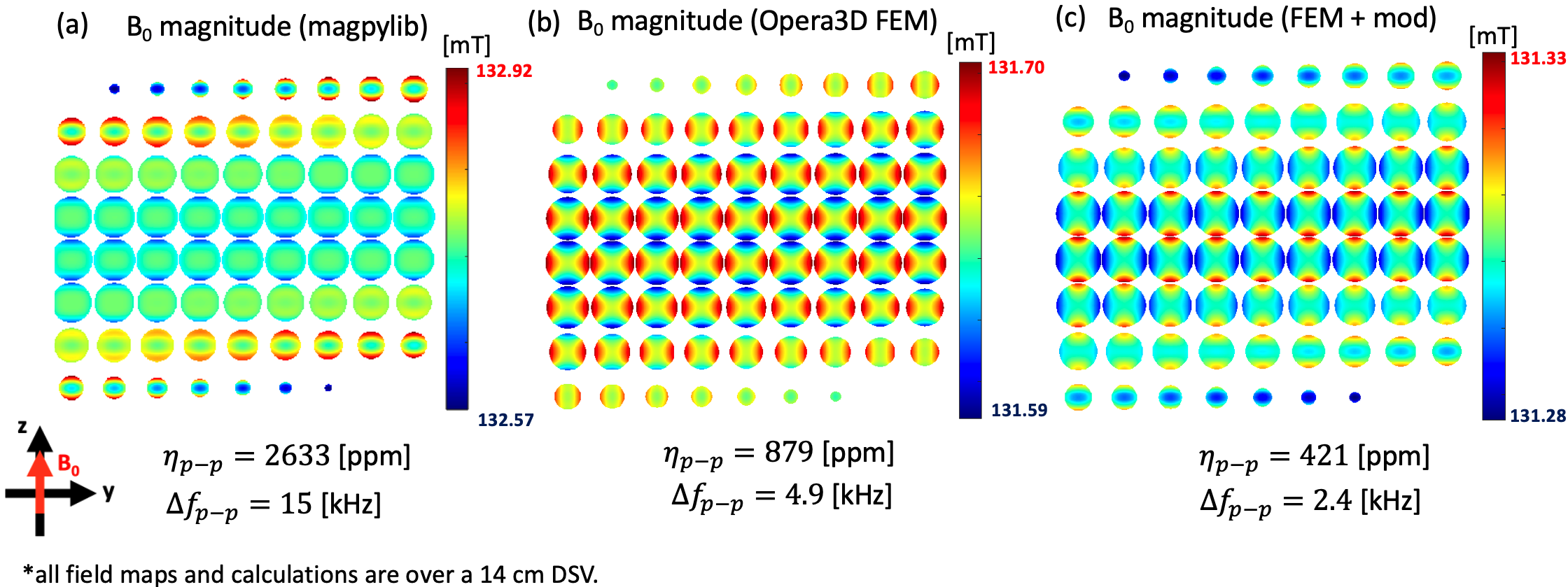
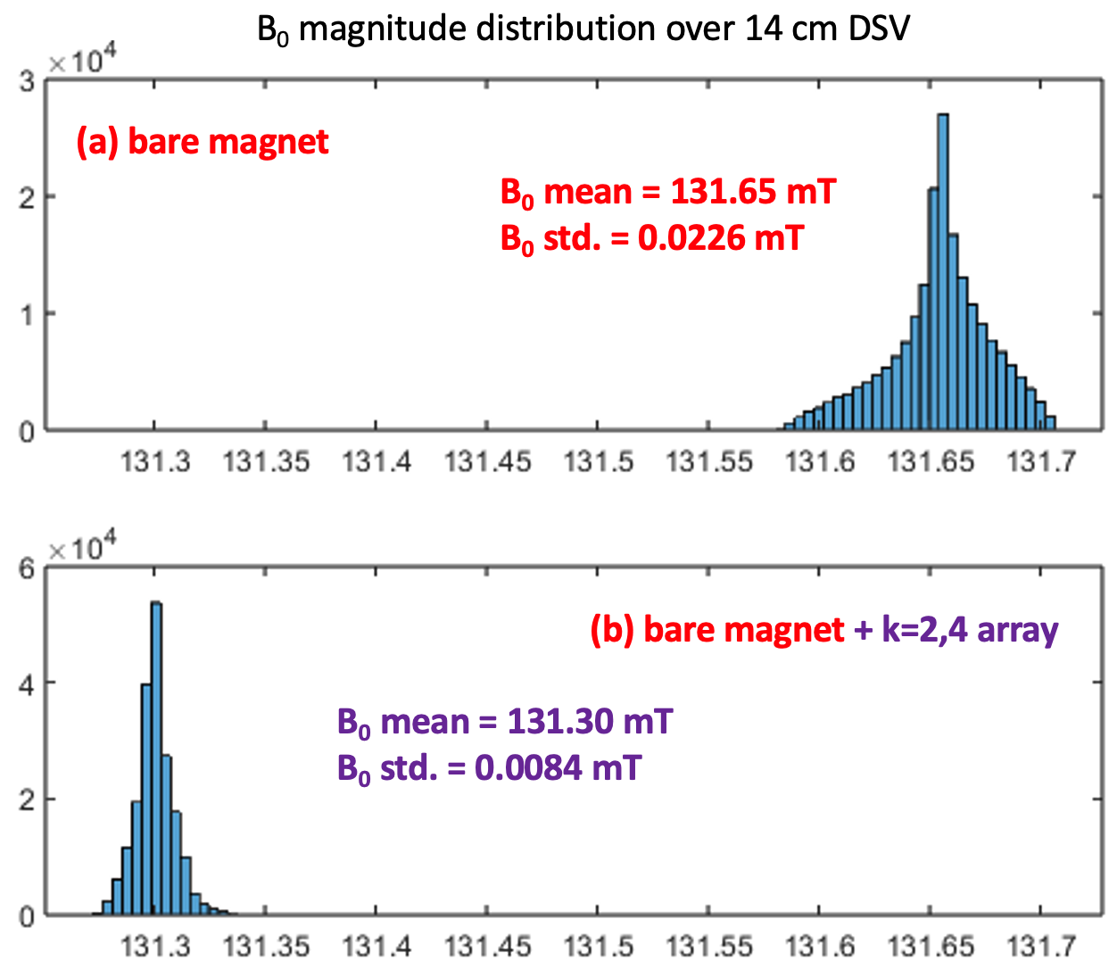
Figure 3b shows the simulated B0 maps in a 14cm DSV of the most optimal main magnet design (without the 3mm block inner array), achieving 131.3mT flux density and peak-to-peak homogeneity (ηFEM) of 879ppm(4.9kHz). For the same optimal geometry, the field was also simulated using the open-source magpylib Python package21, producing 132.6mT and ηmagpy = 2633ppm(15kHz) (Fig.3a). Furthermore, an optimized 3 mm block array (Halbach k=2,4 orientations) was achieved using a target-field approach against the main magnet’s inhomogeneities. When both arrays are combined, the total field is 131.3mT with ηFEM,mod = 421ppm(2.4kHz) (Fig.3c). The B0 distribution throughout the DSV without and with the additional k=2,4 array are shown in Figure4a,b.Discussion
In the design of Halbach magnets, it is common to utilize fast analytical methods for optimizing field homogeneity; however, they neglect demagnetization and interaction effects. As shown here, magpylib and Opera3D FEM output can be vastly different for the same geometry and material remanence. Therefore, we used FEM directly with a GA optimization framework to accurately search for the ring diameters and spacings that produce the most homogeneous field and highest possible field strength. Nevertheless, we found the optimal bare magnet to be limited by field perturbations that appear dipolar near the magnet ends and sextupolar near the geometric isocenter, so we optimized an additional array of 3mm magnets that improved the simulated bare magnet homogeneity by 2x.Conclusion
We introduce a 0.13T Halbach magnet designed specifically for bedside brain MR imaging in the NICU. The ongoing construction of this design involves utilizing high intrinsic coercivity permanent magnets (N40UH), 3D printing of formers, and implementing a k=2,4 array during construction. In addition, custom inner and outer passive shimming inserts and attachments will account for fabrication errors and impact of field inhomogeneity after construction. Notably, compared to other portable MRI systems, our magnet design enables a more than double the field strength, a lighter overall MRI system design, yet has similar overall dimensions11,12.Acknowledgements
Research was supported by NICHD of the National Institutes of Health under award number R01HD104649.References
[1] Douglas-Escobar, M. et al. (2015). JAMA Pediatr., vol. 169, no. 4, pp. 397–403.
[2] Finer, N.N. et al. (1981). J. Pediatr., vol. 98, no. 1, pp. 112–117.
[3] Edwards, A.B., et al. (2018). Brain Sci., vol. 8, no. 8, Apr. 2018, doi: 10.3390/brainsci8080147.
[4] Black R.E. et al. (2010). Lancet, vol. 375, no. 9730, pp. 1969–1987.
[5] Oorshchot, D.E. et al. (2020). Int. J. Mol. Sci., vol. 21, no. 4, Apr. 2020, doi: 10.3390/ijms21041487.
[6] Bano, S. et al. (2017). J. Pediatr. Neurosci., vol. 12, no. 1, pp. 1–6.
[7] Chao, C.P. et al. (2006). RadioGraphics, vol 26, pp. S159-S172.
[8] Barkovich, A.J. et al. (2006). Am. J. Neuroradiol. 27:533-547.
[9] Biochot, C. et al. (2011). Magn. Reson. Imaging. 29:194-201.
[10] Cooley, C.Z. et al. (2020). Nat. Bio. Eng. 5:229-239.
[11] Liu, Y. et al. (2021). Nature Communications. 12:7238.
[12] Hyperfine Swoop. hyperfine.io
[13] BINIC Magnet Co. Ltd. http://www.binicmagnet.com/PR01.asp
[14] Purchase, A.R. et al. (2021). IEEE Access, vol. 9, pp. 95294-95303.
[15] O’Reilly, T et. al. (2019). JMR. 307:106578.
[16] https://www.cdc.gov/growthcharts/html_charts/hcageinf.htm
[17] Cooley, C.Z. et al. (2021). Int. Soc. Magn. Reson. Med. 0559
[18] Merher, S.L. et al. (2017). Pediatr. Radiol., vol. 47, no. 8, pp. 1001-1011.
[19] “Aspect Imaging” Available: https://www.aspectimaging.com/.
[20] Sabir H. (2023). Critical Care. 27:134.
[21] Magpylib. https://magpylib.readthedocs.io/en/latest/
Figures