0904
Connectome 2.0: Performance evaluation and initial in vivo human brain diffusion MRI results1Athinoula A. Martinos Center for Biomedical Imaging, Massachusetts General Hospital and Harvard Medical School, Charlestown, MA, United States, 2Siemens Healthineers, Erlangen, Germany, 3Institute of Medical Physics and Radiation Protection, Mittelhessen University of Applied Sciences, Giessen, Germany, 4Siemens Medical Solutions USA, Boston, MA, United States, 5Eunice Kennedy Shriver National Institute of Child Health and Human Development, National Institutes of Health, Bethesda, MD, United States, 6Q Bio Inc, San Carlos, CA, United States, 7Department of Diagnostic and Interventional Radiology, University Hospital Marburg, Philipps University of Marburg, Marburg, Germany
Synopsis
Keywords: Gradients, Gradients, Diffusion Acquisition, Neuro
Motivation: Current human MR scanners cannot resolve the full range of length scales needed to study the brain's microscopic and mesoscopic structure.
Goal(s): To construct and validate the next-generation human connectomics and microstructure MRI scanner known as Connectome 2.0.
Approach: The 3T Connectome 2.0 scanner incorporates a peripheral nerve stimulation-optimized asymmetric head gradient driven by dual gradient power amplifiers. Custom-built high-sensitivity 72-channel (in vivo imaging) and 64-channel (ex vivo imaging) receive coils were integrated.
Results: The Connectome 2.0 scanner achieves Gmax=500 mT/m and SRmax=600 T/m/s, demonstrates 2x improved SNR for diffusion MRI over Connectome 1.0, and enables high-resolution tractography.
Impact: The Connectome 2.0 scanner will allow the exploration of new microstructure properties and connectional anatomy in the living human brain with unprecedented spatial and diffusion resolution.
Introduction
We present the Connectome 2.0 scanner1, developed for next-generation human connectomics and microstructure imaging of human brain circuits across scales. Here, we report on the hardware’s design, construction, and evaluation, and initial results for in vivo human brain diffusion MRI.Methods
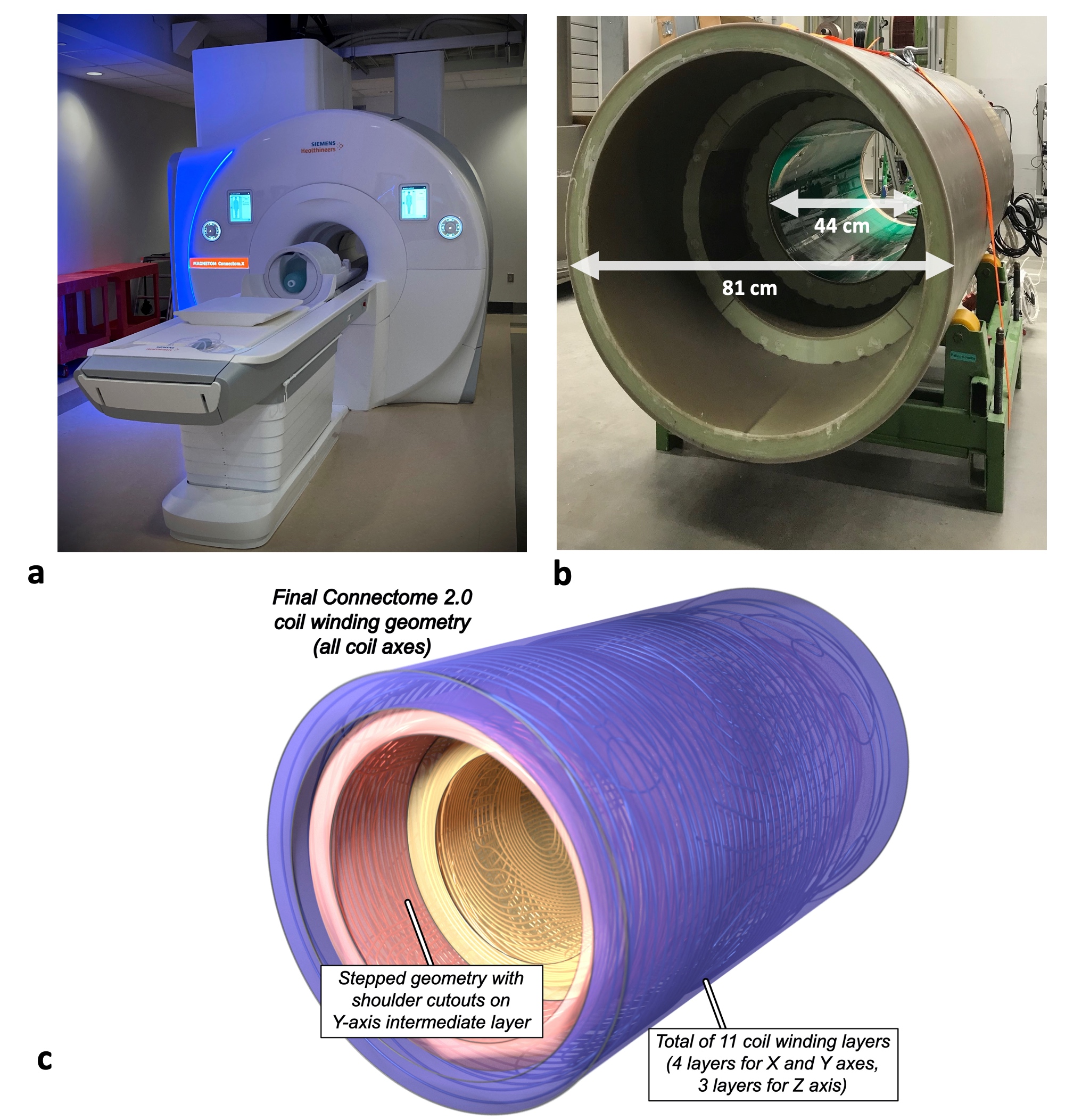
Scanner and gradient coil designThe Connectome 2.0 system was designed on a 3T scanner platform and targeted in vivo human brain imaging using a maximum gradient strength (Gmax) of 500 mT/m and a maximum slew rate (SRmax) of 600 T/m/s. The gradient coil is designed as an asymmetric head gradient following the stepped geometry of the Siemens 7T Impulse head gradient coil (Gmax = 200 mT/m, SRmax = 900 T/m/s),2 but modified to include additional winding layers (four instead of three layers for the X and Y axes) to achieve Gmax = 500 mT/m (Fig. 1c). The gradient coil design was iterated upon using peripheral nerve stimulation (PNS) modeling to raise the PNS thresholds and maximize the usable gradient parameter space.3-5
Radiofrequency coils
A 72-channel in vivo head receive (Rx) coil6 and a 64-channel ex vivo whole brain coil7 were designed and constructed for high-sensitivity diffusion MRI acquisitions. Each Rx array was outfitted with its own dedicated local transmit (Tx) coil and 16-channel 19F clip-on field probe system (Skope, Inc., Zurich, Switzerland) for concurrent field monitoring.8
In vivo diffusion MRI experiments
All in vivo diffusion MRI experiments were performed on the Connectome 2.0 scanner using the 72-ch in vivo head coil.
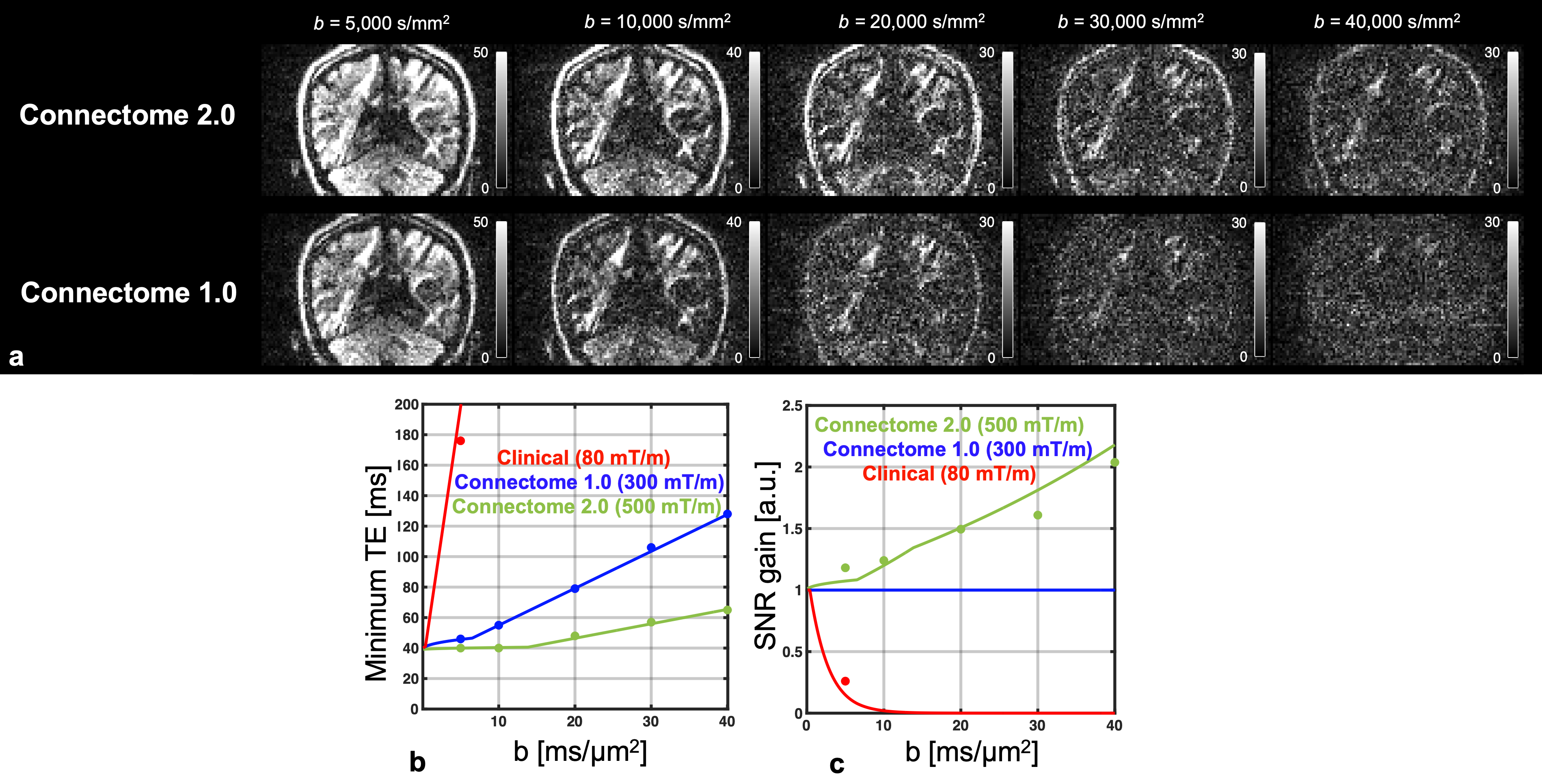
a) High b-value diffusion MRI: Signal-to-noise ratio (SNR) performance was evaluated on a healthy volunteer (43F) using a fixed pulse width $$$\delta$$$=8 ms, diffusion weighting b, diffusion time $$$\Delta$$$, echo time (TE), and Gmax of a state-of-the-art research scanner (80 mT/m), Connectome 1.0 (300 mT/m), and Connectome 2.0 (500 mT/m).
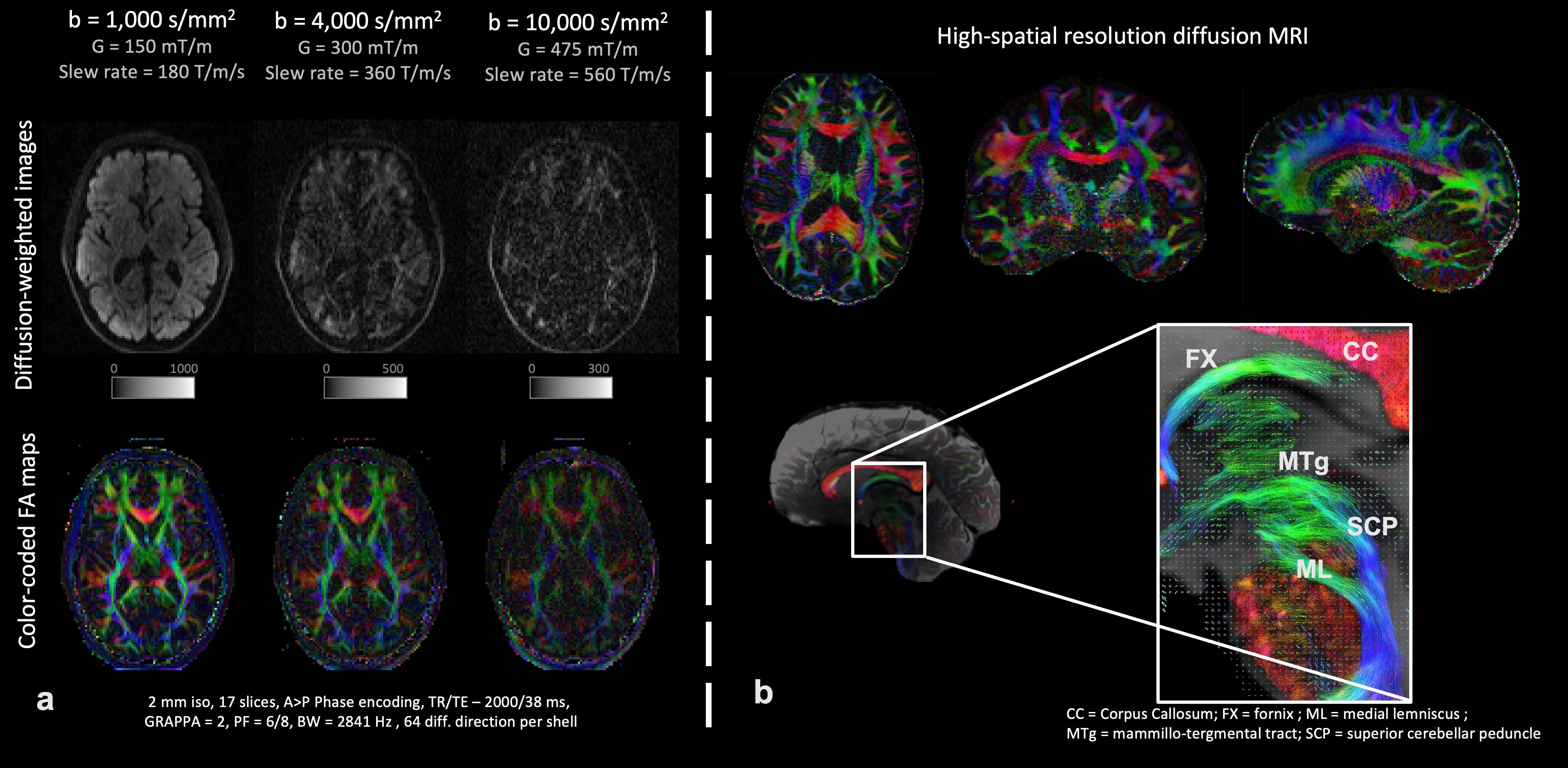
b) First in vivo human brain diffusion MRI scan was performed on a healthy volunteer (68M) on Connectome 2.0 (see caption of Fig. 5 for acquisition parameters).
c) High-resolution tractography was performed on a healthy volunteer (34F) using Connectome 2.0 and Connectome 1.0 protocols (see caption of Fig. 5). Whole-brain tractograms were generated, seeding local probabilistic tractography9 in every voxel within a white matter mask (5 seeds per voxel).
Results
The Connectome 2.0 scanner (MAGNETOM Connectom.X, Siemens Healthineers, Erlangen, Germany) was installed at the MGH Martinos Center in July 2023 and has been operational since August 2023 (Fig. 1a).Gradient system
The Connectome 2.0 gradient coil is shown in Fig. 1b.
Geometry: The stepped design with shoulder cutouts and outer gradient coil diameter of 81 cm provides access for subjects of varying sizes. The inner gradient coil diameter is 44 cm, and the free bore diameter is 40 cm.
Gradient performance: The scanner achieves Gmax=500 mT/m and SRmax=600 T/m/s driven by 2 GPAs (1200A/2250V) per axis compared to the 4 GPAs (900A/2000V) of the original Connectome scanner.10 The gradient coil has higher efficiency (0.42 mT/m/A) and lower inductance per axis than the original Connectome gradient coil (X=2250, Y=2450, Z=1800 $$${\mu}H$$$) with direct current resistance of 0.28 $$$\Omega$$$.
Non-linearity: Maximum field linearity deviation in a 20cm sphere is X=6.7%, Y=8.2%, and Z=11.7%.
Cooling: The gradient coil incorporates direct liquid cooling technology to maximize thermal energy transfer and dissipation.
Active E-shims include 1st and 2nd-order harmonics.
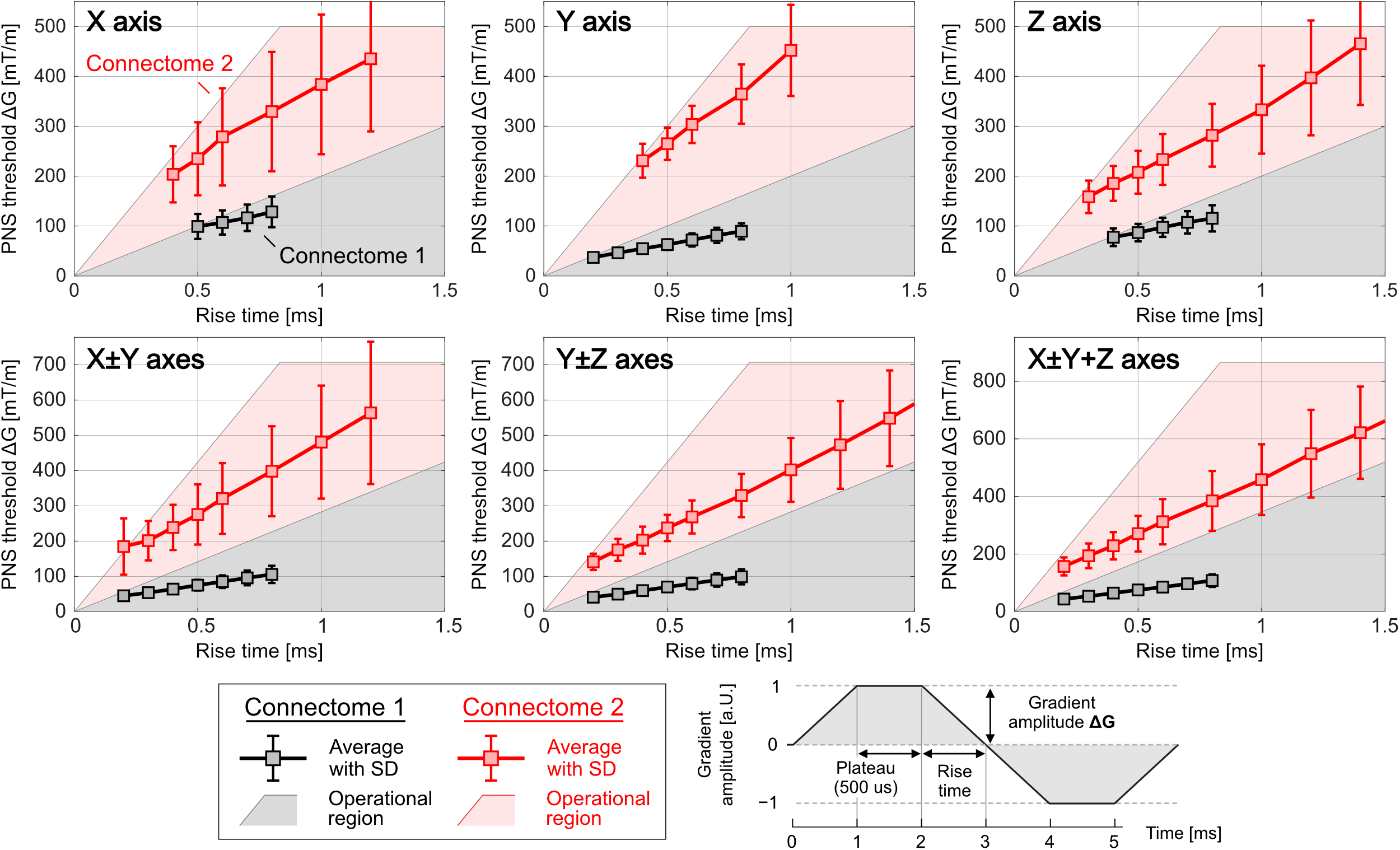
The gradient achieves 2.4–4.2x greater PNS thresholds than the Connectome 1.0 gradient coil (Fig. 2).
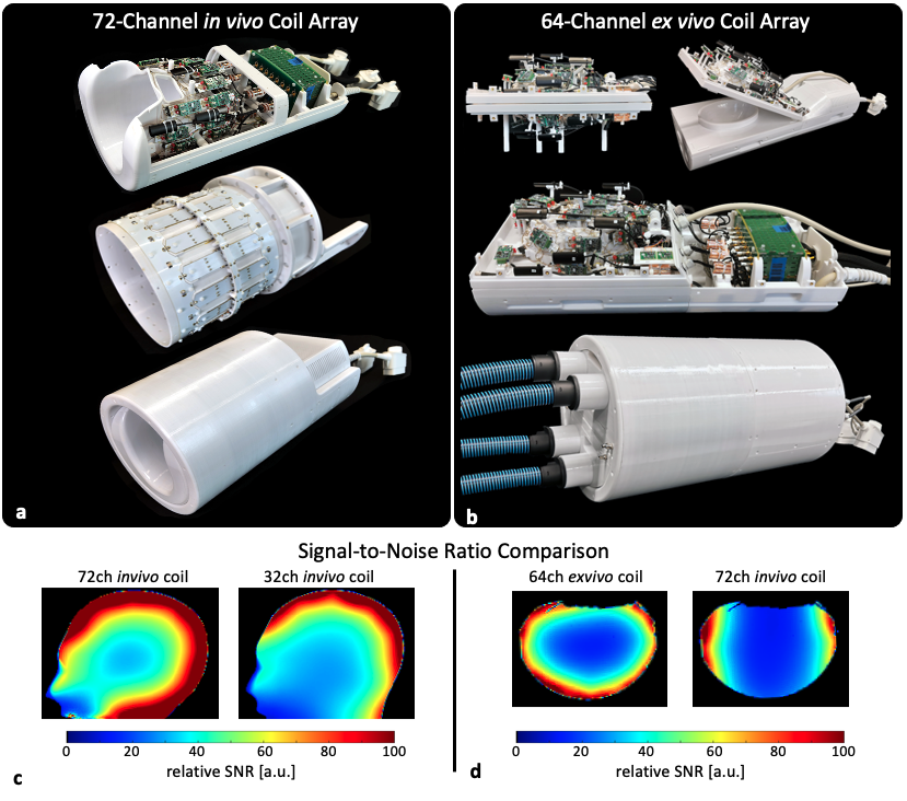
Radiofrequency coils:The 72-channel in vivo coil (Fig. 3a) provides a 1.5x improvement in SNR in the peripheral regions of the phantom compared to a standard 32-channel head coil (Fig. 3c). The 64-channel ex vivo coil (Fig. 3b) outperforms the larger 72-channel in vivo coil by 1.73x in average SNR (Fig. 3d).
Improvements in SNR for diffusion imaging:The stronger gradients of the Connectome 2.0 scanner enable significant reductions in diffusion time $$$\Delta$$$ and pulse width $$$\delta$$$ compared to Connectome 1.0, resulting in shorter TE by up to 50% (Fig. 4b). SNR gains are at least 4x those of a clinical scanner and up to double the SNR over the Connectome 1.0 scanner (Fig. 4c) at high b-values (Fig. 4a).
Initial in vivo human brain imaging: Fig. 5a shows the first in vivo diffusion MRI in the human brain acquired on Connectome 2.0. Fig. 5b presents high-resolution tractography delineating fine fiber tracts like the mammillo-tegmental tract on Connectome 2.0, which could not be seen with a matched protocol using Connectome 1.0 parameters.11
Conclusion
The Connectome 2.0 scanner, equipped with Gmax=500mT/m and SRmax=600T/m/s and the latest RF coil technology, achieves unprecedented sensitivity and resolution for mesoscale diffusion MRI in the living human brain.Acknowledgements
The research reported in this abstract was supported by the National Institute of Biomedical Imaging and Bioengineering of the National Institutes of Health under award number U01EB026996.References
1Huang SY, Witzel T, Keil B, et al. Connectome 2.0: Developing the next-generation ultra-high gradient strength human MRI scanner for bridging studies of the micro-, meso- and macro-connectome. Neuroimage. 2021 Nov;243:118530.
2Feinberg, DA, Dietz, P, Liu C, et al. Design and Development of a Next-Generation 7T human brain scanner with high-performance gradient coil and dense RF arrays. Proc. Intl. Soc. Mag. Reson. Med. 29 (2021), 0562
3Davids, M, Dietz, P, Ruyters G, et al. Peripheral nerve stimulation informed design of a high‐performance asymmetric head gradient coil." Magnetic Resonance in Medicine (2023).
4Davids, M, Guerin B, Klein V et al. Optimizing selective stimulation of peripheral nerves with arrays of coils or surface electrodes using a linear peripheral nerve stimulation metric. Journal of neural engineering 17.1 (2020): 016029.
5Davids, M, Guerin, B, Klein V et al. Optimization of MRI gradient coils with explicit peripheral nerve stimulation constraints. IEEE transactions on medical imaging 40.1 (2020): 129-142.
6Mahmutovic M, Shrestha M, Ramos-Llordén G, et al. A 72-channel Head Coil with an Integrated 16-Channel Field Camera for the Connectome 2.0 Scanner. Submitted to ISMRM 2024.
7Scholz A, Mahmutovic M, Alem M, et al. Design of a 64-channel ex vivo Brain Rx Array Coil with field monitoring and temperature control for DWI at 3T, Proc. Intl. Soc. Mag. Reson. Med. 31 (2023) 0215
8Wilm BJ, Nagy Z, Barmet C, et al. Diffusion MRI with concurrent magnetic field monitoring. Magn Reson Med. 2015;74(4):925-933. doi:10.1002/MRM.25827
9Tournier, J.-D, Calamante, F. and Connelly, A. Improved probabilistic streamlines tractography by 2nd order integration over fibre orientation distributions. Proceedings of the International Society for Magnetic Resonance in Medicine, 2010, 1670
10Setsompop, K., Kimmlingen, R., Eberlein E. et al. Pushing the limits of in vivo diffusion MRI for the Human Connectome Project. Neuroimage 80 (2013): 220-233.
11Maffei C, Ma Y, Ramos-Llordén, G et al. Visualization of fine white matter bundles in the living human brain with diffusion MRI using 500 mT/m gradient strength. Submitted to ISMRM 2024.
Figures