0902
Quantitative assessment of tongue tissue structure with 3D partially spoiled gradient echo1NMR Laboratory, Neuromuscular Investigation Center, Institute of Myology, Paris, France, 2Siemens Healthcare SAS, Saint-Denis, France
Synopsis
Keywords: Muscle, MSK
Motivation: Fat fraction and water T2 have been identified as biomarkers of muscle tissue alterations in neuromuscular diseases. Tongue muscles are involved in several muscular disorder, but due to swallowing motion, quantitative MRI is almost never applied at this level.
Goal(s): Assess the feasibility of a water T2 mapping in the tongue.
Approach: In this feasibility study, we proposed an RF phase-modulated gradient-echo acquisition with 3D radial encoding to obtain FF and water T2 maps of the tongue.
Results: The proposed method enables 3D mapping. The values found in the tongue are consistent with those found with the same methods in the leg muscles.
Impact: Performing quantitative imaging in the tongue is challenging due to swallowing motion. Applying a 3D radial sequence appears the appropriate strategy for water T2 imaging.
Introduction
Swallowing difficulties, or dysphagia is a symptom of several neuromuscular diseases (NMDs) which can lead to malnutrition, deterioration in general condition and inhalation pneumonitis (1). Tongue muscles are the primary links in the swallowing chain (2) and are affected in pathologies such as oculopharyngeal muscular dystrophy (OPMD) and Pompe disease, where significant increases of fat fraction (FF) have been reported using quantitative MRI (3)(4). In particular, tongue muscles are often altered in early stages of OPMD (5).Water T2 (T2H2O) relaxometry using multi spin echo sequences (MSE) provides a highly sensitive imaging biomarker of active muscle damage, that often occur before muscle fatty replacement in NMDs (6). We hypothesize that tongue T2H2O values could also be altered at early stage, before dysphagia is installed. The current limitations of standard MSE sequences (7) for exploring the tongue is their high sensitivity to motion during swallowing and their restriction to 2D imaging. Recent works demonstrated that 3D FF and T2H2O maps can be simultaneously obtained from a partially spoiled-gradient echo sequence (p-SPGR), by an optimal selection of RF-phase increments (φinc) and flip angles (α) (8). Here, we proposed a radial version of the p-SPGR sequence and assessed the feasibility of quantitative tongue measurements in healthy volunteers.
Methods
Acquisitions were performed at 3T (Magnetom PrismaFit, Siemens Healthineers, Germany) using a 20-channel head and neck coil on three healthy volunteers.The simultaneous FF and T2H2O measurement method is based on the acquisition of a series of 10 3D p-SPGR volumes (TE/TR = 1.94/6.6ms for radial and 2.25/5.5 for cartesian) with different φinc and α (φinc =[0.3, -1.1, 0.3, 2.1, -2.1, 3, -2.6, 2.8, -2.6, 0.3], α = [7, 25, 26, 8, 8, 17, 13, 16, 16, 6.5]) (8). The complex signal was fitted by exhaustive search into a two-component dictionary using a phase-constrained least squares linear regression for the weights of the water and fat components. The Cartesian version previously proposed was compared to a 3D stack-of star radial version in order to decrease its sensitivity to motion. Radial data were reconstructed offline using the BART toolbox (9).
For reference, we also acquired a 3D multi-TE GRE sequence (3 TEs, 64 slices, Tacq = 1min34s) from which 3-point Dixon was applied and FF extracted (10) and a MSE sequence (17 TEs, 5 slices, Tacq =5min29s) from which water T2 was extracted using a bi-component EPG fitting method (11). For all acquisitions, FOV was 200x200x192 mm3 and in plane resolution 0.8x0.8x3 mm3.
Tongue muscles were manually delineated on anatomical images. We extracted the mean FF and T2H2O values as well as coefficients of variation with all methods.
Results
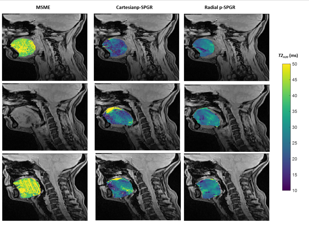
Figure 1 displays FF maps obtained from the tongue muscles of a subject using the three different methods: the reference Dixon method, the Cartesian p-SPGR and the radial p-SPGR methods, at various slice levels. Figure 2 depicts the T2H2O maps obtain in the same subject with the MSE method, the Cartesian p-SPGR and the radial p-SPGR methods. While motion artefacts were clearly visible with the first two methods, the radial approach was less affected.Mean FF values were comparable between the three methods (Table 1). Water T2 values were significantly lower with both p-SPGR methods than the reference MSE approach.
Conclusions/Discussion
In this preliminary study, we presented the feasibility of simultaneous FF and T2H2O mapping of the tongue muscles using a 3D stack-of-star radial p-SPGR method. FF measures were coherent with the literature (12) and the reference Dixon method. T2H2O values were significantly lower with the p-SPGR approach than the reference MSE sequence but this was consistent with what has been recently observed in the lower limb muscles of healthy volunteers (8). The 3D stack of star encoding scheme improved the robustness of the maps in these muscles affected by motion. A recent study reported a significant correlation between tongue pressure and tongue FF in the elderly (13), but little is known about the structure-function relationship in these muscles in the context of NMDs. With the simultaneous measure of FF and T2H2O, our proposed method could be used to establish new biomarkers for studying swallowing muscles involvement in various NMDs and confront them with functional outcomes.Acknowledgements
This study was funded by ANR-20-CE190004References
1. Trollet C, Boulinguiez A, Roth F, et al. Oculopharyngeal Muscular Dystrophy. . GeneReviews® [Internet]. Seattle (WA): University of Washington, Seattle; 1993-2021. October 8, 2001; https://www.ncbi.nlm.nih.gov/books/NBK1126/.
2. Matsuo K, Palmer JB. Anatomy and Physiology of Feeding and Swallowing: Normal and Abnormal. Physical Medicine and Rehabilitation Clinics of North America. 2008;19(4):691–707. doi: 10.1016/j.pmr.2008.06.001.
3. Carlier R-Y, Laforet P, Wary C, et al. Whole-body muscle MRI in 20 patients suffering from late onset Pompe disease: Involvement patterns. Neuromuscular Disorders. 2011;21(11):791–799. doi: 10.1016/j.nmd.2011.06.748.
4. Melkus G, Sampaio ML, Smith IC, et al. Quantitative vs qualitative muscle MRI: Imaging biomarker in patients with Oculopharyngeal Muscular Dystrophy (OPMD). Neuromuscular Disorders. 2022;S0960896622006940. doi: 10.1016/j.nmd.2022.09.010.
5. Alonso-Jimenez A, Kroon RHMJM, Alejaldre-Monforte A, et al. Muscle MRI in a large cohort of patients with oculopharyngeal muscular dystrophy. J Neurol Neurosurg Psychiatry. 2019;90(5):576–585. doi: 10.1136/jnnp-2018-319578.
6. Locher N, Wagner B, Balsiger F, Scheidegger O. Quantitative water T2 relaxometry in the early detection of neuromuscular diseases: a retrospective biopsy-controlled analysis. Eur Radiol. 2022;32(11):7910–7917. doi: 10.1007/s00330-022-08862-9.
7. Marty B, Baudin P-Y, Reyngoudt H, et al. Simultaneous muscle water T 2 and fat fraction mapping using transverse relaxometry with stimulated echo compensation: SIMULTANEOUS MUSCLE WATER T 2 AND FAT FRACTION MAPPING. NMR Biomed. 2016;29(4):431–443. doi: 10.1002/nbm.3459.
8. Vermeulen E, Baudin P-Y, Lapert M, Marty B. Quantification of muscle fat fraction and water T2 via RF phase-modulated 3D gradient-echo imaging. Proc 32nd ISMRM. Toronto, Canada; 2023.
9. Uecker M, Ong F, Tamir J. Berkeley Advanced Reconstruction Toolbox. 2015.
10. Glover GH. Multipoint dixon technique for water and fat proton and susceptibility imaging. J Magn Reson Imaging. 1991;1(5):521–530. doi: 10.1002/jmri.1880010504.
11. Marty B, Baudin PY, Reyngoudt H, et al. Simultaneous muscle water T2 and fat fraction mapping using transverse relaxometry with stimulated echo compensation. NMR in Biomedicine. John Wiley and Sons Ltd; 2016;29(4):431–443. doi: 10.1002/nbm.3459.
12. Song R, Hwang SN, Goode C, et al. Assessment of Fat Fractions in the Tongue, Soft Palate, Pharyngeal Wall, and Parapharyngeal Fat Pad by the GOOSE and DIXON Methods. Invest Radiol. 2022;57(12):802–809. doi: 10.1097/RLI.0000000000000899.
13. Nakao Y, Yamashita T, Honda K, et al. Association Among Age-Related Tongue Muscle Abnormality, Tongue Pressure, and Presbyphagia: A 3D MRI Study. Dysphagia. 2021;36(3):483–491. doi: 10.1007/s00455-020-10165-4.
Figures
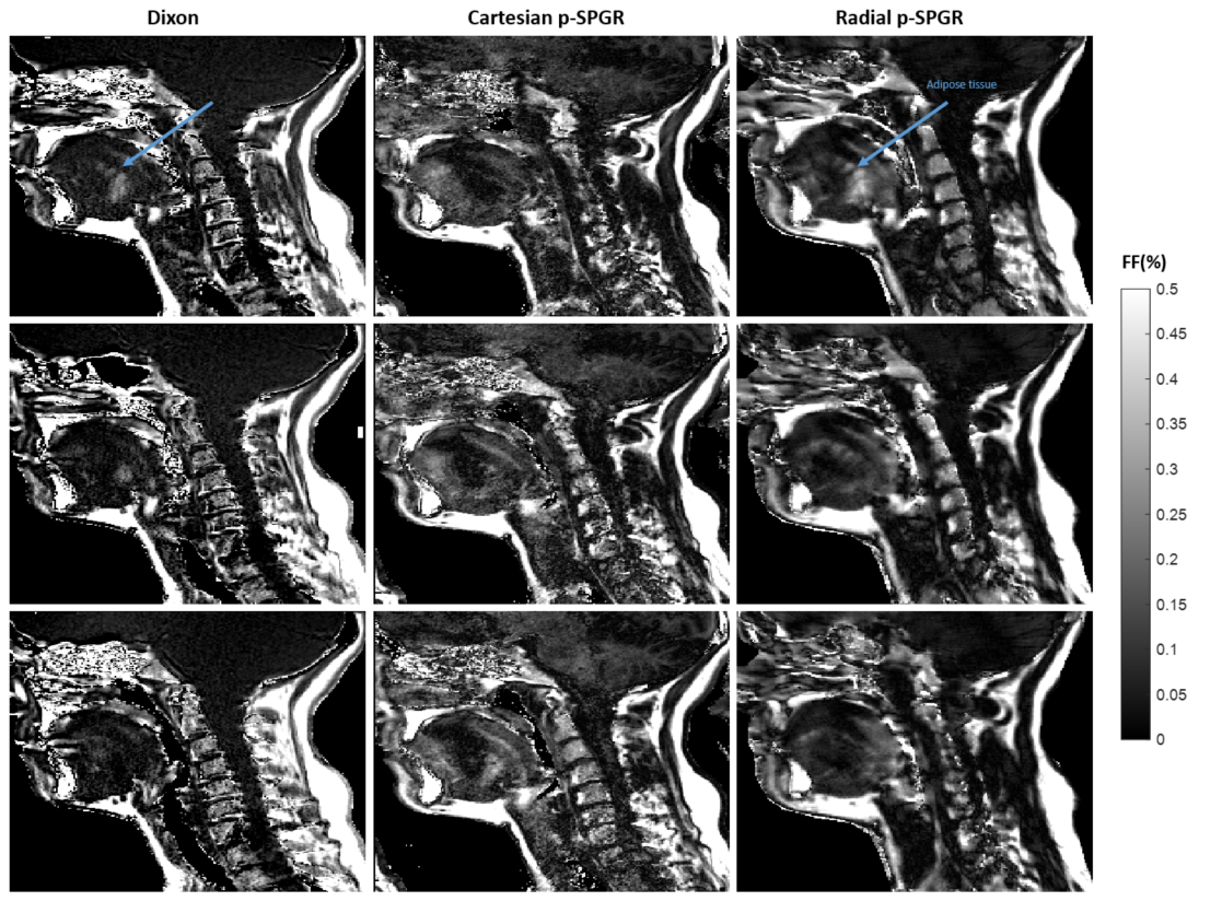
Figure 1: Fat fraction maps obtained by: On the left/middle/right: DIXON/ p-SPGR Cartesian/Radial methods. The blue arrows indicate structures that can be seen on the reference method and on the radial sequence, but that don't show in Cartesian.