0900
Combined Diffusion-Relaxation MRI to Assess Muscle Microstructure and Composition1Centre for Medical Image Computing, University College London, London, United Kingdom, 2Cardiff University Brain Research Imaging Centre, School of Psychology, Cardiff, United Kingdom, 3School of Computer Science and Informatics, Cardiff University, Cardiff, United Kingdom, 4Istituto di Sistemi e Tecnologie Industriali Intelligenti per il Manifatturiero Avanzato, Consiglio Nazionale delle Ricerche, Milano, Italy
Synopsis
Keywords: Muscle, Diffusion/other diffusion imaging techniques, diffusion-relaxation
Motivation: Quantifying muscle tissue properties is crucial for understanding physio-pathological changes occurring in skeletal muscle (SM). However, current methods measure T2 and diffusion separately, and hence conflate them.
Goal(s): Demonstrate a combined diffusion-relaxation MRI approach for disentangling T2 and diffusivity properties in SM.
Approach: We devise and implement a combined T2-diffusion sequence in the leg muscles in five healthy volunteers after exercise. DTI and an advanced diffusion model were implemented and compared.
Results: We calculated disentangled T2 and diffusion-related parameter maps. Our maps capture muscle tissue differences in specific muscle groups highlighting differences related to muscle involvement during exercise.
Impact: Combined diffusion-relaxation MRI can provide detailed non-invasive estimation of muscle tissue properties by mitigating T2 effects on diffusion parameters. These approaches could reduce the need for invasive biopsies for evaluating muscle changes related to neuromuscular diseases, exercise, and rehabilitation.
Introduction
MRI assessment of skeletal muscle (SM) properties with non-invasive techniques could enable quantification of the effects of physical activity, rehabilitation treatments and pharmacological interventions on muscle tissue.Diffusion and relaxation MRI are sensitive to changes in microstructural properties of SM. Previous studies have used separate relaxation and diffusion MRI to investigate SM perfusion1, microstructure2 and fibre composition3. However, complex microstructural environments can only be comprehensively characterised by combined diffusion-relaxation MR4. This is likely the case in SM, as its hierarchical organisation impacts diffusion across various length scales, and there is added complexity due to diverse relaxation and diffusion properties in different muscle structures and conditions3,5-6. Here we demonstrate that combined T2-diffusion MRI quantifies microstructural changes in SM after exercise.Methods
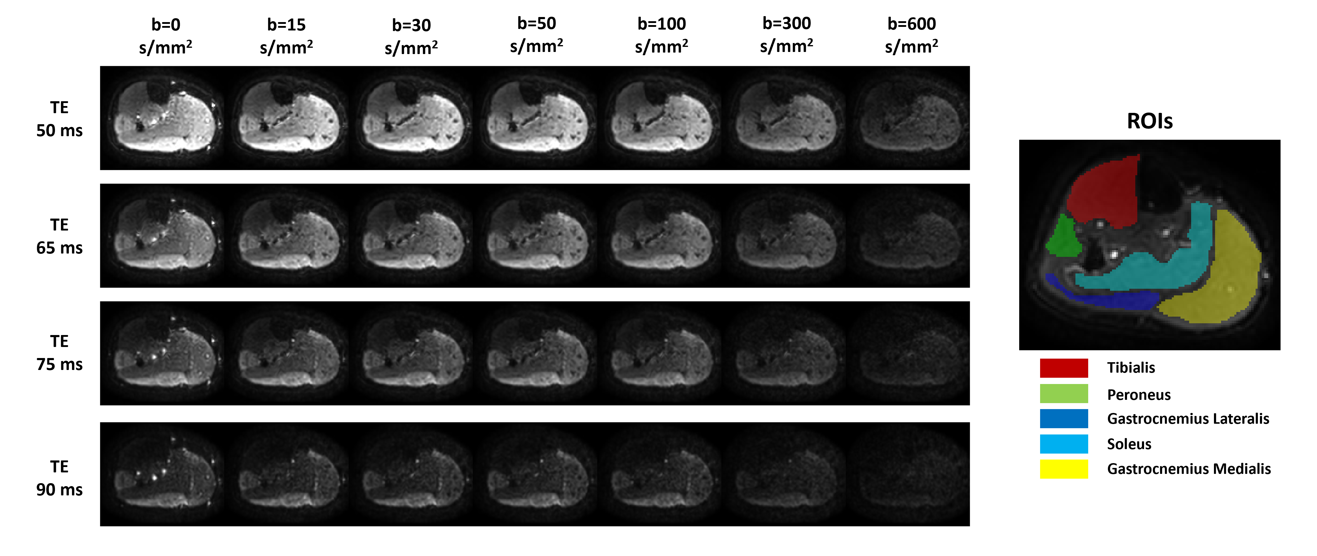
Five healthy participants (2 Males, 3 Females; age: 32±6 yo) were enrolled in this study and gave written informed consent. MRI scans were performed on a 3T scanner (Philips Achieva) with a T2-diffusion protocol with b = 15, 30, 50, 100, 300, 600 s/mm2 and TE = 50, 65, 75, 90 ms. For each pair of b and TE, images were acquired with 6 directions of the diffusion-sensitising gradients; additionally a pair of b=0 images with opposite phase encoding were acquired for each TE. A single slice (thickness: 5 mm; matrix size: 352x352; in-plane resolution:1.2 mm) was acquired on the dominant calf. Total acquisition time was 12 minutes. In Figure 1 an example of the acquired images is shown in one single direction.Prior to the scans, volunteers were instructed to perform repetitive tiptoe standing exercises for a duration of 15 minutes.MR images were denoised and Gibbs ringing artefacts removed using MRtrix7-8. Susceptibility, eddy current-induced distortions and subject movements were corrected and all volumes were coregistered to the reference (b=0, TE=50ms) using FSL9-10.Diffusion tensor fitting was carried out utilising MRtrix11; Axial, Radial and Mean Diffusivity (AD, RD, MD), and Fractional Anisotropy (FA) were extracted.
We used a deep-learning approach to fit the following T2-Ball-Zeppelin model to the diffusion-T2 data:
E = e-TE/T2 . [fVASC . e-bDv + (1 - fVASC) . Zeppelin(AD,RD,𝜃,𝜙)]
where fVASC is the signal fraction in the vascular compartment, modelled as isotropic pseudo-diffusion with diffusivity Dv; the rest of the muscle tissue is modelled by an axially-symmetric diffusion tensor (Zeppelin) with AD, RD and main direction identified by the angles 𝜃 and 𝜙;.
A training set was built by simulating dMRI signals from 106 combinations of the model parameters, equally spaced in physically meaningful ranges. A Multi-Layer Perceptron with 3 hidden layers of size 150 and ReLu activation function was trained in scikit-learn using the Adam optimizer and an initial learning rate of 0.001; the loss function was the mean square error between predictions and ground truth parameters. The trained model was applied voxel-wise to each subject’s data to obtain model parameter maps.
Diffusion-related parameters were computed within five Regions of Interest (ROIs), manually delineated on b0 images at the lowest TE for each subject, in the Tibialis (TIB), Peroneus (PER), Gastrocnemius Lateralis (GL), Gastrocnemius Medialis (GM), and Soleus (SOL) muscles (as illustrated in Figure 1).
Results
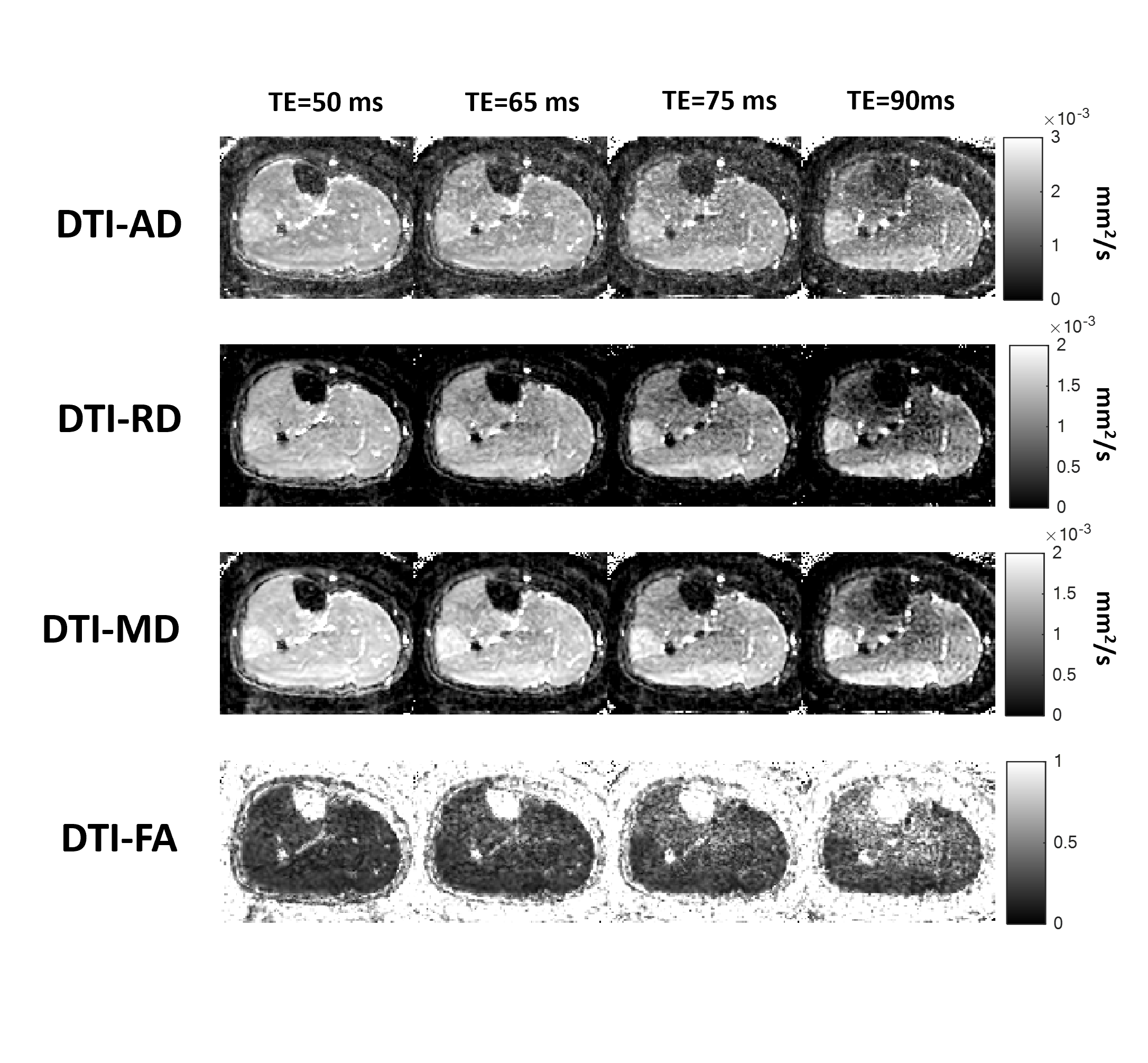
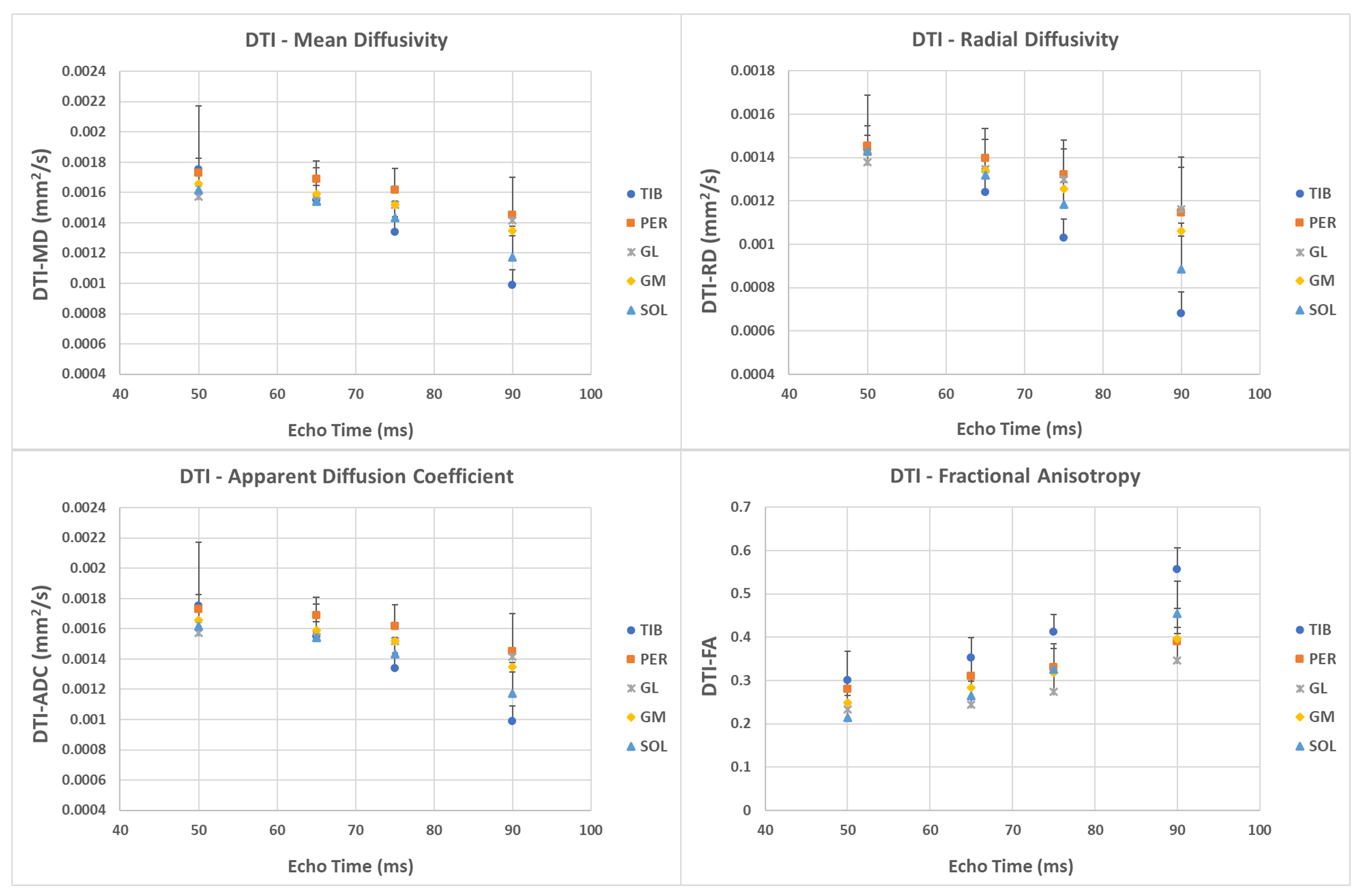
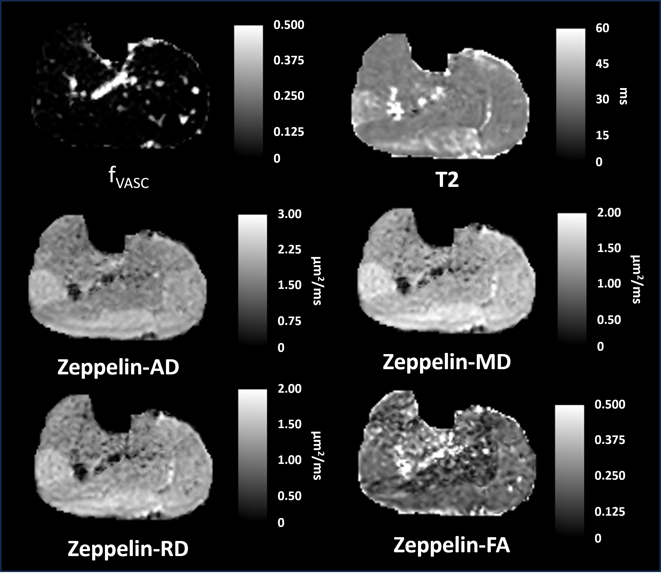
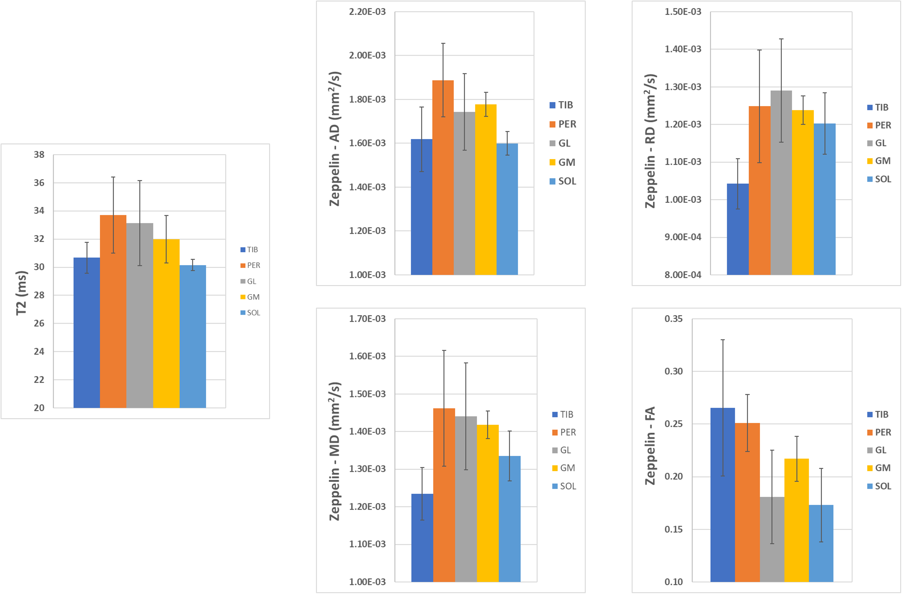
Figure 2 shows that TE significantly impacts estimated DTI parameters. Notably, AD, RD, and MD decrease with increasing TE, while FA increases. These TE-induced alterations are particularly pronounced within the TIB and SOL muscles, as illustrated in Figure 3. Figure 4 shows representative maps of the parameters of the T2-Ball-Zeppelin model. fVASC values were very low except for areas with large vessels, so the Zeppelin compartment characterises almost all the signal in the muscles. Figure 5 shows mean parameters in each muscle ROI averaged across subjects. Both T2 and all the diffusivities (AD, RD and MD) were higher in PER, GL and GM (the muscles most activated during the exercise) than in TIB and SOL.Discussion
Our results demonstrate that the estimation of diffusion parameters is significantly influenced by T2 effects in SM. This is particularly important for post-exercise scenarios, where changes in T2 are observed within the activated muscles. Muscles with short T2 seem particularly susceptible to significant changes in DTI (figures 2 and 3), underscoring the critical importance of selecting the appropriate TE to effectively distinguish between muscle groups. Figures 4 and 5 show that measuring diffusion and T2 simultaneously and pairing with an appropriate model can enhance SM microstructure quantificationConclusion
We introduce and demonstrate a novel combined T2-diffusion acquisition and analysis approach that can mitigate the impact of T2 variations and ensure a reliable estimation of diffusion-related parameters in SM. This approach mitigates T2-related challenges, ensuring reliable diffusion parameter estimation, especially in post-exercise conditions.Acknowledgements
This work received support from the Royal Society International Exchanges scheme (IEC\R2\212086). The authors extend their heartfelt appreciation to Dr. Valeria Contarino and Mr. Luciano Lombardi of Policlinico di Milano for their technical assistance in conducting MRI scans.References
1. Mastropietro, A., Porcelli, S., Cadioli, M., Rasica, L., Scalco, E., Gerevini, S., ... & Rizzo, G. (2018). Triggered intravoxel incoherent motion MRI for the assessment of calf muscle perfusion during isometric intermittent exercise. NMR in Biomedicine, 31(6), e3922.
2. Oudeman, J., Nederveen, A. J., Strijkers, G. J., Maas, M., Luijten, P. R., & Froeling, M. (2016). Techniques and applications of skeletal muscle diffusion tensor imaging: a review. Journal of Magnetic Resonance Imaging, 43(4), 773-788.
3. Bonny, J. M., Zanca, M., Boespflug-Tanguy, O., Dedieu, V., Joandel, S., & Renou, J. P. (1998). Characterization in vivo of muscle fiber types by magnetic resonance imaging. Magnetic resonance imaging, 16(2), 167-173.
4. Slator, P. J., Palombo, M., Miller, K. L., Westin, C. F., Laun, F., Kim, D., ... & Hutter, J. (2021). Combined diffusion‐relaxometry microstructure imaging: Current status and future prospects. Magnetic resonance in medicine, 86(6), 2987-3011.
5. Ochi, E., Tsuchiya, Y., & Nosaka, K. (2016). Differences in post-exercise T2 relaxation time changes between eccentric and concentric contractions of the elbow flexors. European journal of applied physiology, 116, 2145-2154.
6.Cameron, D., Reiter, D. A., Adelnia, F., Ubaida‐Mohien, C., Bergeron, C. M., Choi, S., ... & Ferrucci, L. (2023). Age‐related changes in human skeletal muscle microstructure and architecture assessed by diffusion‐tensor magnetic resonance imaging and their association with muscle strength. Aging Cell, e13851.
7. Veraart, J.; Novikov, D.S.; Christiaens, D.; Ades-Aron, B.; Sijbers, J.; Fieremans, E. (2016). Denoising of diffusion MRI using random matrix theory. Neuroimage, 142, 394-406.
8. Kellner, E.; Dhital, B.; Kiselev, V.G.; Reisert, M. (2016). Gibbs-ringing artifact removal based on local subvoxel-shifts. Magn Reson Med, 76, 1574-1581.
9. Andersson, J.L.R., Skare, S., Ashburner, J. (2003). How to correct susceptibility distortions in spin-echo echo-planar images: application to diffusion tensor imaging. NeuroImage, 20(2):870-888.
10. Smith, S.M., Jenkinson, M., Woolrich, M. W., Beckmann, C. F., Behrens, T.E.J., Johansen-Berg, H., Bannister, P. R., … & Matthews, P. M. (2004). Advances in functional and structural MR image analysis and implementation as FSL. NeuroImage, 23(S1):208-219.
11. Veraart, J.; Sijbers, J.; Sunaert, S.; Leemans, A. & Jeurissen, B. Weighted linear least squares estimation of diffusion MRI parameters: strengths, limitations, and pitfalls. NeuroImage, 2013, 81, 335-346.
Figures