0894
Creatine CH2 and PCr dynamics closely correlate in dynamic interleaved MRS of exercising muscle1Division of Endocrinology and Metabolism, Department of Internal Medicine III, Medical University of Vienna, Vienna, Austria, 2High-Field MR Center, Department of Biomedical Imaging and Image-Guided Therapy, Medical University of Vienna, Vienna, Austria, 3CD Laboratory for MR Imaging Biomarkers (BIOMAK), Vienna, Austria, 4Austrian Cluster for Tissue Regeneration, Ludwig Boltzmann Institute for Experimental and Clinical Traumatology, Vienna, Austria, 5Institute for Clinical Molecular MRI in the Musculoskeletal System, Karl Landsteiner Society, Vienna, Austria, 6High-Field MR Center, Center for Medical Physics and Biomedical Engineering, Medical University of Vienna, Vienna, Austria
Synopsis
Keywords: Muscle, Spectroscopy
Motivation: To explore quantification of skeletal muscle oxidative metabolism by 1H MRS.
Goal(s): We used the increased accuracy of 7T MRS with a dedicated RF-coil, interleaved acquisition and localization of 31P information to compare Creatine-CH2 and Phosphocreatine time courses during exercise and recovery.
Approach: Eight volunteers were measured on a 7T MR system with RF-coil and ergometer dedicated for exercise. 1H and 31P MR spectra were acquired interleaved during exercise and recovery.
Results: Exercise led to disappearance of the Creatine-CH2 resonance, while the CH3 resonance remained stable during exercise. The recovery time constants were similar (τPCr=37±9s and τCr=34±6s) and positively correlated.
Impact: The time course of the Creatine-CH2 resonance in skeletal muscle can be accessed via dynamic 1H MRS. If accurately reflecting oxidative metabolism, this technique has the potential to render non-invasive metabolic studies broadly accessible, without needing multi-nuclear MRI capabilities.
Introduction
Localized 1H and 31P MR spectra of human muscle are strongly affected by exercise. Phosphocreatine (PCr) levels measured by 31P MRS decrease during exercise, reach steady state, and recover towards basal values after exercise. 1H MRS exhibits two Creatine (Cr) signals, CH3 (3.02 ppm) and CH2 (3.92 ppm). Exercise-induced PCr depletion is known to influence also the spectral appearance of the Cr-CH2 3.92 ppm resonance1. We used the increased accuracy of 7T MRS with a dedicated RF-coil, interleaved acquisition and localization of 31P information to compare Cr-CH2 and PCr time courses during exercise and recovery. Furthermore, we investigated skeletal muscle oxidative metabolite kinetics from time-resolved 1H MRS.Methods
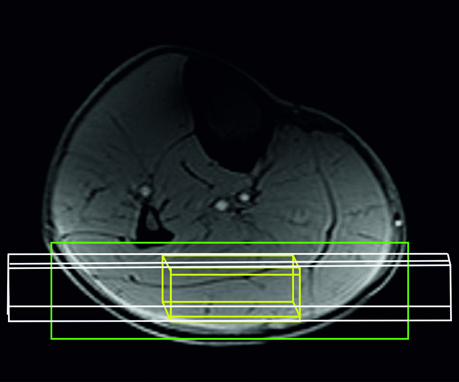
Twelve healthy, recreationally active volunteers (age: 30±7 years, BMI: 25.2±7.9kg/m2, sex: 5f/7m) participated in the study on a 7T whole-body MR system (Terra-DotPlus, Siemens Healthineers, Erlangen, Germany). A dedicated 1H(2-channel)/31P(3-channel) surface coil transceiver array2 was used on the subject’s right calf. An MR-compatible ergometer (Trispect, Ergospect, Innsbruck, Austria) was used for plantar flexion exercise. Data from four subjects were excluded in the analysis due to either poor shim (> 70 Hz, n=2), water-suppression (n=1) or baseline distortion (n=1), all presumably caused by motion. Measurements were performed in the morning after overnight-fasting in a single exercise-recovery session. Volunteers were positioned supine with the right calf on the coil and ergometer, inside the scanner. The VOI for 1H MRS Cr detection (15x40x53 mm3) and 18-mm slab for 31P MRS PCr and inorganic phosphate (Pi) detection were carefully placed predominantly within the gastrocnemius medialis and lateralis muscles (Fig 1). Water was shimmed to 35–45 Hz (magnitude spectra).Dynamic semi-LASER-localized3,4 1H MR spectra (TE=30ms) (Cr) and DRESS-slab-localized5 31P MR spectra (PCr, Pi) were acquired interleaved in one exercise/recovery session (3min rest, 5min exercise at 30% of maximal voluntary contraction force, 10min recovery) with a TR of 6 s, similarly as published6. Volunteers were pushing the pedal twice per TR, audio-cued by gradient, to ensure that data were acquired always in relaxed muscle. 1H/31P spectroscopy data were processed from raw data using in-house-developed Python scripts (http://www.python.org), phasing signals to the highest peak magnitude of PCr, water, or lipids in the frequency domain for channel combination. Peak amplitudes were quantified with AMARES, using jMRUI v6.0 alpha. For fitting the 3.9 ppm Cr-CH2 resonance, we used HLSVD peak removal over the lipid resonances at 1.5 ppm (HLSVD no-max-2048-points filter, 5 components). For absolute quantification of the visible fraction of Cr-CH₂ resonance, the fully-relaxed water signal was measured separately. Concentrations were calculated in mmol/L (tissue volume) units, by: Cm=CH2O x (Sm/SH2O) x (nH2O/nm) x (CFH2O/ CFm) x WH2O x ρmuscle
with S the signal intensity of water or Cr, nH2O and nm the number of equivalent protons (= 2), CF the correction factors for T1 and T2 relaxation, CH2O = 55.56 mol/L the water concentration, CFm the approximate water content in skeletal muscle tissue, (0.77 L/kgww), and ρmuscle the specific weight of skeletal muscle tissue (1.06 kg/L). 31P MRS yielded PCr and Pi concentrations, relative PCr depletion and depletion-rate during exercise (τPCr on-kinetics) and resynthesis-rate during recovery (τPCr recovery), the maximal oxidative phosphorylation rate, i.e., mitochondrial capacity (Qmax), and the time course of intracellular pH7,8 . Data are presented as mean ± standard deviation.
Results
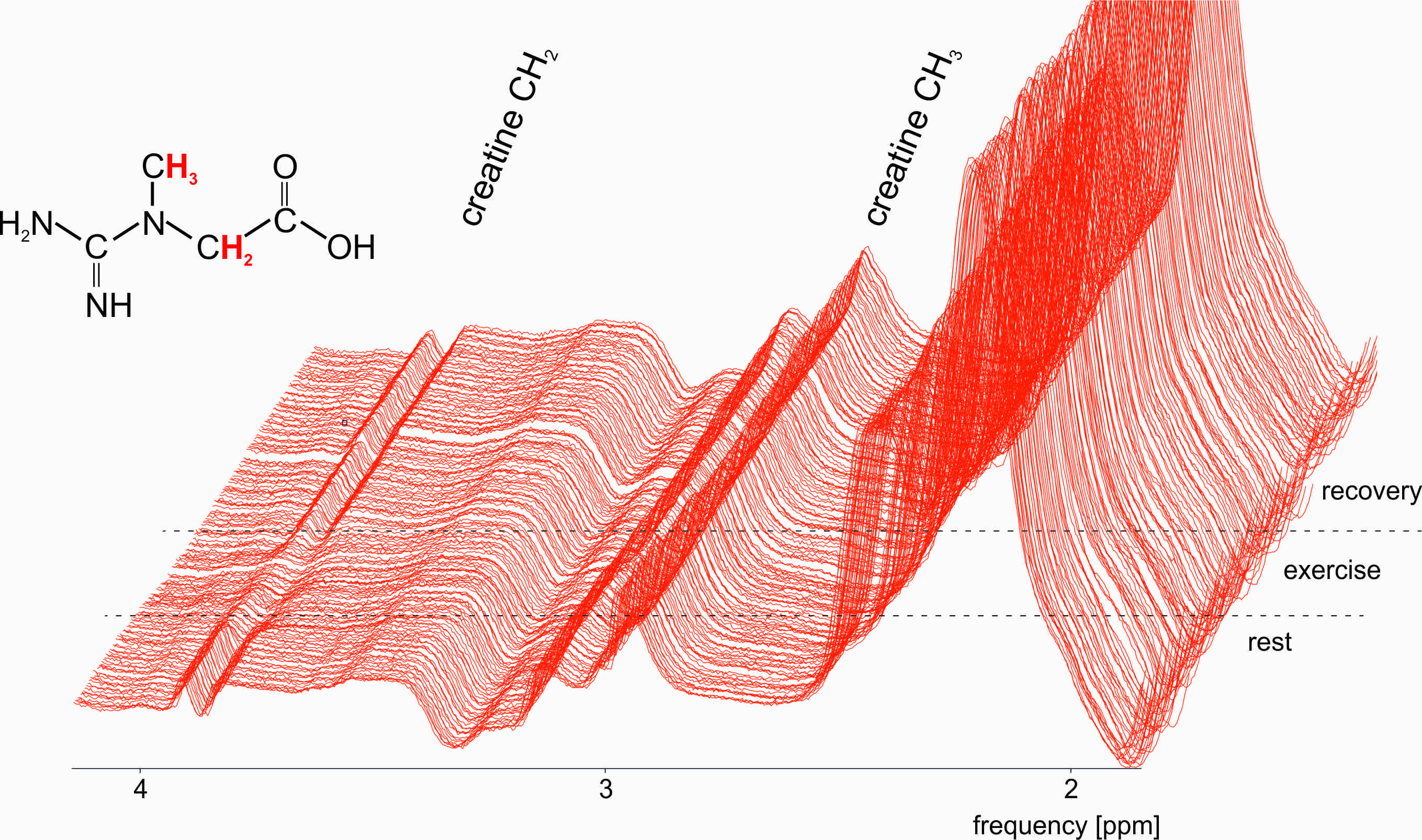
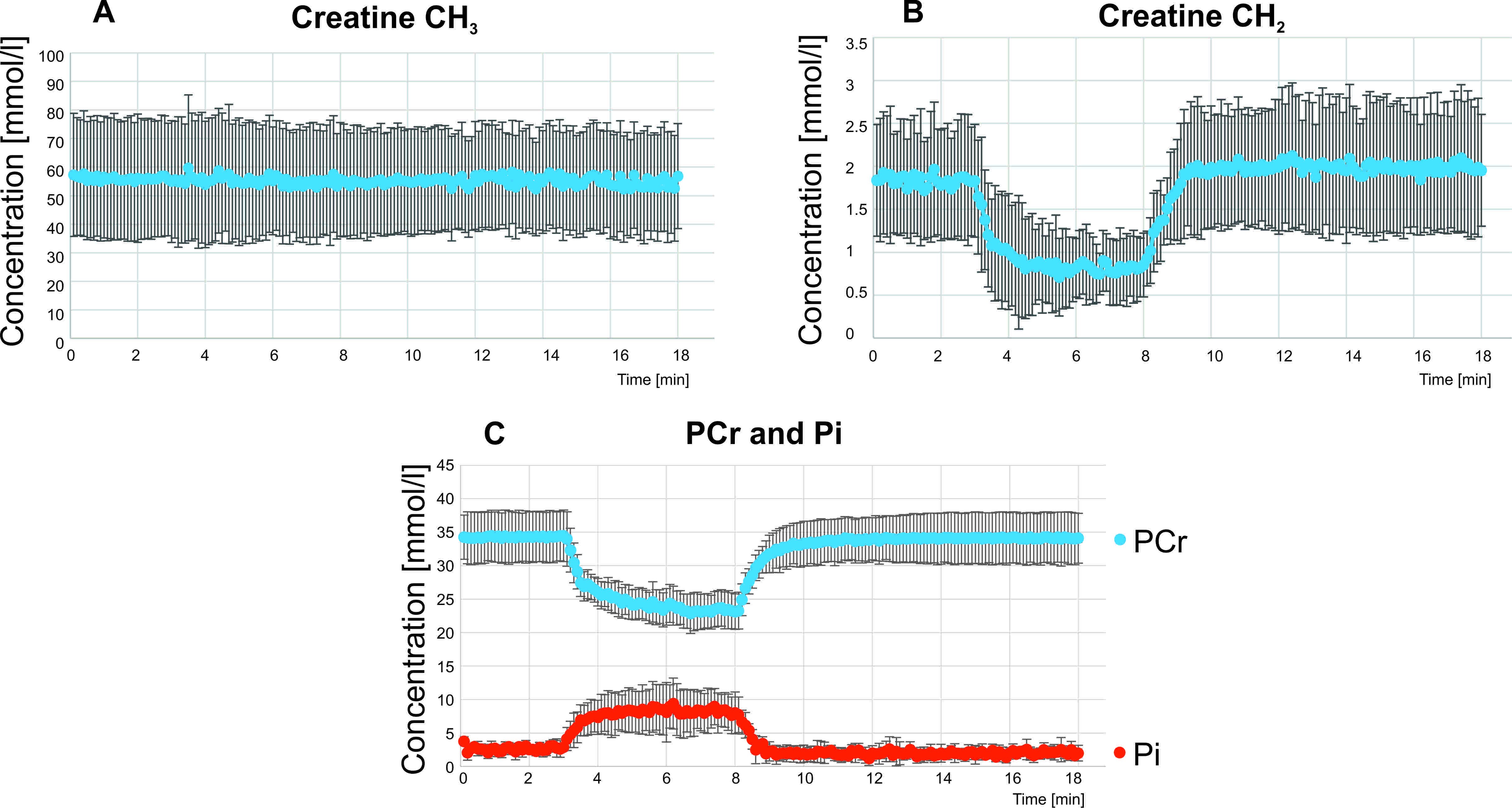
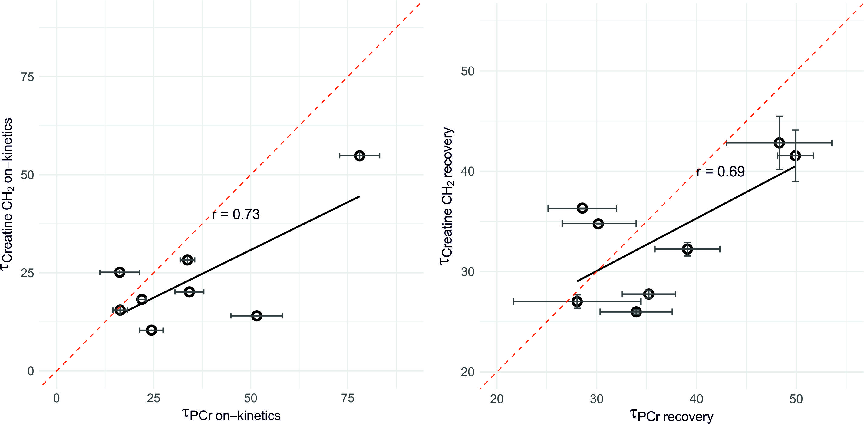
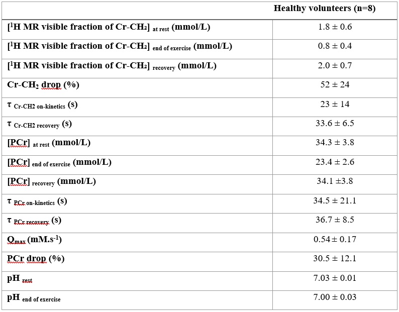
Submaximal exercise leads to disappearance of the Cr-CH2 resonance at 3.92ppm in 1H MR spectra (Fig 2, Fig 3) whereas the Cr-CH3 resonance at 3.02ppm remained stable during exercise. Detailed numerical results regarding concentrations and time constants are shown in Table 1, individual data in Fig 4.Most strikingly, the mean time constants of PCr and Cr-CH2 recovery dynamics were not significantly different, τPCr = 37±9 s and τCr = 34±6 s, p=0.2, showing marginally positive correlation (r=0.69, p=0.058). τPCr on-kinetics and τCr-CH2 on-kinetics were positively correlated (r=0.73, p=0.039).
Discussion and Conclusion
We assessed the skeletal muscle Cr-CH2 resonance together with PCr during a single dynamic session using interleaved 1H/31P MRS at 7T. Despite limited visibility of the Cr-CH2 resonance, we were able to quantify both time courses in a single experiment in eight subjects. Therefore, we were able to directly compare Cr-CH2 and PCr time courses during exercise and recovery. We observed remarkably similar kinetics of Cr-CH2 and PCr time courses in absolute numbers, and positive correlations between τPCr and τCr-CH2. The discrepancy between end-exercise depletion measured with 1H and 31P MRS may be explained by differing volumes of interest9. Studies including higher numbers of subjects will be necessary to explain quantitative differences, especially found in exercise-on kinetics, and verification assessing skeletal muscle oxidative metabolism by 1H MRS.Acknowledgements
Colleagues at the High-Field MR Center, this study was supported by FWF (#KLI 904 to MKrss)References
1. Kreis R, Jung B, Slotboom J, Felblinger J, Boesch C. Effect of Exercise on the Cr Resonances in1H MR Spectra of Human Skeletal Muscle. Journal of Magnetic Resonance. 1999;137(2):350-357. doi:10.1006/JMRE.1998.1683
2. Goluch S, Kuehne A, Meyerspeer M, et al. A form-fitted three channel phosphorus 31P, two channel 1H transceiver coil array for calf muscle studies at 7 T. Magn Reson Med. 2015;73(6):2376-2389. doi:10.1002/mrm.25339
3. Meyerspeer M, Scheenen T, Schmid AI, Mandl T, Unger E, Moser E. Semi-LASER localized dynamic 31P magnetic resonance spectroscopy in exercising muscle at ultra-high magnetic field. Magn Reson Med. 2011;65(5):1207-1215. doi:10.1002/MRM.22730
4. Scheenen TWJ, Klomp DWJ, Wijnen JP, Heerschap A. Short echo time 1H-MRSI of the human brain at 3T with minimal chemical shift displacement errors using adiabatic refocusing pulses. Magn Reson Med. 2008;59(1):1-6. doi:10.1002/mrm.21302
5. Bottomley PA, Foster TB, Darrow RD. Depth-resolved surface-coil spectroscopy (DRESS) for in Vivo 1H, 31P, and 13C NMR. Journal of Magnetic Resonance (1969). 1984;59(2):338-342. doi:10.1016/0022-2364(84)90179-3
6. Niess F, Schmid AI, Bogner W, et al. Interleaved 31P MRS/1H ASL for analysis of metabolic and functional heterogeneity along human lower leg muscles at 7T. Magn Reson Med. 2020;83(6):1909-1919. doi:10.1002/MRM.28088
7. Valkovič L, Chmelík M, Ukropcová B, et al. Skeletal muscle alkaline Pi pool is decreased in overweight-to-obese sedentary subjects and relates to mitochondrial capacity and phosphodiester content. Sciientific Reports, Nature. 2016;(December 2015):1-9. doi:10.1038/srep20087
8. Valkovič L, Chmelík M, Krššák M. In-vivo 31P-MRS of skeletal muscle and liver: A way for non-invasive assessment of their metabolism. Anal Biochem. Published online January 2017. doi:10.1016/j.ab.2017.01.018
9. Meyerspeer M, Robinson S, Nabuurs CI, et al. Comparing localized and nonlocalized dynamic 31P magnetic resonance spectroscopy in exercising muscle at 7T. Magn Reson Med. 2012;68(6):1713-1723. doi:10.1002/MRM.24205
Figures