0889
Human fMRI at 10.5T: new regimes of high resolutions1CMRR, University of Minnesota, Minneapolis, MN, United States
Synopsis
Keywords: Task/Intervention Based fMRI, fMRI (task based), Neuroscience
Motivation: Submillimeter fMRI allows imaging the human brain noninvasively at the mesoscopic scale, targeting layers and columns. Standard submillimeter resolution however may be inadequate to fully capture these ensembles. Consequently, the Brain Initiative challenged the MR community to achieve 0.1µL, and subsequently, 0.01µL voxel resolution
Goal(s): Achieve and surpass the goal of human functional mapping at 0.1µL, towards the goal of 0.01µL using a 10.5T scanner.
Approach: We recorded human functional BOLD responses at 10.5T, during visual experiments, at different spatial resolutions.
Results: Using 10.5T and NORDIC denoising we demonstrate functional imaging the human brain with <0.1µL resolution.
Impact: We demonstrate functional mapping at 10.5 T with unprecedented spatial resolutions, moving towards the 0.01µL voxel volume goal (Brain Initiative 2.0). At these resolutions single voxels contain a few thousand neurons, heralding major new opportunities in human neuroscience.
Introduction
Functional MRI is a key tool to study the living human brain non-invasively. Recent technological developments have allowed the acquisition of functional images with spatial resolutions in the submillimeter range, reaching into human mesoscopic scale functional organizations. To date such studies typically utilize ~0.512µL voxels (0.8x0,8x0,8mm3) but are limited by SNR and only marginally adequate to study layers and columns. This limitation is reflected in the Brain Initiative Working group goals, which challenged the MR community to achieve functional resolutions of ≤0.1µL voxels initially and 0.01µL subsequently. Here we exploit the higher SNR conferred by 10.5T together with NORDIC1 denoising to demonstrate significantly larger coverage and SNR with typical 0.512µL (0.8x0,8x0,8mm3) voxels, functional imaging at resolutions that go beyond 0.1µL voxel volume (0.4x0.4x0.6mm3 – 0.096µL volume; and 0.4x0.4x0.4mm3 – 0.064µL volume) and BOLD amplitude differences across cortical depths indicative of attentional modulations in early visual cortex, at the resolution of 0.35x0.35x0.35mm3 voxels (0.0428µL volume).Methods
Using a human whole body 10.5T scanner, T2*-weighted images were collected with 2D GE EPI for the 0.4x0.4x0.6mm3 data and 3D GE EPI for the isotropic 0.8mm, 0.4mm, and the 0.35mm images. The isotropic 0.4mm protocol was replicated at 7T in the same participant. Functional preprocessing included slice-time correction for the 2D acquisition, rigid body motion correction, and low drift removals. We implemented a 24 seconds on/off block design paradigm with a face perception task for the isotropic 0.8mm data (modified from 2) and with a center (i.e. target) and a surround checkerboard counterphase flickering as in 1 for the remaining protocols. Each run lasted approximately 7 minutes for the face perception (8 blocks) and 5 minutes (3 trials x 2 conditions) for the gratings task. Moreover for the 0.35mm isotropic voxel image, we performed equivolume cortical-depths segmentation using LAYINII3Results
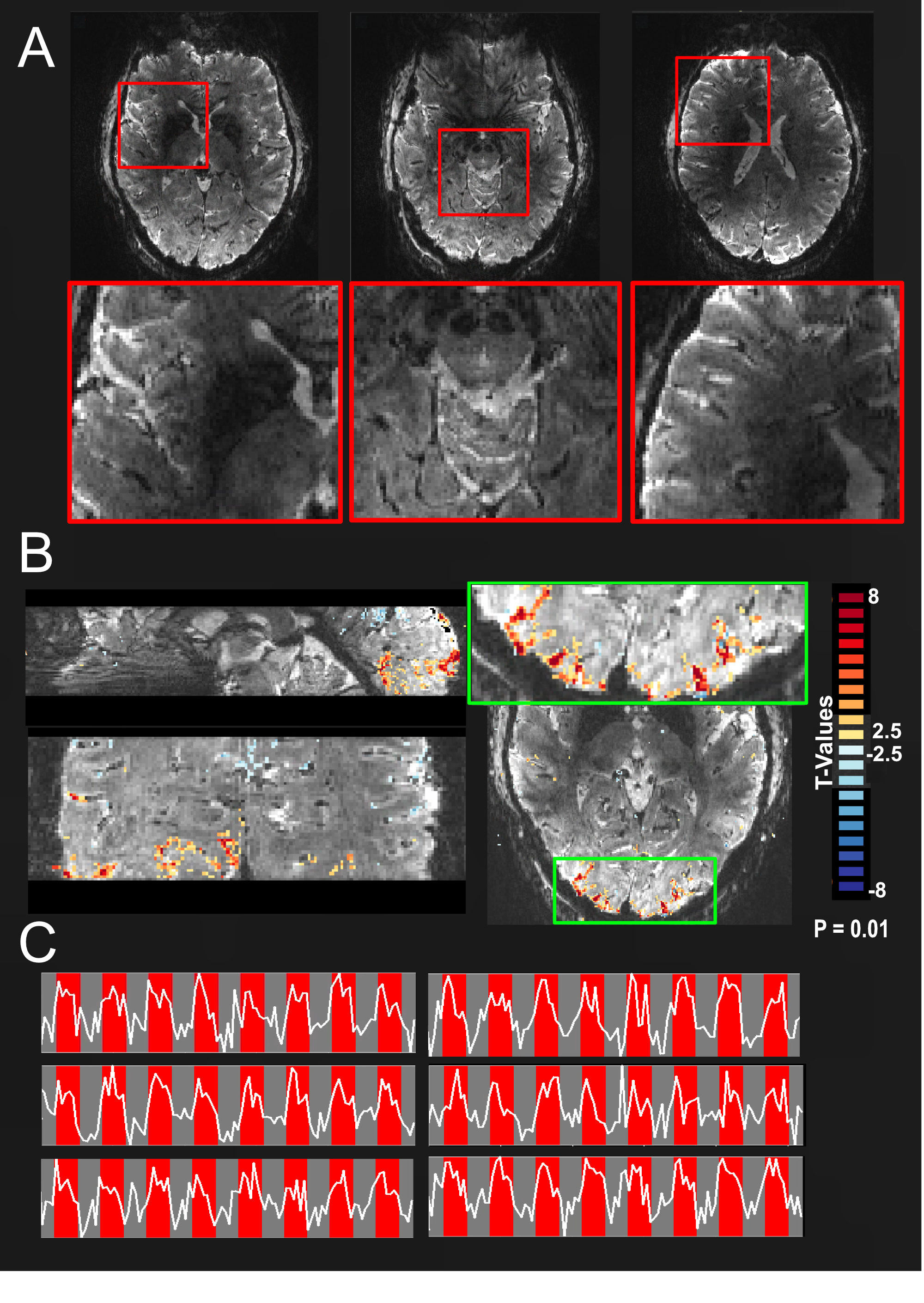
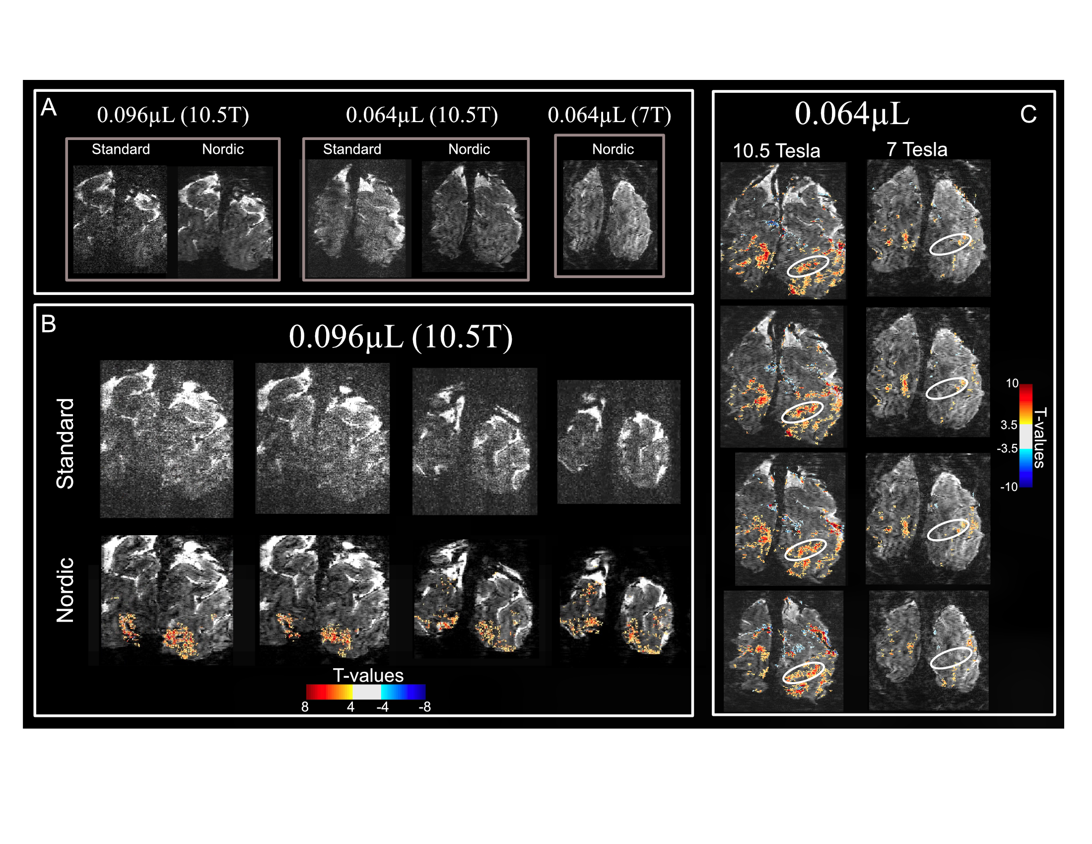
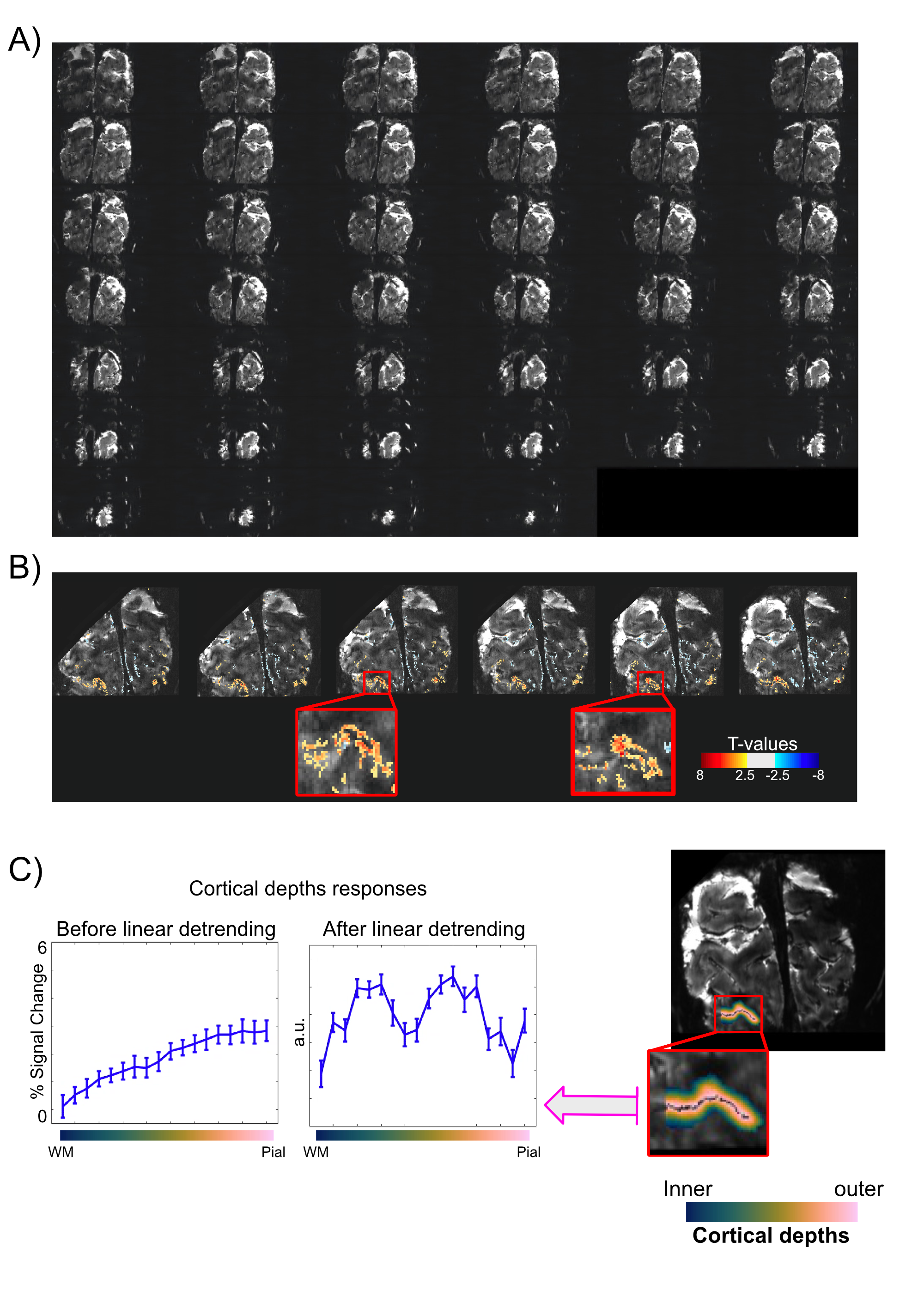
Figure1 shows an example of individual slices of a single EPI image (A); t-maps for the contrast faces < 0 (B); and examples of single voxel timecourses (C). Note the larger axial coverage and the quality of single-run single-voxels time courses and functional mapping achieved with as little ~30 minutes of data. Figure 2 illustrates single EPI images for the 10.5T 0.096µL and 0.064µL voxel acquisitions before and after NORDIC denoising; and for the 7T 0.064µL voxel after NORDIC only (A). For a ~5 minute run of the 0.096µL acquisition, no activation could be detected before NORDIC denoising at a t-threshold >3.5, even at 10.5T (B). We further demonstrate reliable functional mapping with 0.064µLvoxels across multiple subjects, with as little as 20 minutes of data (Figure 2D). Moreover, consistent with increases in SNR and BOLD contrast at 10.5T, at this resolution and t-threshold, significant portions of the expected retinotopic activation in the visual cortex (circled regions in figure 2C) for the 0.064µL acquisitions are only visible in the 10.5T images. Finally, we demonstrate functional mapping at an unprecedented resolution of 0.0428µL voxel volume in humans (Figure3) with as little as ~20 minutes of data, and report that BOLD response amplitudes in early visual cortex are most pronounced in inner and outer depths, indicative of attentional modulations.Discussion
In this work, we demonstrate human functional brain mapping at 10.5T with unprecedented resolutions (<0.1µL voxel volume), as well as higher coverage and SNR at the typical 0.8x0.8x0.8mm3 resolution. We successfully deal with the thermal noise barrier, dominant at these resolutions, using NORDIC denoising1. We used a higher-level face perception task2 as well as a low-level gratings1 to exploit the retinotopic properties of V1, which provided a ground truth for functional activation, allowing evaluation of the quality of evoked BOLD responses in space and time. Performing human fMRI at such high resolutions was enabled by the simultaneous use of NORDIC denoising and 10.5T. Moreover, as predicted by animal models, we report preliminary single condition GE BOLD fMRI data on depth-dependent suggestive of attentional modulations in inner and outer depths in visual cortex at 0.35mm isotropic resolution, without resorting to upsampling, after linear detrending4 (Figure3).Conclusion
We demonstrate the feasibility of functional mapping at 10.5 T with unprecedented spatial resolutions. We achieve the Brain Initiative goal of <0.1µL voxel volume fMRI in humans and move towards the 0.01µL goal set by the Brain Initiative 2.0 by recording the first ever human functional images with ~0.04µL voxel volume. At these ultra-high resolutions, a single voxel only contains a few thousand neurons, further bridging the gap with invasive optical imaging and heralding a major new opportunity for human fMRI applications.Acknowledgements
this work was supported by the NIH P41 EB027061 "Technology to Realize the Full Potential of UHF MRI" awarded to K.U.References
1. Vizioli, L., Moeller S., Dowdle, L., Akçakaya M., De Martino F., Yacoub E., Ugurbil K. (2021) Lowering the thermal noise barrier in functional brain mapping with magnetic resonance imaging. Nature Communications
2. Dowdle, L., Ghose, G., Moeller, S., Ugurbil, K., Yacoub, E., Vizioli, L. (2022). Characterizing top-down microcircuitry of complex human behavior across different levels of the visual hierarchy. Bioarchive
3. Huber, L. et al. (2021) LayNii: A software suite for layer-fMRI. NeuroImage
4. Fracasso, A., Petridou, Dumoulin, S., N. (2104) Distinct BOLD laminar profiles elicited by retino-cortical and inter-hemispheric sources in human early visual cortex. ISMRM
Figures