0888
Enabling brain-wide mapping of directed functional connectivity at 3T via layer-dependent fMRI with draining-vein suppression1Radiology, UNC at Chapel Hill, Chapel Hill, NC, United States, 2Psychology, UNIV OF NORTH CAROLINA AT CHAPEL HILL, Chapel Hill, NC, United States
Synopsis
Keywords: fMRI Acquisition, fMRI, Layer-dependent fMRI
Motivation: Mapping brain-wide directed functional connectivity demands techniques with high spatiotemporal resolution yet current methods fall short.
Goal(s): To improve spatial specificity of GE-BOLD EPI by reducing draining-vein contamination without compromising speed.
Approach: Incorporating velocity-nulling (VN) gradients into a GE-BOLD fMRI sequence at 3T. We also integrated NORDIC denoising to enhance signal sensitivity.
Results: The VN fMRI method demonstrated decent spatial specificity, evidenced by identifying double-peak activation patterns within the M1 area during a finger-tapping task. This technique showed enhanced robustness across participants compared to conventional fMRI. Our findings on directed functional connectivity reveal layer-specific relationships that closely align with the existing literature.
Impact: Leveraging its comprehensive brain coverage and efficient scan time, VN fMRI has yielded promising results in directed FC studies. Given the widespread accessibility of 3T scanners, we anticipate this development will have a significant impact across multiple neuroscience research domains.
Introduction
Layer-dependent fMRI is an emerging field that allows for the non-invasive measurement of layer-specific activity in human brains, distinguishing between feedforward and feedback responses in cortical units. Although BOLD contrast has been the gold standard for fMRI for almost three decades, it faces challenges from the draining-vein contamination, which diminishes the spatial specificity. Various methods have been explored to mitigate this issue, including model-based draining-vein removal, phase regression, and vascular space occupancy (VASO) fMRI. The VASO fMRI has superior spatial specificity than BOLD contrast but the scan time for the whole brain is long (> 8s). In this study, we investigate the depth-dependent spatial specificity of sub-millimeter fMRI with velocity-nulling gradients at 3T to suppress the draining-vein contamination, demonstrating the potential for brain-wide directed functional connectivity studies at this resolution.Method
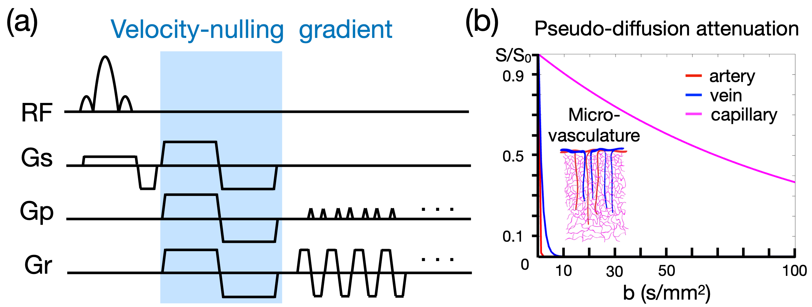
The draining-vein effect causes spatial blurring and bias towards superficial layers in BOLD measurements at cortical layers. To suppress the signal from draining veins, we incorporated a velocity-nulling (VN) gradient into GE-EPI at 3T. In our study, we utilized the Intravoxel Incoherent Motion (IVIM) process to model the collective motion of water molecules in blood within a vessel network as a pseudo-diffusion process. The pseudo-diffusion coefficient (D*) was calculated for capillaries, cortical penetrating veins, and cortical arteries based on their respective average displacements and mean velocities. Our results showed that a relatively small b value could effectively suppress the signal from draining veins while causing minimal attenuation of signals from capillaries. Additionally, we adjusted the b value to account for the varying orientation of penetrating veins within the human cortex. For instance, using a b value of 30, equivalent to a single-axis b value of 10, can suppress the signal from veins to less than 0.1%, with a 26% loss of the signal from capillaries.Results
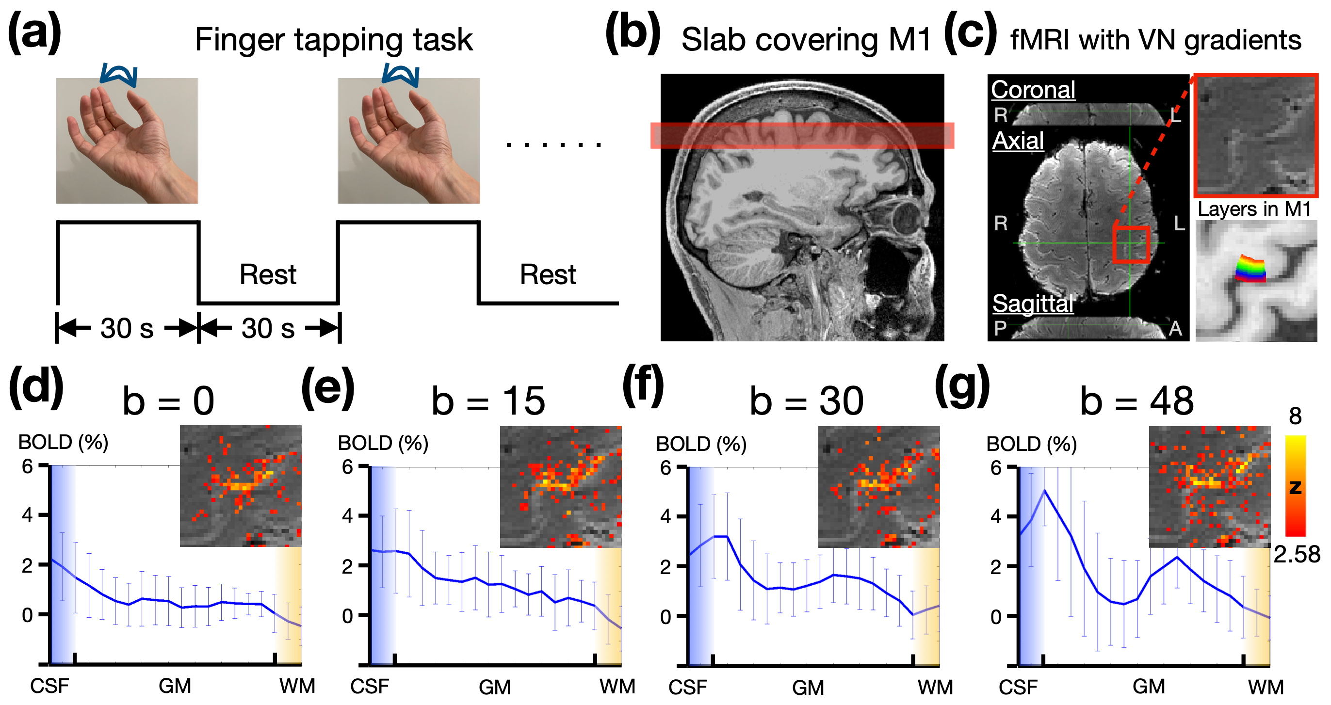
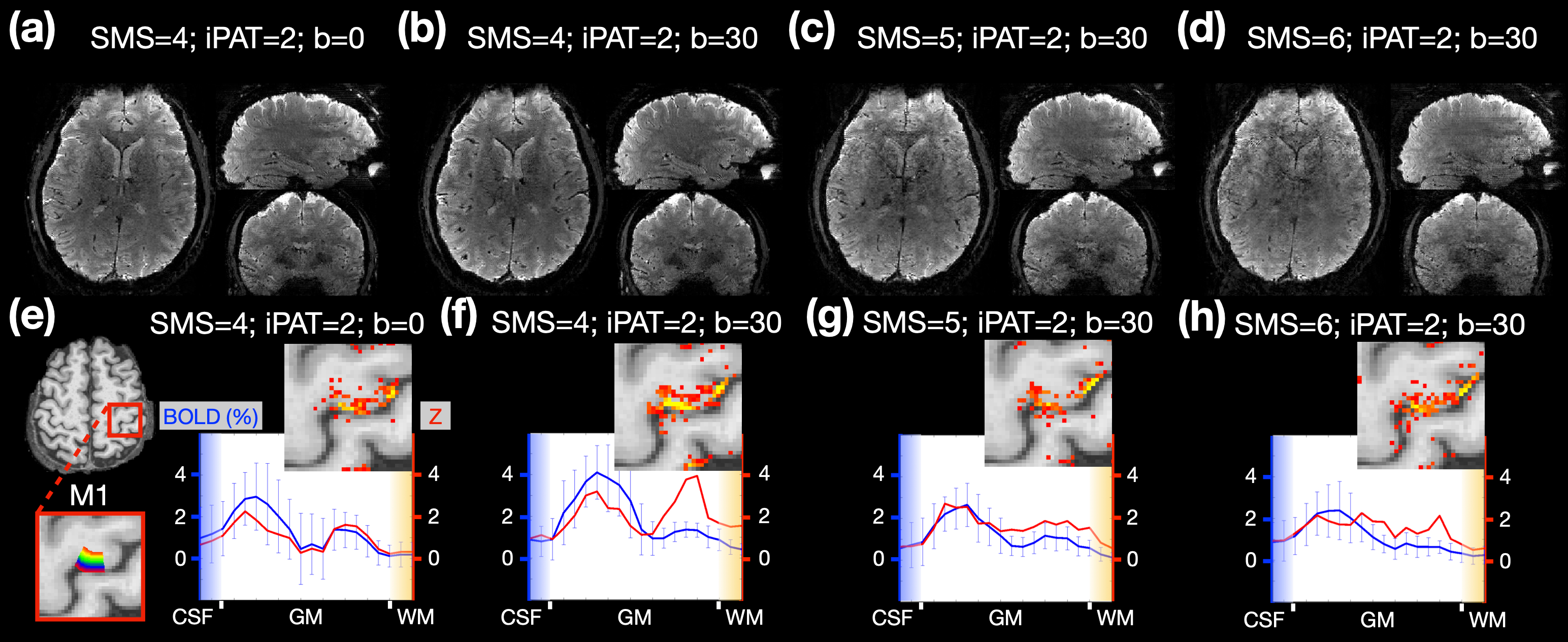
We assessed the effectiveness of our draining-vein suppression technique by analyzing BOLD activation profiles during a finger-tapping task at the primary motor cortex (M1). Our results, displayed in Figure 2, show that the velocity-nulling (VN) gradient successfully minimized the draining-vein effect, enhancing laminar specificity at 3T. Both b values of 30 and 48 s/mm2 showed double-peak response patterns; however, the spatial variance was lower with b = 30 s/mm2. As the b value of 48 may overly suppress capillary signals, we opted for a b value of 30 s/mm2 in subsequent experiments.To transition from slab to volume imaging, we used simultaneous multi-slice (SMS) factors of 4, 5, and 6 with a velocity-nulling (VN) gradient (b=30 s/mm2), assessing their effects on signal quality and depth-dependent profiles. While an SMS factor of 4 provided optimal image quality, increasing the SMS factor led to minor image artifacts. The depth-dependent profiles at M1 revealed a double-peak pattern with greater spatial stability and statistical significance in deeper layers when using an SMS factor of 4 with a VN gradient, as opposed to higher SMS factors that resulted in spatial instability due to excessive feature loss.
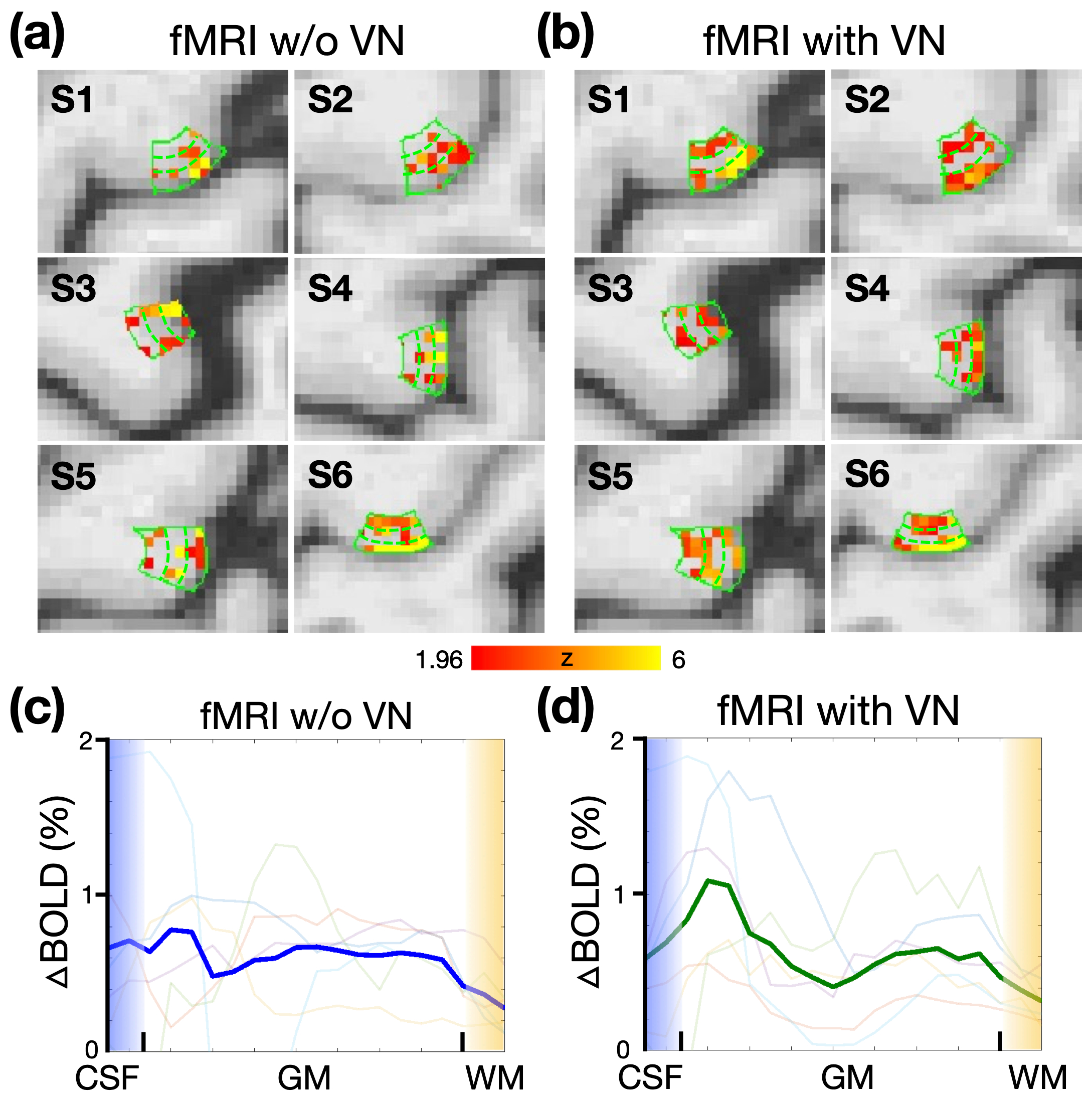
While a double-peak pattern was observed in the depth-dependent profile of a single participant, its consistency across a larger participant base remained unconfirmed. As shown in Figure 4, the analysis of six participants showed that without the velocity-nulling (VN) gradient, most depth-dependent profiles were spatially unstable, and the group-averaged profile lacked a clear double-peak pattern. However, with the VN gradient, the middle layers showed reduced activation, and the double-peak pattern was evident in the group-averaged profile, demonstrating the effectiveness of the VN gradient in achieving consistent and stable depth-dependent profiles across participants.
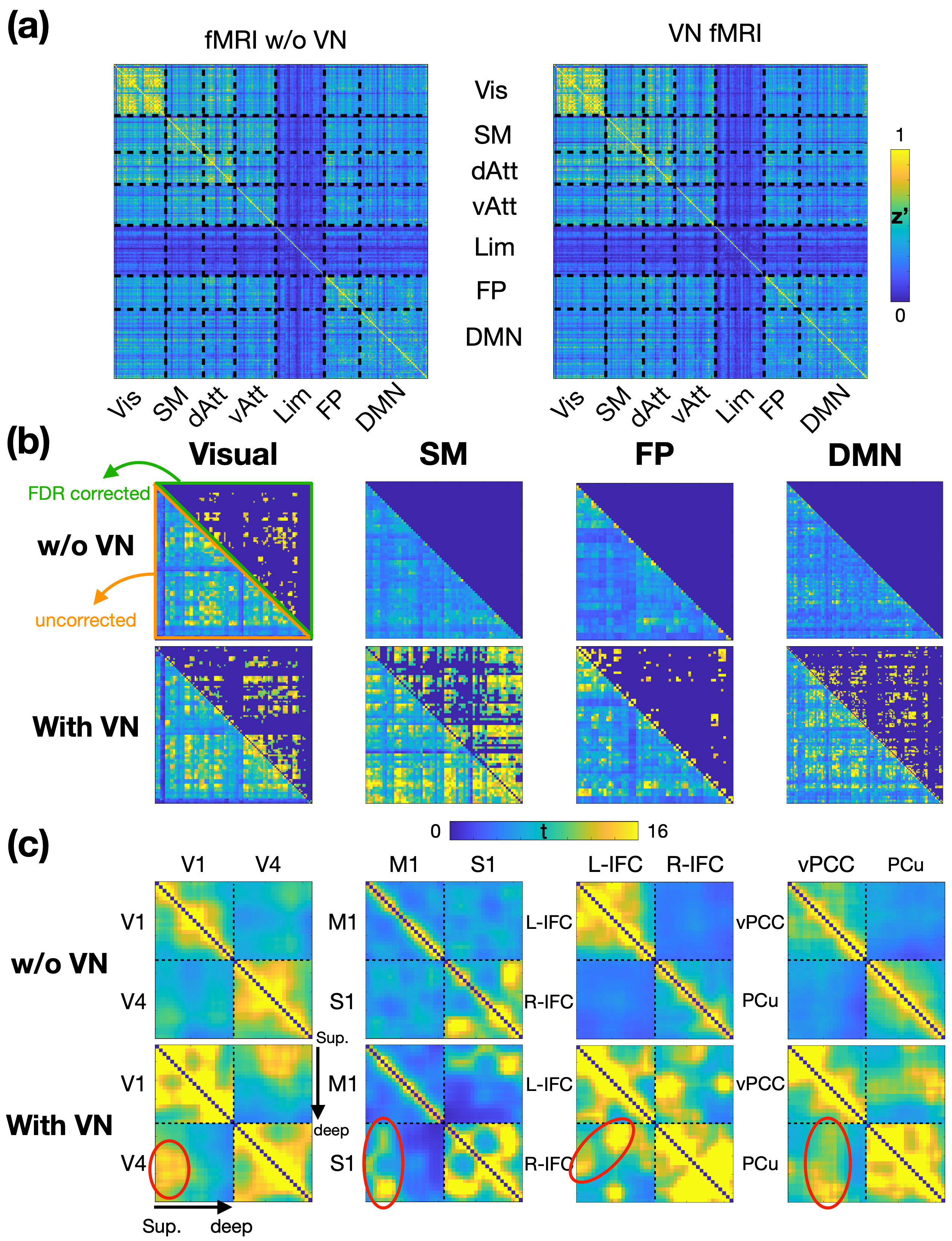
The application of velocity-nulling (VN) gradients in functional magnetic resonance imaging (fMRI) significantly enhances statistical power and reduces inter-participant variability, as evidenced by the group-averaged functional connectivity (FC) matrices and directed FC matrices as shown in Figure 5. VN fMRI demonstrated statistically significant FCs across various functional networks, whereas conventional fMRI without VN gradients showed limited significant FCs. Moreover, VN fMRI enabled the identification of layer-specific directed brain connectome, revealing specific feedforward and reciprocal connectivity patterns within visual, sensorimotor, frontoparietal, and default-mode networks. These findings underscore the importance of VN gradients in achieving more accurate and reliable FC analyses in fMRI studies.
Discussion
In this study, we have developed a brain-wide directed functional connectivity at 3T using velocity-nulling (VN) gradients, simultaneous multi-slice (SMS) acceleration, and NORDIC denoising techniques to achieve sub-mm spatial resolution and high functional sensitivity. Our VN fMRI sequence provided a 0.9-mm isotropic spatial resolution, a TR of 3.84 seconds, and brain-wide coverage. The VN fMRI revealed layer-specific functional relationships and was more robust across participants than conventional fMRI methods.Acknowledgements
This work was supported in part by NIH grants R21AG060324.References
1. Setsompop, K. et al. High-resolution in vivo diffusion imaging of the human brain with generalized slice dithered enhanced resolution: Simultaneous multislice (gSlider-SMS). Magn Reson Med 79, 141–151 (2018).
2. Dai, E. et al. A 3D k‐space Fourier encoding and reconstruction framework for simultaneous multi‐slab acquisition. Magn. Reson. Med. mrm.27793 (2019) doi:10.1002/mrm.27793.
3. Mani, M. et al. SMS MUSSELS: A navigator‐free reconstruction for simultaneous multi‐slice‐accelerated multi‐shot diffusion weighted imaging. Magn. Reson. Med. 83, 154–169 (2020).
4. Haldar, J. P. Low-Rank Modeling of Local $k$-Space Neighborhoods (LORAKS) for Constrained MRI. IEEE Trans. Med. Imaging 33, 668–681 (2014).
5. Vizioli, L. et al. Lowering the thermal noise barrier in functional brain mapping with magnetic resonance imaging. Nat. Commun. 12, 5181 (2021).
Figures