0885
T1234: A distortion-matched structural scan solution to misregistration of high resolution fMRI data1NIH, Bethesda, MD, United States, 2German Center for Neurodegenerative Diseases (DZNE), Bonn, Germany, 3Maastricht University, Maastricht, The Netherlands, & Brain Innovation, Maastricht, The Netherlands, Maastricht, Netherlands
Synopsis
Keywords: fMRI Acquisition, fMRI, layer-fMRI, Ultra High Field, Structural scan
Motivation: High-resolution fMRI at 7T is limited by misregistration of functional data with structural scans.
Goal(s): We aim to provide a fast acquisition method that provides distortion matched, artifact mitigated structural reference data.
Approach: T1234: T1-weighted 2-inversion 3D-EPI with 4 directions for high-resolution fMRI. A forward Bloch model is implemented for T1 quantification and protocol optimization.
Results: Our protocol is fast (3:40 min) and provides whole-brain segmentations in EPI-space. It is robust across sessions, participants, and scanners.
Impact: This structural mapping approach allows precise registration with fMRI data. T1234 is implemented, validated, and tested to serve users of our sequence (locally and 43 centers worldwide).
Purpose
High-resolution fMRI promises to reveal fine-scale neural information flow across layers and columns. A survey by the ISMRM brain function study group found that the most limiting factor of high-resolution fMRI is image registration1. This is despite the fact that researchers commonly invest significant time (≈10 min) in acquiring high-quality anatomical data with MP2RAGE2.In this study, we build on previous works2-14 and present a novel acquisition method: T1234, involving T1-weighted acquisition with 2 inversions 3D-EPI and 4 directions, which provides distortion-matched, high anatomic contrast, and artifact mitigated structural reference data in 3:40 min.
Methods
We tested T1234 in twelve MRI sessions (7 participants) across three 7T scanners in our center (2x SIEMENS Terra & 7T plus).Scan parameters: 0.8mm, Skipped-CAIPI 1x3z1, segmentation factor 14(Stirnberg)11, matrix 232x232x186, phase PF=6/8. We used a relatively high segmentation factor to minimize distortions and T2*-blurring. Full parameter list:
https://github.com/layerfMRI/Sequence_Github/tree/master/T1234. The looping structure of this sequence3,12, is depicted in Fig. 1 for the proposed protocol.
A single volume of a bias-field corrected T1-weighted signal is reconstructed from pairs of TIs (comparable to UNI-image in MP2RAGE, (Eq3, Marques2)). Besides increased SNR through averaging, the following corrections are facilitated:
- Two read directions allows mitigation of EPI typical low spatial frequency shadows “Fuzzy Ripples” that arise from imperfection of k-space trajectories and off-resonance effects13,14.
- Two images in reverse phase encoding directions have opposite distortions. The adjustable level of segmentation and phase bandwidth in T1234 can then be freely matched to the effective echo spacing of the functional data, thus matching distortions on the acquisition level. Furthermore, having these distortions in opposite directions allows the researcher to estimate the distortion field to retrospectively synthesize any desired level of geometric warping (like topup, here in AFNI).
Furthermore, we acquired two functional experiments to explore the usability of this tool for functional T1-mapping. Three 12min runs of checkerboard stimulation with four TIs and IR-TRs (Fig. 1A) of 3.8 sec. Functional layer-profiles were extracted in LayNii16.
To predict the optimal distribution of variable flip angles across the inversion-recovery evolution dependent on the employed looping structure (Fig. 1), we implemented a forward Bloch simulation that allowed us to find the maximum contrast-to-noise ratio (CNR) of gray matter and white matter (Fig. 2).
This Bloch solver was also used to generate a look-up table (Fig. 3A) converting relative signal changes to physical units of T1 in ms.
Simulation and analysis scripts: https://github.com/layerfMRI/repository/tree/master/T1234
Results
Fig. 2C depicts the CNR optimization and image combination framework.Fig. 3 shows similar quantitative T1-values of a T1234 scan (3:40 min) compared with estimated T1-values from a MP2RAGE (10 min). Slight deviations might arise from MT-effects and incomplete inversion.
We find that T1234 is highly robust across participants, sessions, and scanners (Fig. 4A). And it can be used in conventional fMRI analysis pipelines (Fig. 4B).
As an incidental finding of this study, we conclude that EPI-based multi-volume combination approach in T1234 is less sensitive to head motion compared to conventional single volume approaches (Fig. 4C). Inter-volume motion is mitigated with conventional retrospective motion correction (here SPM), and intra-volume motion can be mitigated by censoring individual averages (at small cost of higher noise level).
Fig. 5A shows that distortion-matched acquisition of the proposed method can provide Freesurfer segmentation directly in the native EPI space (2D or 3D). Only one phase encoding direction is needed, if the phase bandwidths are matched.
We find that this method can also be used for simultaneous imaging of functional of T1 and T2* weighted signals (Fig. 5B). This can be useful to study structural plasticity with dynamic changes of T1 due to different tissue-type partial-voluming or signal origin of VASO with respect to signal conversion between CBV and T1w signals17.
Discussion and Conclusion
EPI-based acquisition of structural reference data has the potential to solve registration challenges for high-resolution fMRI studies3-10. However, aside from VASO/VAPER studies that get these data for free, as the functional scans are also used for the structural scan, such approaches have not caught on widely yet, as long acquisition times and EPI artifacts associated with functional scanning, reduce the desire to use these for structural reference. In this abstract, we propose a multi-directional EPI readout approach that solves two important limitations: distortion and artifact level. And it is fast (3:40 min). This sequence is available for sharing on 7T SIEMENS scanners and is already actively shared with 43 sites worldwide.Acknowledgements
The research was conducted as part of the NIMH Intramural Research Program (#ZIAMH002783). We thank Hoan Le and Vinai Roopchansingh for help with computing hardware used for the simulation work conducted here.References
- BSFG ISMRM, L., 2018. ISMRM Study Group Current Issues in Brain Function Survey results: biggest challenges of high-resolution fMRI, in: Proc. Intl. Soc. Mag. Res. Med. https://doi.org/10.7490/f1000research.1115658.1
- Marques, J.P., Kober, T., Krueger, G., van der Zwaag, W., Van de Moortele, P.-F., Gruetter, R., MP2RAGE, a self bias-field corrected sequence for improved segmentation and T1-mapping at high field. NeuroImage 2010;49(2):1271–1281. https://doi.org/10.1016/j.neuroimage.2009.10.002
- Huber, L., Poser, B.A., Bandettini, P.A., Arora, K., Wagstyl, K., Cho, S., Goense, J., Nothnagel, N., Morgan, A.T., van den Hurk, J., Müller, A.K., Reynolds, R.C., Glen, D.R., Goebel, R., Gulban, O.F., 2021. LayNii: A software suite for layer-fMRI. NeuroImage 2021;237:118091. https://doi.org/10.1016/j.neuroimage.2021.118091
- Huber, L., Ivanov, D., Krieger, S.N., Streicher, M.N., Mildner, T., Poser, B.A., Möller, H.E., Turner, R., Slab-Selective, BOLD-corrected VASO at 7 Tesla provides measures of cerebral blood volume reactivity with high signal-to-noise ratio. Magnetic Resonance in Medicine 2014;72(1):137-148. https://doi.org/10.1002/mrm.24916
- Renvall, V., Witzel, T., Wald, L.L., Polimeni, J.R., Automatic cortical surface reconstruction of high-resolution T1 echo planar imaging data. NeuroImage 2016;134:338-354. https://doi.org/10.1016/j.neuroimage.2016.04.004
- Van der Zwaag, W., Buur, P.F., Fracasso, A., van Doesum, T., Uludağ, K., Versluis, M.J., Marques, J.P., Distortion-Matched T1 maps and unbiased T1-weighted images as anatomical reference for high-resolution fMRI. NeuroImage 2018;176:41-55. https://doi.org/10.1016/j.neuroimage.2018.04.026
- Sanchez Panchuelo, R.M., Mougin, O., Turner, R., Francis, S.T., Quantitative T1 mapping using multi-slice multi-shot inversion recovery EPI. NeuroImage 2021;234:117976. https://doi.org/10.1016/j.neuroimage.2021.117976
- Kashyap, S., Ivanov, D., Havlicek, M., Poser, B.A. Uludağ, K., Impact of acquisition and analysis strategies on cortical depth-dependent fMRI. NeuroImage 2018;168:332-344. https://doi.org/10.1016/j.neuroimage.2017.05.022
- Chai, Y., Li, L., Wang, Y., Huber, L., Poser, B.A., Duyn, J., Bandettini, P.A., Magnetization transfer weighted EPI facilitates cortical depth determination in native fMRI space. NeuroImage 2021;242:118455. https://doi.org/10.1016/j.neuroimage.2021.118455
- Malekian, V., Graedel, N.N., Hickling, A., Aghaeifar, A., Dymerska, B., Corbin, N., Josephs, O., Maguire, E., Callaghan, M., Mitigating susceptibility-induced distortions in high-resolution 3D EPI fMRI at 7T. NeuroImage 2023;279:120294. https://doi.org/10.1016/j.neuroimage.2023.120294
- Stirnberg, R., Stöcker, T., Segmented K-space blipped-controlled aliasing in parallel imaging for high spatiotemporal resolution EPI. Magnetic Resonance in Medicine 2021;85(3):1540-1551. https://doi.org/10.1002/mrm.28486
- Stirnberg, R., Dong, Y., Bause, J., Ehses, P., Stöcker, T., T1 Mapping at 7T Using a Novel Inversion-Recovery Look_Locker 3D-EPI Sequence. Proceedings of the International Society of Magnetic Resonance in Medicine 2019
- Stirnberg, R., Deistung, A., Stöcker, T., T2*-weighted dual-polarity skipped-CAIPI 3D-EPI: 400 microns isotropic whole-brain QSM at 7 Tesla in 6 minutes. ISMRM 2022: 0594
- Huber, L., Stirnberg, R., Feinberg, D., Ma, S.J., Ehses, P., Gulban, O.F., Polimeni, J.R., Koiso, K., Ma, E., Beckett, A.JS., Stöcker, T., Bandettini, P.A., Poser, B.A., Low spatial-frequency ripple artifacts in layer-fMRI EPI: Identification, cause, and mitigation strategies with Dual-polarity readout. ISMRM 2023:1149
- Clarke, W.T., Mougin, O., Driver, I.D., Rua, C., Morgan, A.T., Asghar, M., Clare, S., Francis, S., Wise, R.G., Rodgers, C.T., Carpenter, A., Muir, K., Bowtell, R., Multi-site harmonization of 7 tesla MRI neuroimaging protocols. NeuroImage 2020;206:116335. https://doi.org/10.1016/j.neuroimage.2019.116335
- Huber, L., Poser, B.A., Bandettini, P.A., Arora, K., Wagstyl, K., Cho, S., Goense, J., Nothnagel, N., Morgan, A.T., van den Hurk, J., Müller, A.K., Reynolds, R.C., Glen, D.R., Goebel, R., Gulban, O.F., LayNii: A software suite for layer-fMRI. NeuroImage 2021;237:118091. https://doi.org/10.1016/j.neuroimage.2021.118091
- Lu, H., Golay, X., Pekar, J.J., Van Zijl, P.C.M.M., Functional magnetic resonance imaging based on changes in vascular space occupancy. Magnetic Resonance in Medicine 2003; 50(2), 263–274. https://doi.org/10.1002/mrm.10519
Figures
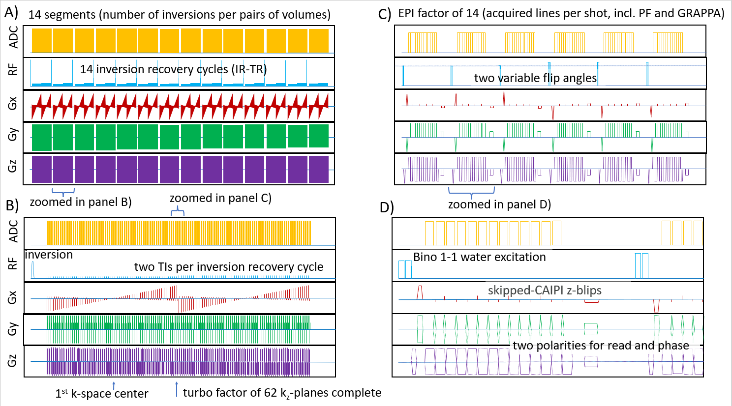
Fig. 1: Sequence looping structure.
Individual panels are zoomed sections of each other and depict the sequence looping structure across interleaved multi-shot segmentats (main loop in A), across inversion recovery cycles (second loop in B showing one IR-TR), across kz planes (third loop in C), and ky lines (fourth loop in D).
This entire cycle is repeated 4 times with all combinations of reversed read and phase directions.
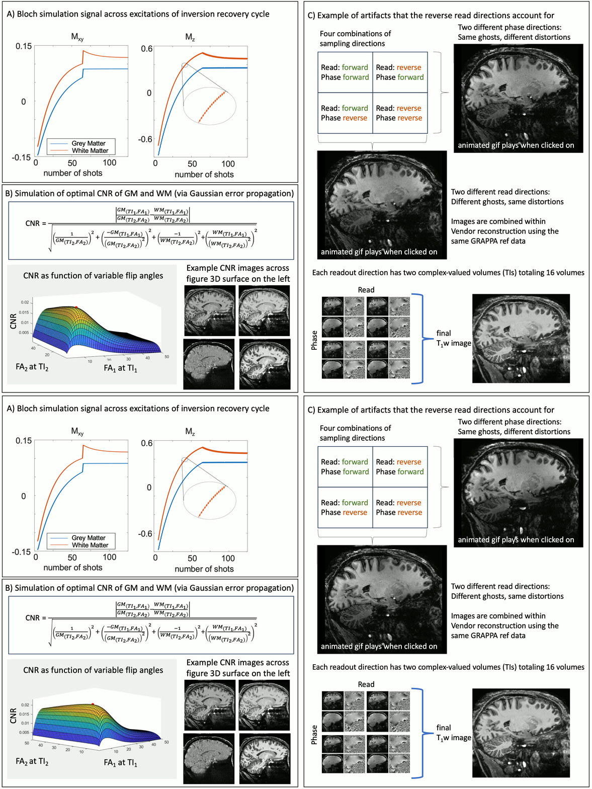
Fig. 2: Flip angle optimization and sampling polarities
A) Bloch simulation of GM and WM magnetization across kz segments of an inversion recovery cycle with two TIs.
B) This Bloch solver predicts optimal variable flip angle distributions with maximal GM/WM CNR.
C) Depiction of reconstruction logistics across reversed sampling directions and complex-valued volumes of two TIs.
Gif animation plays, when clicked on.
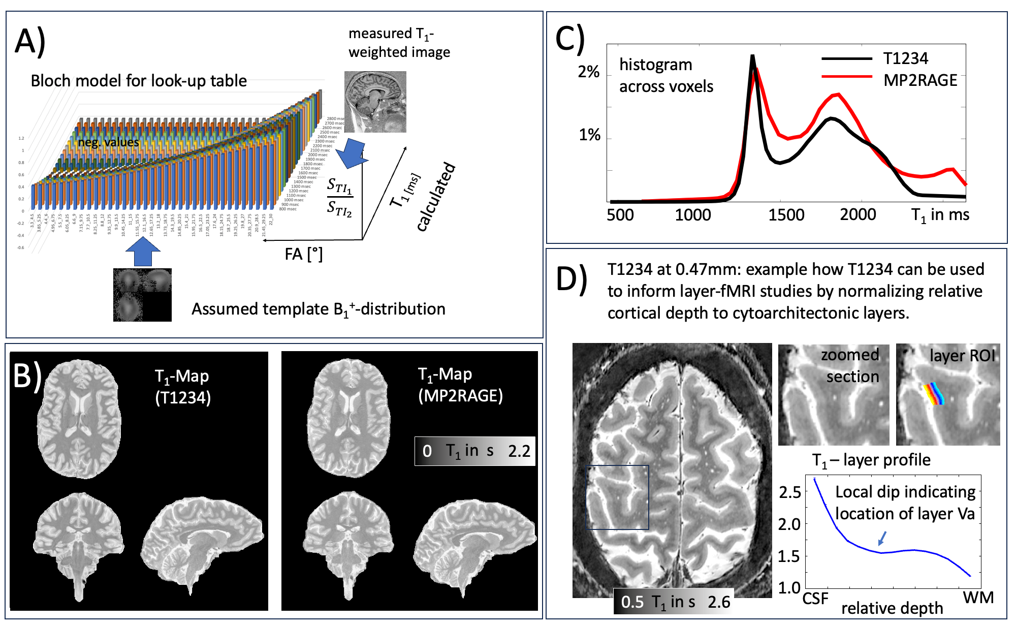
Fig. 3: T1 quantification for the T1234 approach.
A) The Bloch solver is used to generate a lookup table of expected relative MR signal intensities of T1-tissue types of both inversion times (dependent on TR, FA, IR looping structure).
B-C) Representative T1-map and corresponding histogram generated with the proposed approach compared to a T1-map generated with MP2RAGE.
D) Example application of slab-selective T1234 at higher spatial resolution for layer-specific mapping.
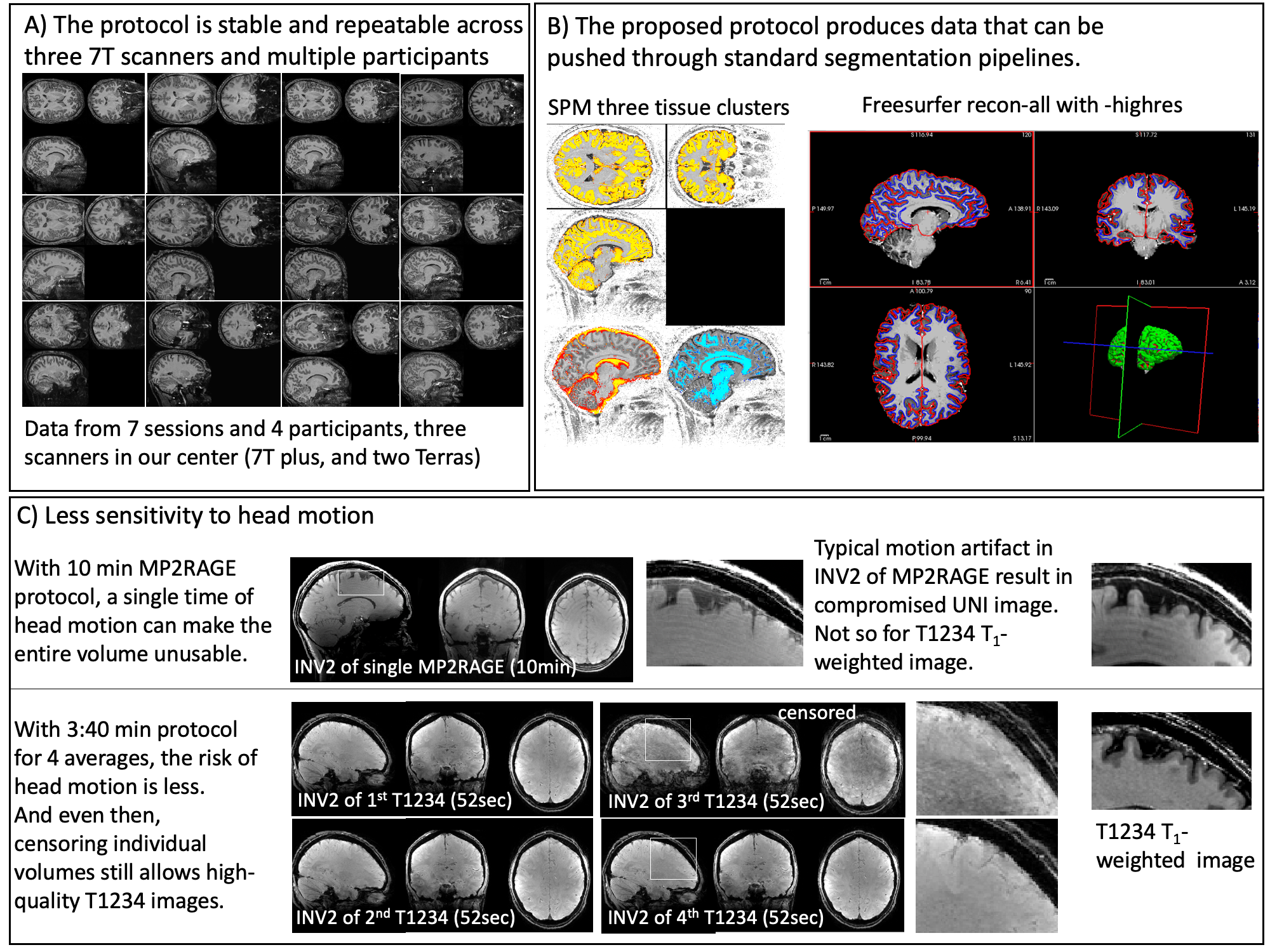
Fig. 4: Applicability of the proposed T1234 protocol in practice.
A) T1234 is stable across participants, scanners and sessions.
B) The T1234 T1w image can be used for automatic segmentation and alignment to atlas space with common software packages.
C) Acquiring structural reference data so fast in multiple averages reduces sensitivity to head motion. Different from conventional single volume anatomical imaging this allows retrospective motion correction and censoring of motion corrupted repetitions.
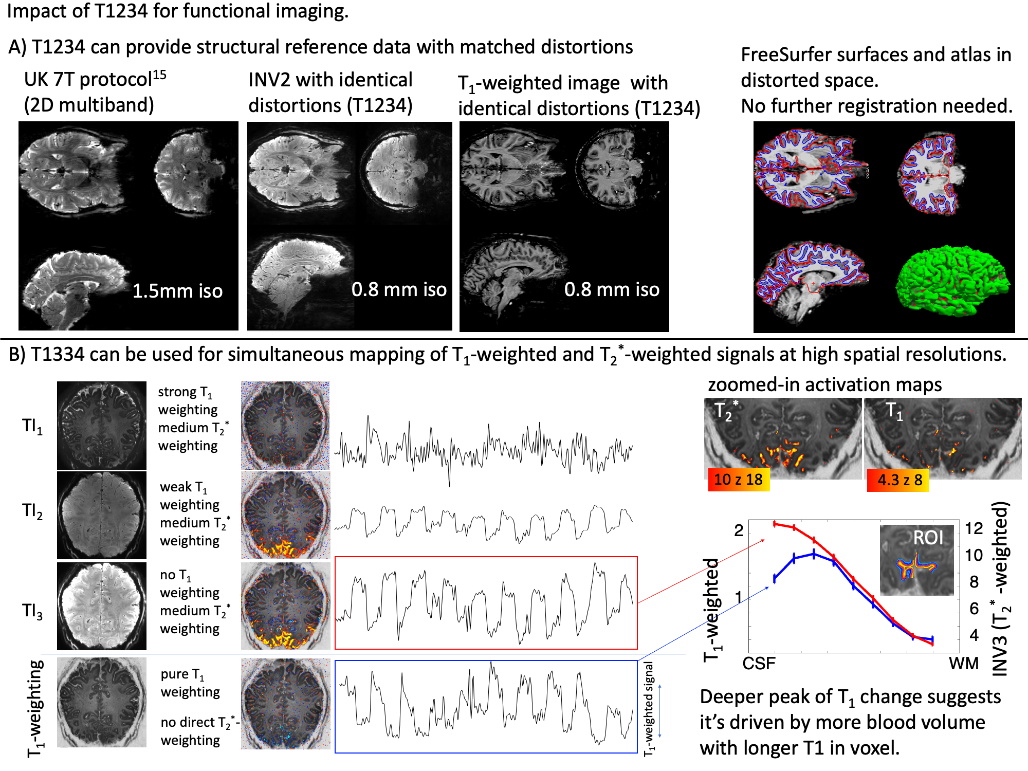
Fig. 5: Utility of T1234 for functional imaging at 7T
A) For fMRI at conventional resolutions at 7T (1.5mm), T1234 can be useful as structural reference data with identical distortions. This circumvents challenges with respect to registrations.
B) Slab-wise scanning of this protocol allows simultaneous measurements of T1-weighted and T2* weighted signals at a fine spatial scale. This can give insights into contrast mechanisms behind VASO.