0883
Metal artifact synthesis: Enabling inclusive Deep learning for patients with implants1GE HealthCare, Bangalore, India, 2GE HealthCare, Niskayuna, NY, United States
Synopsis
Keywords: Analysis/Processing, Artifacts, Metal implants, simulation, augmentation
Motivation: AI medical imaging solutions are impacted by the presence metal implants and a design of appropriate synthesis method can improve robustness of DL models.
Goal(s): Simulation of patient medical condition like metal artifacts in MRI medical images.
Approach: The proposed method blends regions from template images containing metal artifacts into target images by using metal segmentation mask for selection, blending this region into a chosen target image RoI .
Results: Improvement in knee classification accuracy of 8% and decrease in spine plane distance error by 25-40% and plane angle error by 4-30% using the proposed approach.
Impact: A data adaptive metal simulation method in semantically relevant regions in anatomy ensures robust of DL models in patients with metal implants who hitherto would not have benefitted from AI driven tasks .
Introduction
Metal implant artifacts affect performance of AI solutions in MRI data, due to dramatic changes in MR signal intensity in regions around implants. Metal implants only constitute 10-20% of the imaging pool and can have unbalanced representation in AI training which is one of the reasons for inferior performance in patients with implants. To address this lacuna, a framework for synthesis of metal implants for AI application is developed for enriching the training data and developing robust AI models. This data is subsequently used in the development of MR based AI solution for two different anatomies and impact demonstrated.Methods
Subjects:Data for our experiments came from various clinical sites with 1.5T and 3T GEHC MRI scanners.
A. Metal template: MRI imaging data from 25 knee and 10 spine subjects across different orientations wer used to generate metal region templates.
B. Knee Exams: SSFSE and fGRE three-plane localizer images were used. 9017 slices for training /validation, equal number produced using proposed pasting technique and 1299 images were used for testing.
C. Spine Exams: Axial T2 spine images from different sites were obtained for a total of 638 subjects with 416 in train, 60 in valid and 158 exams in test pool. Using proposed metal augmentation method 292 cases were simulated and added to the training set.
D. Ground-truth (GT):
GT masks for metal regions were created by manually annotating 25 knee volumes and 10 spine volumes in three view planes (coronal, sagittal and axial).
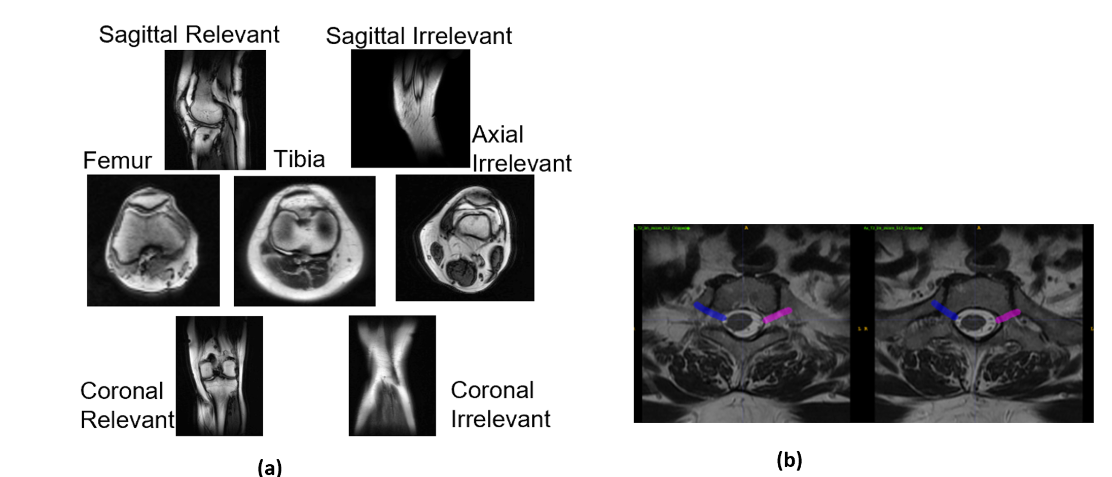
For knee classification tasks, we used methodology described in [1] to data for generate 5 classes as shown in Fig 1a .
Similarly in Spine, we identified task of delineating the Pars interarticular plane [2] which is used to improve the assessment of lumbar spondylolysis and a sample is shown in Fig 1b.
Proposed metal simulation method
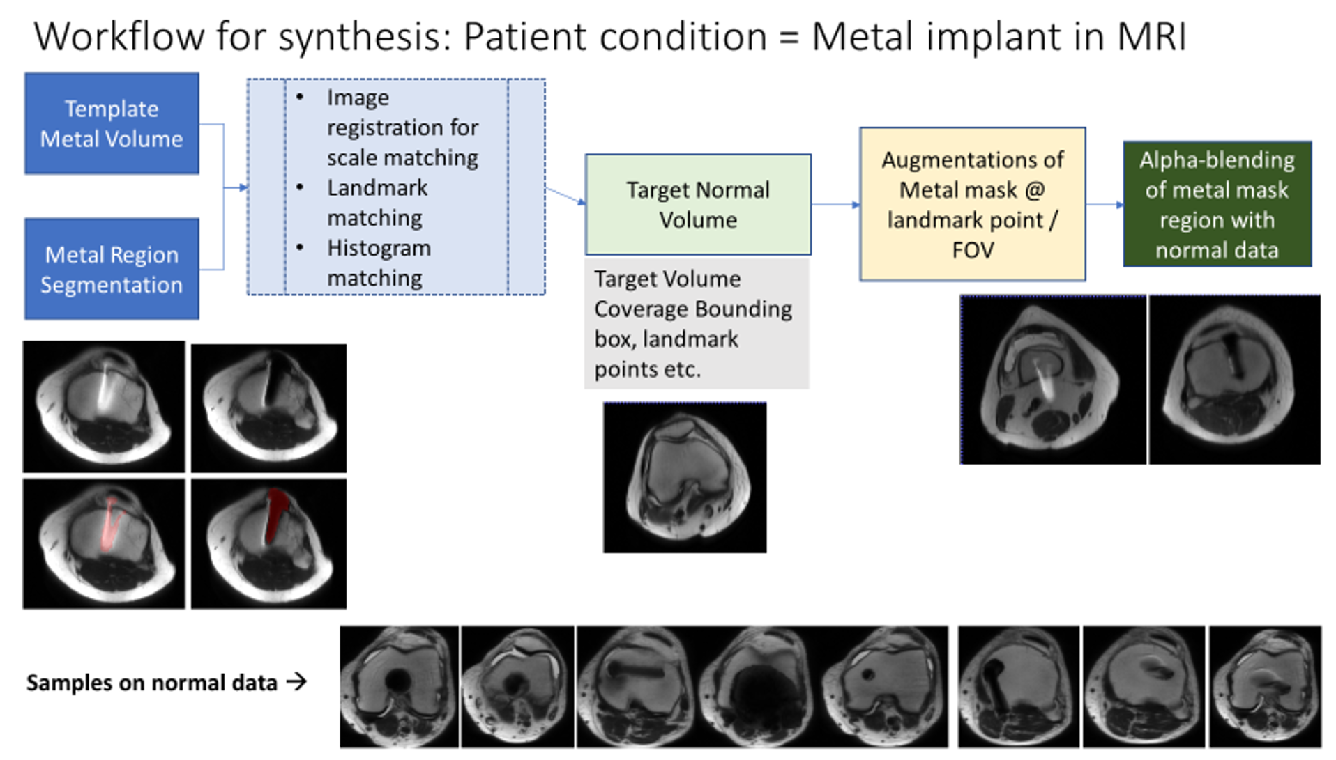
Simulated images are generated by pasting metal affected region template to normal datasets. Due to variations in intensity between template and new data, anatomy size variations, memorization due less number of templates etc., the scheme presented in Fig.2 is implemented to generate realistic simulate metal implant data from normal data. The steps are:
a. Creating a metal region mask from diverse sample metal implant images.
b. A RoI in target is either randomly identified within FOV or based on landmarks pre-disposed to metal implants based on the task needs.
c. To ensure variability of artefact appearance, segmented artefact region from source image undergoes a series of transformations, including, rotation, resizing.
d. Contrast equalization of source and the target image is performed to ensure perceptual smoothness between target image and the pasted artefact region.
e. Finally blending is used to paste the artifact region to target image.
Examples of sample images generated is shown in Fig. 3.
For both knee and spine applications, two models were trained -Model A without any metal augmentation and Model B with the proposed metal augmentation.
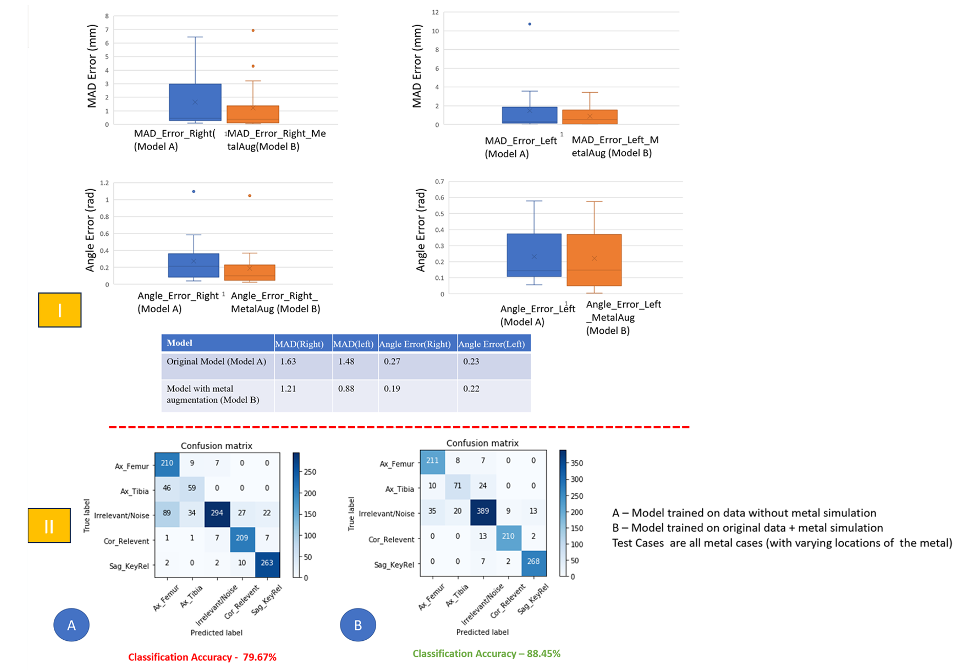
For spine model, evaluation is done using the case-wise mean distance and angle error and for knee data classification confusion matrix and accuracy is used. Qualitative evaluation of both is shown in Fig. 4.
Results
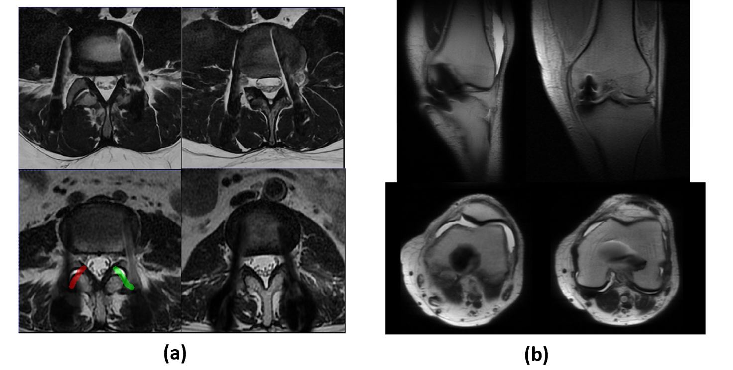
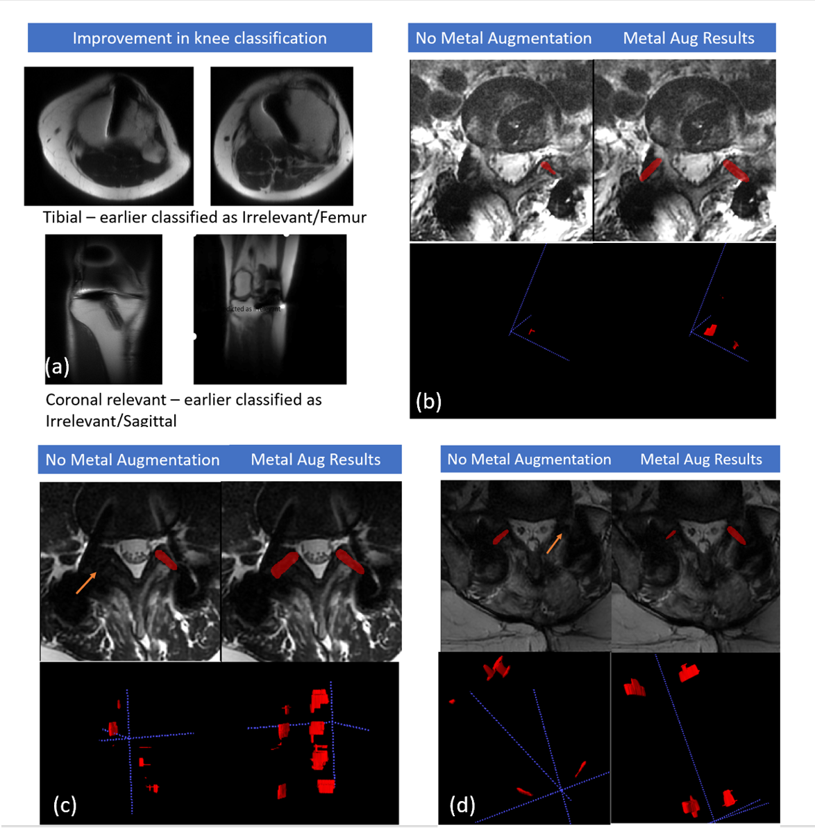
Fig. 4 show the results of running model A and model B on knee and spine test sets. From Fig. 4(I) we observe that spine model B trained with metal outperforms model A both in terms of MAD and angle errors, with around 25-40% improvement in distance error and 4-30% improvement in angle errors. From fig. 5 (b)-(d) we also observe an improvement in the prescribed PI plane using model B as compared to model A since model A fails to predict PI planes where there is heavy metal presence. Similarly, Fig. 4(II) shows the improvement of knee model B as compared to model A for classification performance with >8% improvement with the most significant improvements in irrelevant and axial tibia class. Fig. 5(a) shows examples of metal cases which were earlier misclassified as being correctly predicted and 5(b) improvement of PI plane detection.Conclusions
In this study we propose an augmentation strategy which mitigates the sparsity of metal cases by using blending or in-painting technique for simulating manifestations of metal implants. This helps enrich training set with enough examples of various patient conditions with all variations, thus making the models more robust and generalizable. We demonstrate the efficacy of the proposed method on spine and knee exams for the task of plane prediction and classification respectively and observe that such an augmentation can lead to a significant improvement in model performance.Acknowledgements
No acknowledgement found.References
[1] Anand, D. et al, Data augmentation using features from activation maps improved performance for deeplearning based automated knee prescription, In Proceedings of ISMRM 2021
[2] Chitresh, B . et al, Deep Learning based prediction of the planes for automated planning of MRI imaging of cervical neural foramina and lumbar parsintera, In Proceedings of ISMRM 2023
Figures