0881
Enhancing transcranial focused ultrasound treatment planning with synthetic ct from ultra-short echo time (UTE) MRI: a deep learning approach1Department of Neuroscience, Columbia University, New York City, NY, United States, 2F.M. Kirby Research Center for Functional Brain Imaging, Kennedy Krieger Institute, Baltimore, MD, United States, 3Department of Biomedical Engineering, Columbia University, New York City, NY, United States
Synopsis
Keywords: Analysis/Processing, Focused Ultrasound, UTE MRI, image guided therapy
Motivation: There’s a clinical interest in exploring an alternative option using ultrashort-time-echo MRI to replace CT imaging for accurate transcranial FUS treatment planning.
Goal(s): To employ a deep learning approach to generate synthetic CT images from a limited UTE-MRI dataset.
Approach: A deep learning framework based on 3D Transformer U-net is applied to the paired UTE-CT dataset and acoustic simulation is performed to validate the results.
Results: Utilizing UTE MRI can offer synthetic CT as an alternative to traditional CT imaging. The simulations showed a minimal maximum acoustic pressure difference of less than8% and a focus shift of less than1.5mm compared to CT-based simulations.
Impact: This study introduces a novel multi-task deep learning approach that enables accurate synthetic CT generation from limited UTE-MRI data. This innovation provides a cost-effective and radiation-free alternative to traditional CT imaging, significantly enhancing transcranial focused ultrasound treatment planning.
INTRODUCTION
Transcranial focused ultrasound has emerged as a promising non-invasive therapy for neurological and psychiatric disorders[1-2]. Current research focused on modulating neural activity in brain regions like amygdala, anterior cingulate cortex and striatum, which play pivotal roles in various neurological disorders[3-5]. The efficacy of tFUS therapy relies heavily on precise image-guided treatment planning. However, the presence of the skull complicates this planning due to ultrasound wave attenuation and aberration, leading to inaccurate pressure estimation and focus shifting. While CT imaging provides reliable skull acoustic property measurement, it comes with radiation exposure, increased hospital time and higher costs. To address these challenges, researchers have turned to deep learning techniques to generate synthetic CT (sCT) images from MRIs. Various deep learning architectures, such as 2D and 3D CNNs and GANs, have proven effective in producing high-fidelity sCT images[6-9]. Recent studies show that deep learning models, especially those using specified MRI sequences like ultrashort echo-time (UTE) or zero-echo time (ZTE), closely replicate CT-based simulations for ultrasound treatment planning, reducing errors in acoustic pressure estimation and focus accuracy[10-11]. This study aims to employ a multi-task deep learning approach to generate sCT images from a limited UTE-MRI dataset, with the goal of evaluating their performance in transcranial focused ultrasound simulations.METHODS
T1-weighted MPRAGE (GE Signa Premier 3.0T, 0.41x0.41x0.80 mm³) and UTE MRI images (TR 598.424 ms, TE 0.016 ms, 0.47x0.47x0.4 mm³) were acquired from three subjects (Age: 60-72, 2 females, 1 male), along with corresponding CT images (Siemens Biograph mCT 64, 120 kVp, 110 mAs, 0.59x0.59x1 mm³).After image registration, the data are processed through a 3D Transformer U-Net-based image generation framework[12], successfully transforming UTE images into CT images, as illustrated in Figure 1. a. We randomly sampled 1,000 patches size of 128x128x128 through an embedded 3D patch extraction function within the skull mask region of each subject. These samples were fed into two separate pipelines for precise segmentation of skull structures and prediction of pixel values within the region of interest. The global synthetic CT images were sequential reconstructed by the fused patch predictions. All performance evaluations were proceeded under the leave-one-out validation.To assess synthetic CT models from UTE and T1 images, we performed transcranial acoustic simulations using a single-element focused ultrasound transducer (SEFT) with parameters: 0.2 MPa source pressure, 250 kHz frequency, 64 mm radius, and 64 mm aperture depth. Targets included Anterior Cingulate Cortex (ACC), Precuneus (PCu), and Supplemental Motor Cortex (SMC). Simulations utilized Sim4life software, generating 3D acoustic pressure distributions. Five simulations per target, totaling 135 across subjects and three image-based simulations. Pressure profiles (max pressure and acoustic focus) were compared among real CT, UTE-sCT, and T1-sCT models.RESULTS
Figure 2 displays the synthetic CT images and residual maps between real CT and synthetic CT. The first column represents the synthetic CT obtained from T1-weighted images, while the second column represents the synthetic CT obtained from UTE data. Table 1 presents the performance of our image generation framework across all subjects in the leave-one-out setting. Both visualization and evaluation metrics indicate that UTE-based results outperform those based on T1-weighted images.Pressure distribution generated by SEFT on the models of real CT, UTE-sCT and T1-sCT as shown in Fig.3. The mean differences in focus pressure at ACC, PCu and SMC between UTE-sCT and CT-based simulation models were 7.23% ± 6.13%, 3.44 ± 4.63% and 8.28 ± 5.40% respectively, with corresponding mean difference of the focus positions at these targets being 0.7mm ± 0.4 mm, 0.6 ± 0.5mm and 1.4 ± 1.7 mm respectively. In contrast, the mean difference between T1-sCT and CT based simulation models for ACC, PCu and SMC stimulation were 10.52 ± 12.36%, 9.9± 7.1% and 23.43% ± 21.25% respectively. Furthermore, significant focus shifting compared to CT based simulation were also observed in T1-sCT simulations, with a maximum shift up to 5 mm.DISCUSSION & CONCLUSION
In this study, we investigated the utilization of a multi-task deep learning model for synthesizing CT images from a restricted set of UTE MRI images. By conducting acoustic simulations on various targets, we assessed the reliability of UTE-based synthetic CT (sCT) models and confirmed their utility in transcranial focused ultrasound (tFUS) treatment planning. Our findings highlight the superior performance of UTE-sCT compared to T1-sCT in tFUS simulations, characterized by reduced pressure discrepancies and negligible focus difference between sCT and CT based models. These results hold significant promise for enhancing the precision and effectiveness of tFUS treatments.Acknowledgements
The work is supported by NIH R01-MH133020 and BBRF YI 31298.References
1. H. A. Kamimura, A. Conti, N. Toschi, and E. E. Konofagou, "Ultrasound neuromodulation: Mechanisms and the potential of multimodal stimulation for neuronal function assessment," Frontiers in physics, vol. 8, p. 150, 2020.
2.Blackmore, Daniel G., Daniel Razansky, and Jürgen Götz. "Ultrasound as a versatile tool for short-and long-term improvement and monitoring of brain function," Neuron, vol. 111 no. 8, pp. 1174-1190, 2023
3. L. Verhagen et al., "Offline impact of transcranial focused ultrasound on cortical activation in primates," Elife, vol. 8, Feb 12 2019, doi: 10.7554/eLife.40541.
4 D. Folloni et al., "Manipulation of Subcortical and Deep Cortical Activity in the Primate Brain Using Transcranial Focused Ultrasound Stimulation," Neuron, vol. 101, no.6, pp. 1109-1116 e
5, Mar 20 2019, doi: 10.1016/j.neuron.2019.01.019.5. F. Munoz et al., "Long term study of motivational and cognitive effects of low-intensity focused ultrasound neuromodulation in the dorsal striatum of nonhuman primates," Brain Stimulation, vol. 15, no. 2, pp. 360-372, 2022.
6. S. N. Yaakub, T. A. White, E. Kerfoot, L. Verhagen, A. Hammers, and E. F. Fouragnan, "Pseudo-CTs from T1-weighted MRI for planning of low-intensity transcranial focused ultrasound neuromodulation: An open-source tool," Brain Stimulation: Basic, Translational, and Clinical Research in Neuromodulation, vol. 16, no. 1, pp. 75-78, 2023.
7. H. Koh, T. Y. Park, Y. A. Chung, J.-H. Lee, and H. Kim, "Acoustic simulation for transcranial focused ultrasound using GAN-based synthetic CT," IEEE Journal of Biomedical and Health Informatics, vol. 26, no. 1, pp. 161-171, 2021.
8. Han X. “MR-based synthetic CT generation using a deep convolutional neural network method,” Medical Physics, vol 44, no.4, pp. 1408-1419, 2017 doi: 10.1002/mp.12155.
9. Liu, Han, Michelle K. Sigona, Thomas J. Manuel, Li Min Chen, Benoit M. Dawant, and Charles F. Caskey. "Evaluation of Synthetically Generated CT for use in Transcranial Focused Ultrasound Procedures." arXiv preprint arXiv:2210.14775, 2022
10. M. Miscouridou, J. A. Pineda-Pardo, C. J. Stagg, B. E. Treeby, and A. Stanziola, "Classical and learned MR to pseudo-CT mappings for accurate transcranial ultrasound simulation," IEEE Transactions on Ultrasonics, Ferroelectrics, and Frequency Control, vol. 69, no. 10, pp. 2896-2905, 2022.
11. P. Su et al., "Transcranial MR Imaging-Guided Focused Ultrasound Interventions Using Deep Learning Synthesized CT," AJNR Am J Neuroradiol, vol. 41, no. 10, pp. 1841-1848, Oct 2020, doi: 10.3174/ajnr.A6758.
12. Rao VM, et al. Improving across-dataset brain tissue segmentation for MRI imaging using transformer. Front Neuroimaging. 2022;1:1023481. doi: 10.3389/fnimg.2022.1023481.
Figures
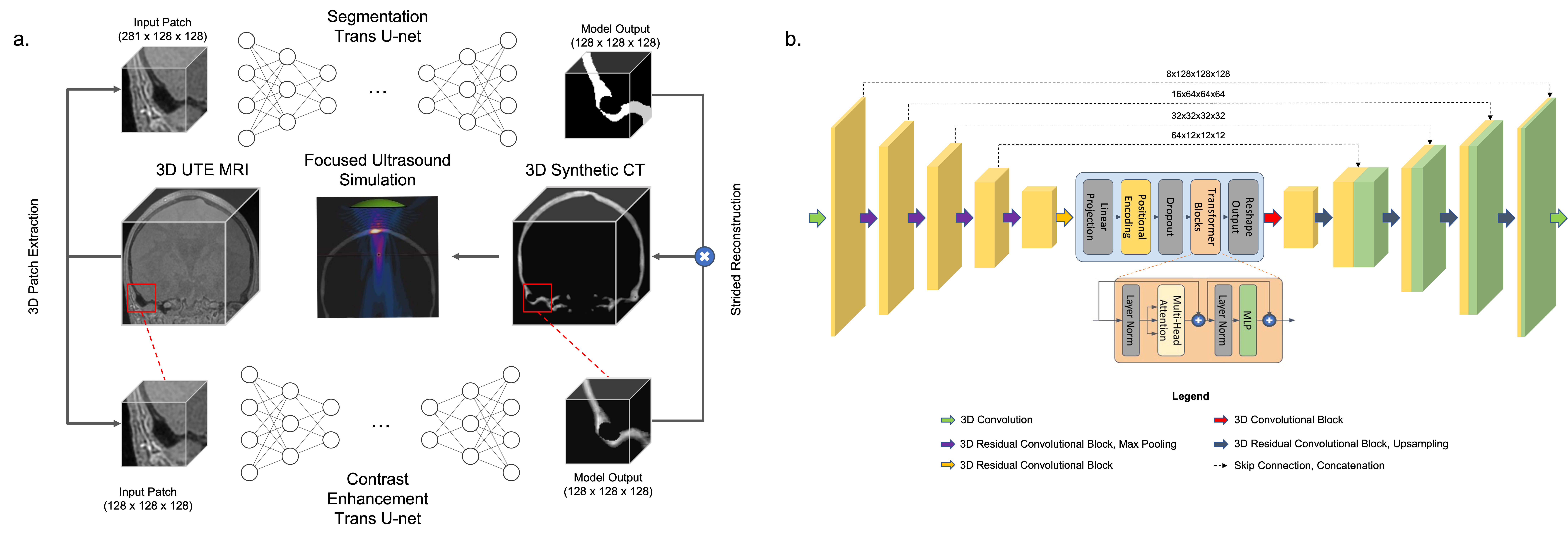
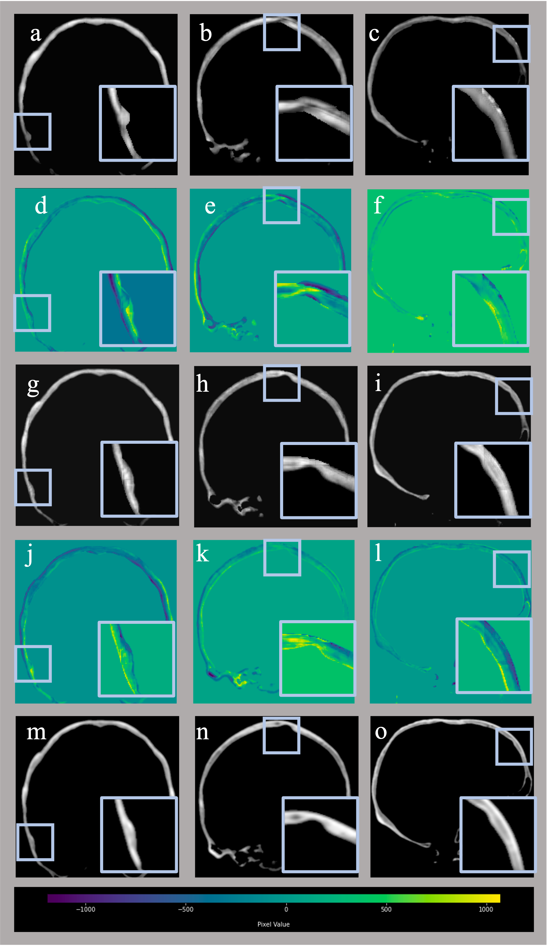
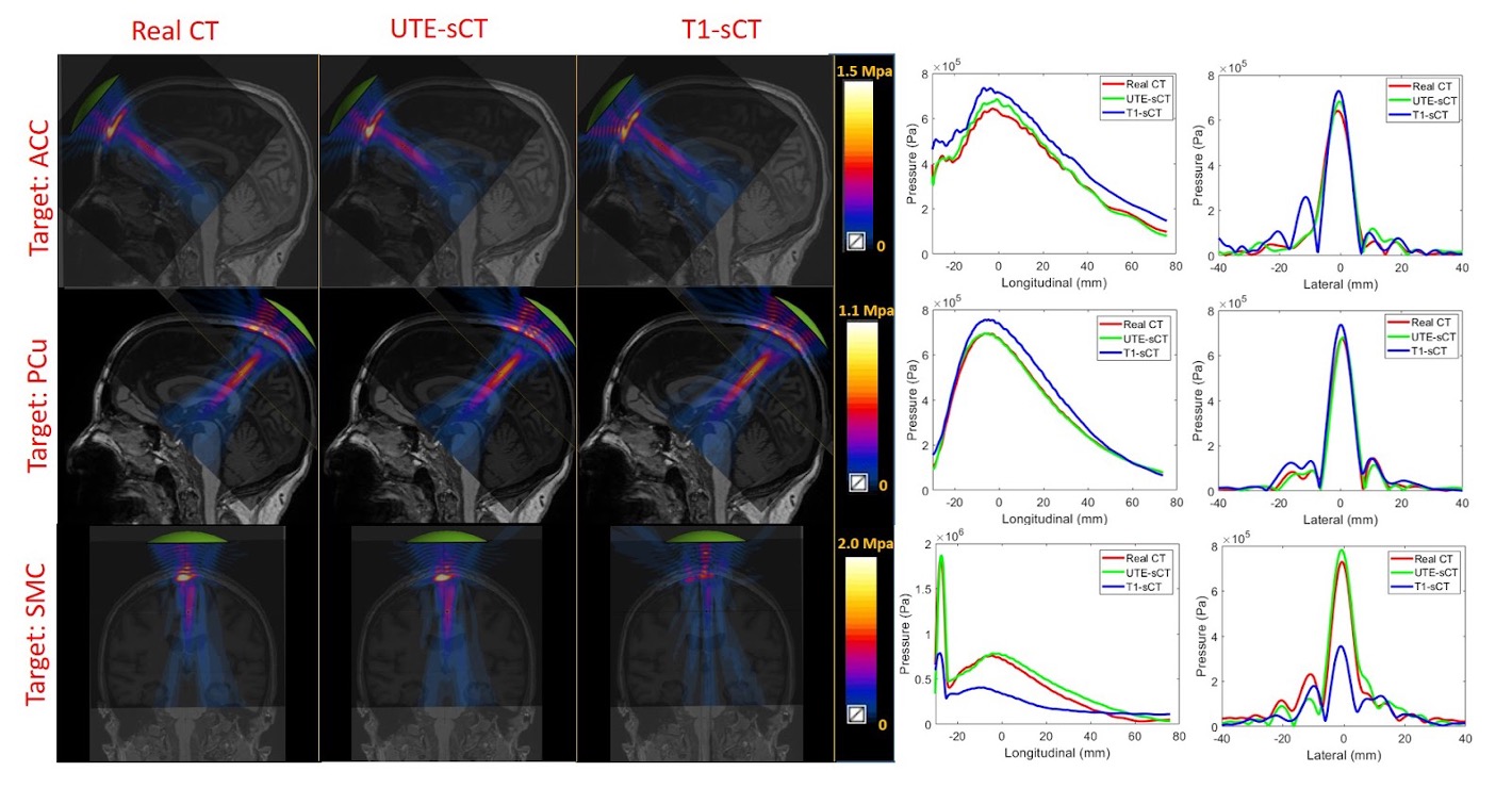
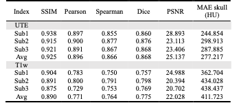
Table1. Performance on the test data set.
* SSIM = Structural similarity index, Pearson = Pearson’s correlation, Spearman = Spearman’s rank correlation, Dice = Sørensen–Dice coefficient, Jaccard = Jaccard similarity coefficient, PSNR = Peark Signal-to-Noise-Ratio, MAE = Mean Average Error, HU = Hounsfield Unit.