0875
Accelerating Low-Field Prostate DWI: Self-Supervised Denoising for Rapid Scan Acquisition1Pattern Recognition Lab, Friedrich-Alexander Universität Erlangen-Nürnberg, Erlangen, Germany, 2Magnetic Resonance, Siemens Healthineers AG, Erlangen, Germany
Synopsis
Keywords: Analysis/Processing, Low-Field MRI, Diffusion-Weighted Imaging
Motivation: Diffusion-weighted imaging (DWI) is crucial for lesion detection but suffers from inherently low signal-to-noise ratio (SNR), especially in low-field settings.
Goal(s): The goal of this work is to accelerate low-field prostate DWI, reducing the number of image repetitions and scan time while maintaining image quality.
Approach: We present a self-supervised denoising method employing Stein's unbiased risk estimator (SURE) and a physics-based noise model and evaluate the denoising results without relying on ground-truth data.
Results: Our method excels in preserving image content, outperforming other denoising techniques. This allows a substantial reduction in scan time, making it a promising advancement in low-field DWI.
Impact: Our proposed denoising approach accelerates low-field prostate DWI via self-supervised denoising, improving scan efficiency without compromising image quality. We further demonstrate how to employ a physics-based noise model to evaluate denoising performance in the absence of noise-free ground-truth data.
Introduction
Diffusion-weighted imaging (DWI) plays a crucial role in the detection and characterization of lesions within the human body. Water diffusion rates are typically measured in multiple directions and subsequently consolidated into a single trace image for diagnostic purposes.A persistent challenge in DWI lies in the inherently low signal-to-noise ratio (SNR), especially at high b-values. The SNR decreases further when imaging smaller organs, such as the prostate, or in low-field (B0 < 1.0 T) settings1,2. In clinical practice, multiple repetitions of the same slice are acquired and averaged to enhance SNR. However, this approach considerably prolongs scan duration and increases sensitivity to patient movement.
We demonstrate a novel deep learning-based approach to accelerate low-field prostate DWI in a self-supervised manner, employing an augmented version of Stein's unbiased risk estimator (SURE)3,4, and a physics-based noise model. Additionally, we demonstrate how to evaluate denoising results without access to clean ground-truth data.
Methods
Data: We trained a neural network using a dataset of 319 DWI prostate scans obtained from volunteers using 1.5 T and 3 T scanners (MAGNETOM, Siemens Healthineers AG, Erlangen, Germany) and standardized protocols. Each scan included two b-values, b=50 s/mm² (b50) and b=800 s/mm² (b800), with four diffusion directions. For b50, four repetitions were collected, whereas b800 involved ten repetitions.We then applied the network to five DWI prostate scans acquired from volunteers using a 0.55 T scanner (MAGNETOM Free.Max, Siemens Healthineers AG, Erlangen, Germany). In the standard low-field DWI scans, we typically perform four repetitions for b50 and 22 repetitions for b800. However, as part of our study, we decreased the repetition count to two for b50 and 15 for b800, resulting in a scan time reduction from 7:00 min to 4:47 min.
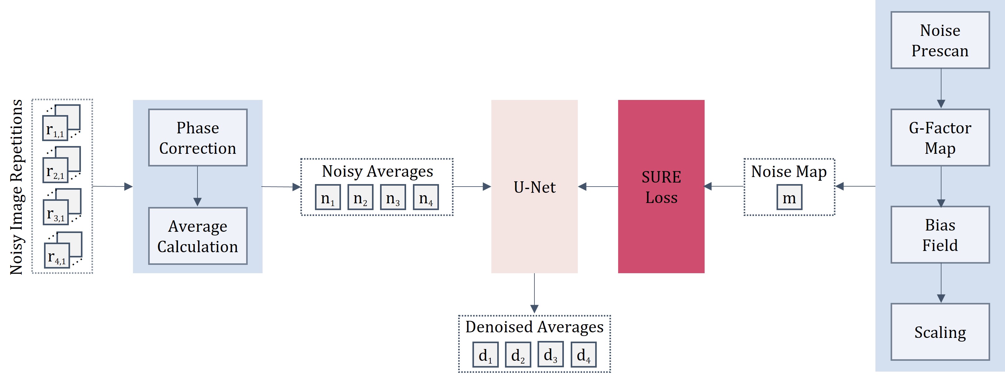
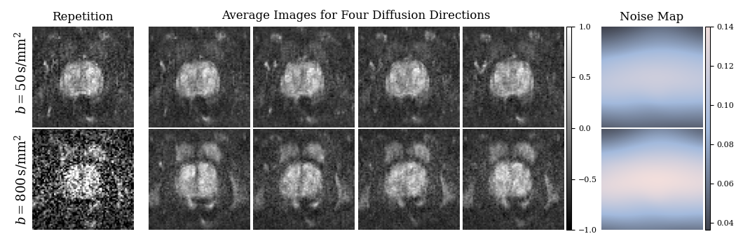
Pipeline: The visualization of our proposed denoising pipeline is presented in Figure 1, while exemplary representations of images and noise maps are visualized in Figure 2. We initiate the process with a phase correction step to enable complex averaging and maintain a zero-mean Gaussian noise distribution5,6, followed by the generation of averaged images for each diffusion direction. These resulting average images are subsequently input to the U-Net architecture7 for denoising. The network is trained in a self-supervised manner leveraging an extended version of SURE for spatially variant Gaussian noise3,4 to estimate the Mean Squared Error (MSE) between the denoised output and the unknown ground truth. To accomplish this, a noise map that characterizes the standard deviation of the noise distribution for each pixel is employed. This noise map is generated by propagating noise data obtained from a noise adjustment scan, a routine element of the scanner calibration, through the reconstruction pipeline. This map is then scaled to account for the effects of phase correction and average calculation.
Experiments: We compare our approach to the learning-based Noise2Noise (N2N)8 method and a denoising technique based on random matrix theory (MPPCA)9. Both methods are advantageous when multiple noisy image realizations are available.
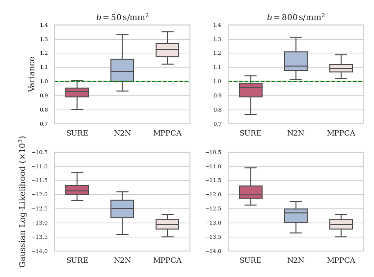
Evaluation: Full repetition count (FRC) images used as ground truth contain inherent noise, potentially causing inaccuracies when calculating quantitative metrics. To mitigate this, we propose a novel approach that leverages the computed noise maps. For optimal denoising results, the residual images should contain only pure noise. Once the residual image is corrected by dividing it with the computed noise map, it should ideally follow a Gaussian distribution10 with a variance of one. To assess this desired state, we compute the Gaussian log-likelihood and the variance of the corrected residual.
Results and Discussion
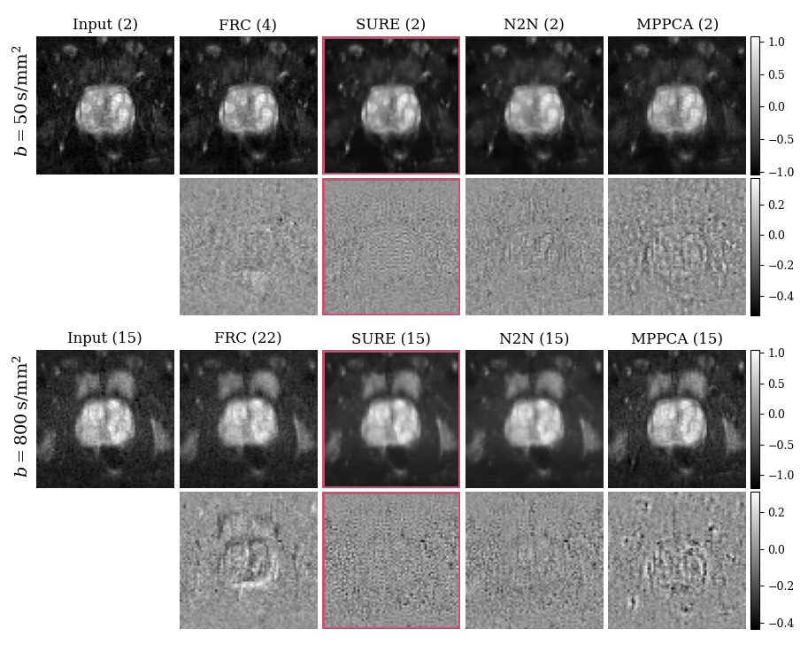
Figure 3 showcases visual representations of exemplary results. An increase in the number of repetitions enhances SNR yet introduces alterations in image content due to patient motion. Despite being trained on high-field data, our method effectively mitigates noise without compromising image content or blurring high-frequency image structures. In contrast, the Noise2Noise residual image and, particularly, the MPPCA result exhibits discernible structural artifacts.The quantitative denoising evaluation is presented in Figure 4. The SURE-based approach generates a corrected residual with variance close to 1.0 and tends to be more conservative in noise removal, whereas the N2N and MPPCA methods, in contrast, tend to overly denoise the images, potentially compromising image content. Further, the Gaussian log-likelihood of the corrected residual is observed to be highest for the SURE-based approach.
Conclusion
Our proposed deep learning-based denoising method excels in comparison to alternative denoising techniques, exhibiting superior performance in both visual assessments and quantitative evaluations. This advancement enables a decrease in the number of image repetitions necessary for low-field prostate DWI, resulting in a considerable reduction in scan time while maintaining high image quality.Acknowledgements
No acknowledgement found.References
- Kitajima K, Kaji Y, Kuroda K et al. High b-value diffusion-weighted imaging in normal and malignant peripheral zone tissue of the prostate: effect of signal-to-noise ratio. Magn. Reson. Med. 2008; Sci. 7, 93–99.
- Marques JP, Simonis FF, and Webb AG. Low-field MRI: An MR physics perspective. Journal of Magnetic Resonance Imaging 2019; 49(6), 1528-1542.
- Stein C. Estimation of the mean of a multivariate normal distribution. The Annals of Statistics 1981; 9(6):1135–1151.
- Pfaff L, Hossbach J, Preuhs E et al. Training a tunable, spatially-adaptive denoiser without clean targets. ISMRM 2022.
- Prah DE, Paulson ES, Nencka AS et al. A simple method for rectified noise floor suppression: Phase-corrected real data reconstruction with application to diffusion-weighted imaging. Magn Reson Med 2010; 64(2):418-29.
- Eichner C, Cauley SF, Cohen-Adad J et al. Real diffusion-weighted MRI enabling true signal averaging and increased diffusion contrast. Neuroimage 2015; 15,122:373-84.
- Ronneberger O et al. U-Net: Convolutional networks for biomedical image segmentation. In International Conference on Medical Image Computing and Computer-Assisted Intervention 2015; 234–241.
- Lehtinen J, Munkberg J, Hasselgren J et al. Noise2noise: Learning image restoration without clean data. arXiv preprint arXiv:1803.04189, 2018.
- Veraart J, Novikov DS, Christiaens D et al. Denoising of diffusion MRI using random matrix theory. Neuroimage 2016; 142, 394–406.
- Cárdenas-Blanco A, Tejos C, Irarrazaval P et al. Noise in magnitude magnetic resonance images. Concepts Magn. Reson. Part A: An Educ. J. 2008; 32, 409–416.
Figures