0872
MIROR: The Clinical Decision Support System with Functional Imaging and Machine Learning1Cancer and Genomic Sciences, University of Birmingham, Birmingham, United Kingdom, 2Oncology, Birmingham Children's Hospital, Birmingham, United Kingdom, 3Radiology, Birmingham Children's Hospital, Birmingham, United Kingdom, 4RRPPS, University Hospital Birmingham NHS Foundation Trust, Birmingham, United Kingdom, 5Engineering, University of Birmingham, Birmingham, United Kingdom
Synopsis
Keywords: Diagnosis/Prediction, Radiomics
Motivation: Radiomics has potential to bring added values to cancer diagnosis and prognosis, whilst a practical tool that is accessible for clinicians and radiologists has not been available.
Goal(s): To design MIROR, a clinical decision support system, that can aid tumour diagnosis with real-time image and spectroscopy analysis.
Approach: The project keeps collecting childhood brain tumour proton MR images and spectroscopy in England and has built a multi-centre database that includes 377 cases.
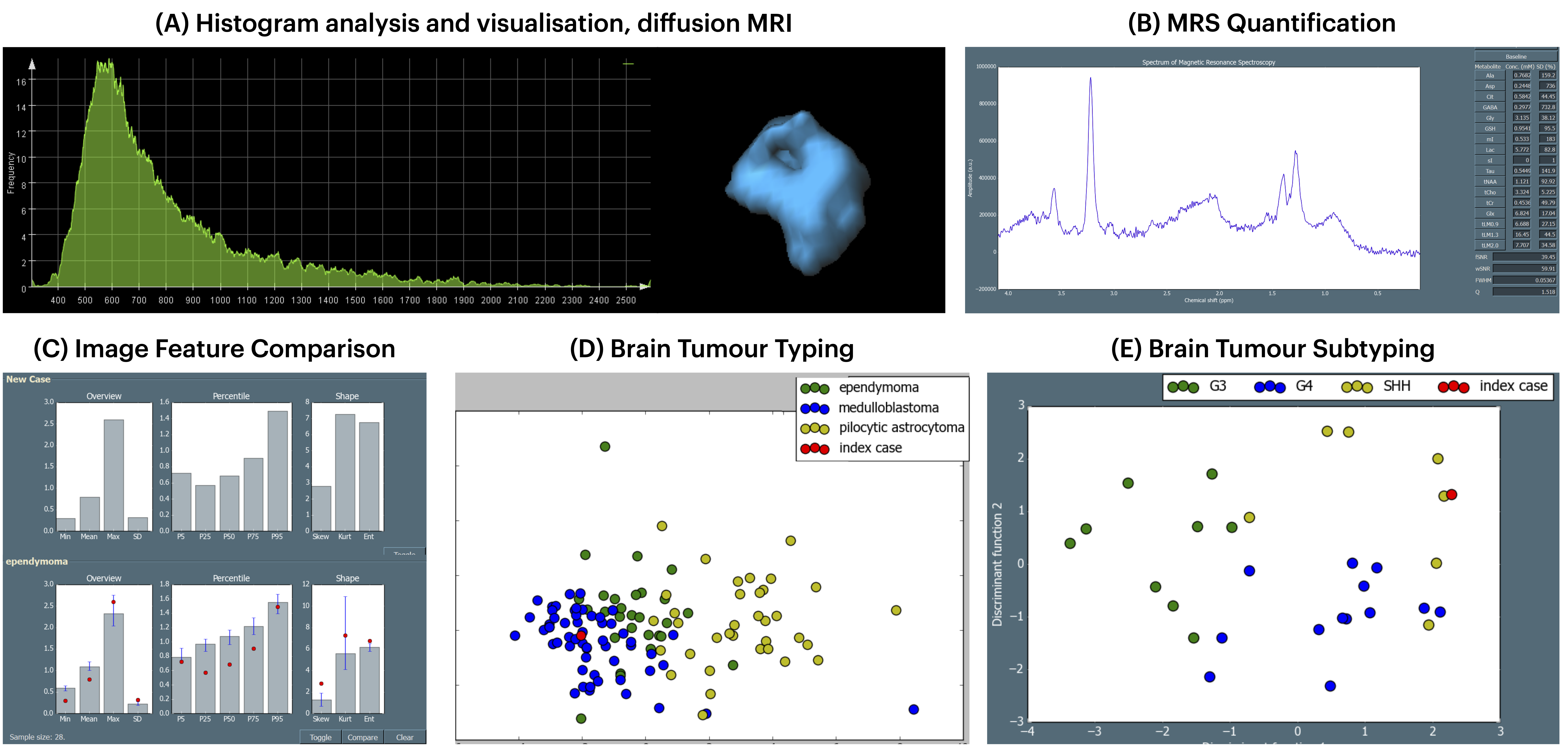
Results: MIROR supports key features including visualisation, image analysis, feature extraction, spectroscopy quantification, and tumour type and subtype prediction through machine learning.
Impact: A practical solution that translates radiomics and machine learning into clinical scenarios can aid tumour diagnosis and treatment planning, bring benefits to patient healthcare, and improve clinical outcomes.
Introduction
Brain tumours remain amongst the most common lethal diseases in children. [1] Early diagnosis through pre-clinical non-invasive MR imaging and spectroscopy can bring benefits to patients by improving treatment planning. [2] Radiomics evolve rapidly in precision oncology. [3] Such as, quantitative imaging biomarkers can aid early-stage brain cancer diagnosis and potentially improve patient outcomes. [4] Multi-modal MR imaging and spectroscopy can provide structural and physiological information that may bring added value to tumour diagnosis. [5] With machine learning, brain tumour types and subtypes could be identified timely and quantitatively. This abstract presents a clinical decision support system, MIROR, where imaging biomarkers can be analysed with machine learning for brain tumour typing and sub-typing, and a multi-centre repository has been available for building classifiers dynamically.Method
MIROR was designed by following an evidence-adaptive modular architecture [6] and considering the needs of clinical scenarios. The supported imaging modalities are conventional imaging, diffusion-weighted MRI, perfusion-weighted MRI, intra-voxel incoherent motion imaging, and single-voxel proton MR spectroscopy. The repository that supports MIROR is continuously updated by collecting patient data nationally, and current partner hospitals are Alder Hey Children’s Hospital (Liverpool), Birmingham Children’s Hospital, Great Ormond Street Hospital (London), Queen’s Medical Centre (Nottingham), and Royal Victoria Infirmary (Newcastle upon Tyne). MIROR’s design was updated based on the questionnaires collected from radiologists who attended tutorials and tried the software. Three main modules, Summary, Image and Spectroscopy Analysis, and Decision Support, are included in MIROR’s workflow. The module summary demonstrates the demographical and clinical variables for the patient as well as the summarised quantitative imaging biomarkers. Structural and functional imaging can be loaded, processed, visualised, and analysed in image and spectroscopy analysis, where imaging features can be automatically extracted from the patient data. Specifically, regions of interest can be drawn, loaded, and saved for structural imaging, and MR spectroscopy can be quantified for estimating metabolite concentration levels. Such quantitative imaging biomarkers can be analysed with the repository and machine learning in decision support. The tumour types or subtypes can be determined with a confidence interval from two or three manually pre-described potential tumour types by the user.Results
The lastly updated MIROR repository includes 124 diffusion MRI cases, 85 perfusion MRI cases, and 192 MR spectroscopy cases, which were acquired in either 1.5 T or 3 T. The brain tumour types, which have at least two cases in MIROR’s repository and were tested for classification, include atypical teratoid rhabdoid tumour, diffuse intrinsic pontine glioma, ependymoma (posterior fossa and supertentforial), medulloblastoma, and pilocytic astrocytoma. An example of evaluating a new medulloblastoma case by using MIROR that was installed in a NHS computer in Birmingham Children’s Hospital shows how diagnosis can be performed and what information can be provided. Initially, patient images can be loaded and preprocessed, followed by image analysis and spectroscopy quantification for extracting imaging biomarkers. When such quantitative imaging biomarkers are ready, they are ranked [7] and selected for determining the tumour type or subtype with machine learning. The visualised classification map can show how reliable the diagnosis is and potentially how the new case may be misdiagnosed.Discussion
MIROR addresses the needs of clinicians and radiologists in real-world scenarios. Its visualisation features allow users to perform image segmentation, reconstruction, and analysis at one station. The analysed imaging biomarkers can be visualised for comparison, and the certainty is demonstrated by the variance generated from the repository. Visualisation also supports MR spectroscopy, through which users can assess the fitting performance of each metabolite.Radiomics are demonstrated in MIROR by allowing users to select the tumour types of interest and dynamically build the classifier. The repository, which is continuously updated by collecting patient data across England, can provide clinical reference evidenced by a larger trial. Computational methods can select quantitative imaging biomarkers and allow users to focus on the most important features that have higher diagnostic ability.
The challenges of moving to practical healthcare system include improving data quality, utilising computational methods, and enhancing clinical expertise [8]. The repository allows building a computational model that considers demographic and clinical variables to better predict clinical information. The work is supported by our previous finding of added value from different modalities [9], and the visualised information is also interpretable for assisting clinical decision-making.
Future development plan of MIROR includes enhancing the repository by expanding collaboration, improving data quality and software usability, ameliorating computational methods in data analysis, designing informative reports, and planning prospective assessment of MIROR’s added value.
Acknowledgements
NIHR Research Professorship, Grant/Award Numbers: NIHR-RP-R2-12-019; Help Harry Help Others; UK National Institute of Health Research’s Nottingham Biomedical Research Centre; Cancer Research UK and EPSRC Cancer Imaging Programme at the Children’s Cancer and Leukaemia Group (CCLG) in association with the MRC and Department of Health (England), Grant/Award Numbers: C7809/A10342; Cancer Research UK and NIHR Experimental Cancer Medicine Centre Paediatric Network, Grant/Award Numbers: C8232/A25261; Children’s Research Fund; Birmingham Women’s and Children’s Hospital Charities; The Children’s Cancer and Leukaemia Group Little Princess Trust, Grant/Award Numbers: 2017/15 and2019/01; Children with Cancer, Grant/Award Numbers: 15/118; Action Medical Research and the Brain Tumour Charity, Grant/Award Numbers:GN2181; Health Data Research UK (HDR UK).References
[1] Mahase E. Cancer overtakes cvd to become leading cause of death in high income countries. BMJ. 2019;366:l5368.
[2] Peet AC, Arvanitis TN, Leach MO, Waldman AD. Functional imaging in adult and paediatric brain tumours. Nature Reviews Clinical Oncology. 2012;9:700-711.
[3] Aerts HJWL. The potential of radiomic-based phenotyping in precision medicine: a review. JAMA Oncology. 2016;2(12):1636–1642.
[4] Waldman AD, Jackson A, Price SJ et al. Quantitative imaging biomarkers in neuro-oncology. Nature Reviews Clinical Oncology. 2009;6:445-454.
[5] Yearley AG, Blitz SE, Patel RV et al. Machine learning in the classification of pediatric posterior fossa tumors: A systematic review. Cancers. 2022;14:5608.
[6] Sim I, Gorman P, Greenes RA et al. Clinical decision support systems for the practice of evidence-based medicine. Journal of the American Medical Informatics Association. 2001;8:527-534.
[7] Zhao D, Grist JT, Rose HEL et al. Metabolite selection for machine learning in childhood brain tumour classification. NMR in Biomedicine. 2022;35:e4673.
[8] Panagiotou OA, Högg LH, Hricak H et al. Clinical Application of Computational Methods in Precision Oncology: A Review. JAMA Oncology. 2020;6:1282-1286.
[9] Manias KA, Gill SK, MacPherson L, Foster K, Oates A, Peet AC. Magnetic resonance imaging based functional imaging in paediatric oncology. European Journal of Cancer. 2017 Feb;72:251-265.
Figures