0870
An artificial intelligence decision tree diagnostic platform helps neuroradiologists reclassify adult-type diffuse gliomas1The First Affiliated Hospital of Chongqing Medical University, Chongqing, China, 2Southwest University, Chongqing, Chongqing, China, 3Shanxi Provincial People's Hospital, Shanxi, Shanxi, China, 4Chongqing Hospital of Traditional Chinese Medicine, Chongqing, China, 5School of Medical and Life Sciences Chengdu University of Traditional Chinese Medicine, Chengdu, China
Synopsis
Keywords: Diagnosis/Prediction, Brain
Motivation: Deep learning networks offers an opportunity for diffuse gliomas classification, which may be help for therapeutic decision making and selection of patient groups suitable for targeted genetic analysis.
Goal(s): The purpose of this study is to develop an artificial intelligence method to reclassify adult-type diffuse gliomas based on the new WHO CNS tumor classification.
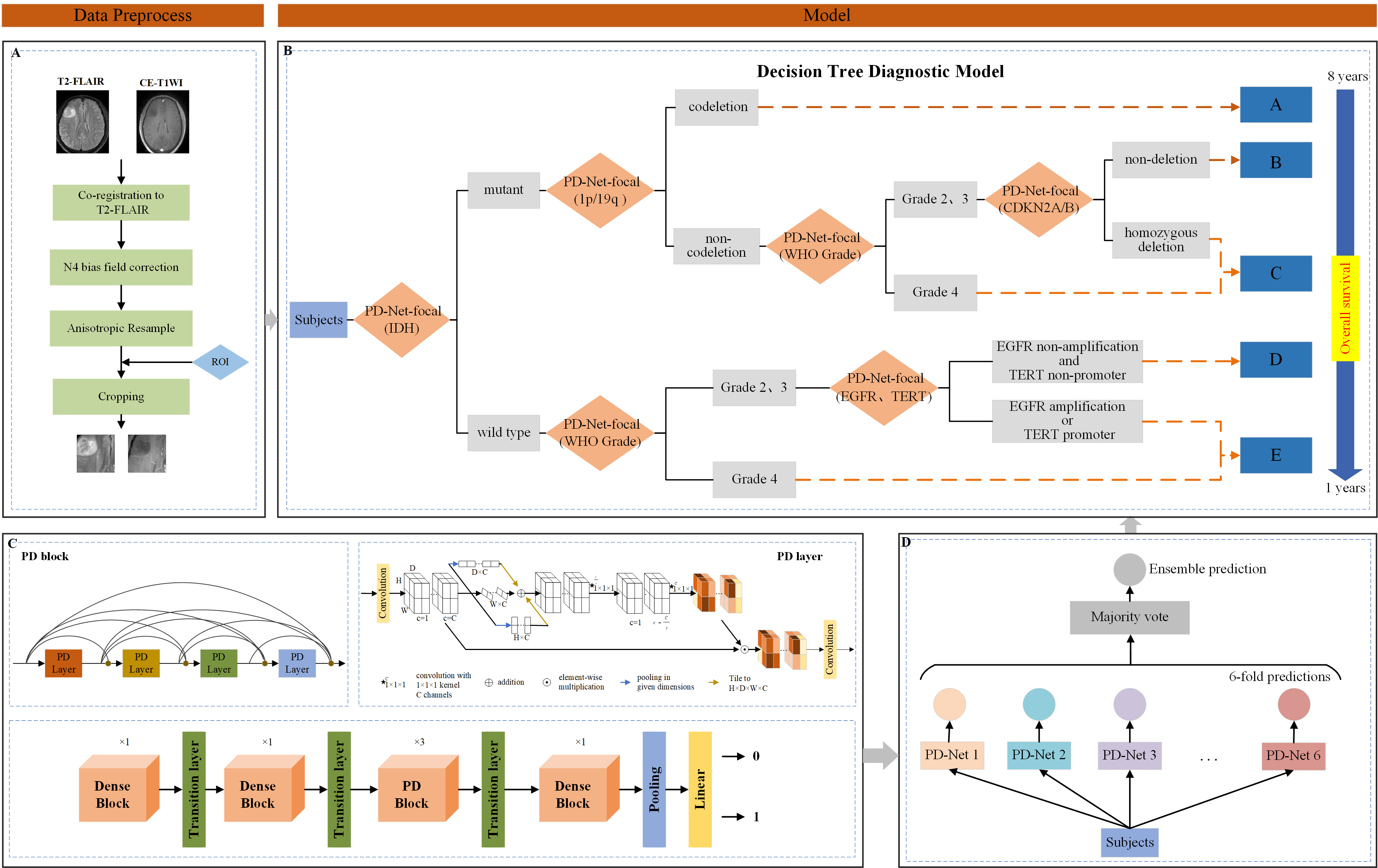
Approach: An artificial intelligence decision tree diagnostic platform(DTDP) based on MRI and deep learning networks was developed by combined 6 individualized CNNs models in series and parallel
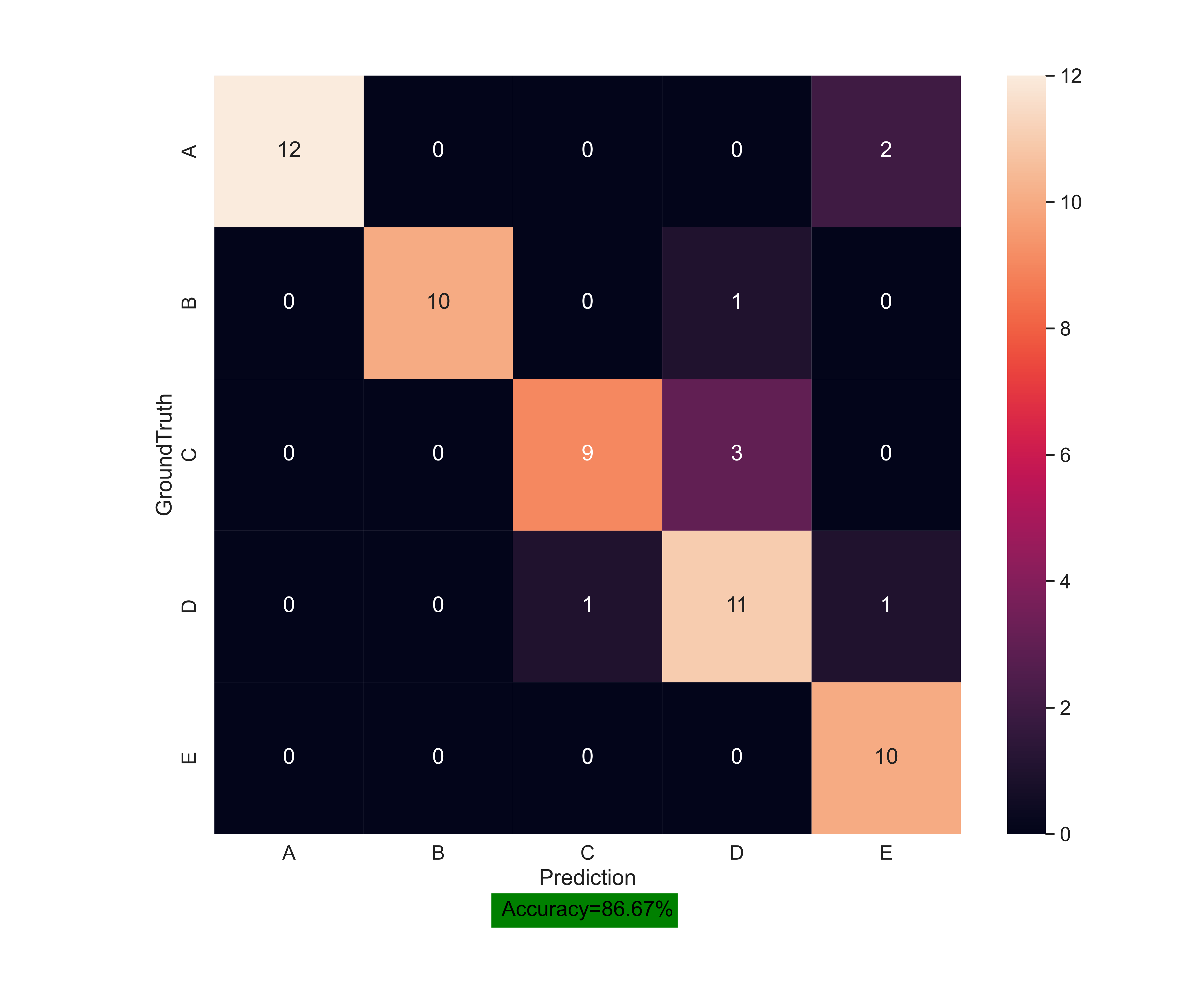
Results: The DTDP performed well with accuracy of 86.67%.
Impact: The DTDP achieved automatic classification and comprehensive diagnosis of adult‑type diffuse gliomas by combining genetic biomarkers and histological grading, and effectively helped neuroradiologists to reclassify adult-type diffuse gliomas.
Introduction
The fifth edition of the WHO classification of CNS tumors published in 2021 once again clarified the status and value of genetic biomarker in the diagnosis and classification of glioma, and emphasizing the combination of histological morphology diagnosis and genetic biomarkers for classification, and genetic biomarkers are superior to histomorphology in evidence.Methods
An artificial intelligence decision tree diagnostic platform(DTDP) based on MRI and deep convolutional neural networks (CNNs) was designed to assist neuroradiologists to reclassify adult-type diffuse gliomas. T2-FLAIR and CE-T1WI images of glioma were obtained from The Cancer Imaging Archive (TCIA) and the First Affiliated Hospital of Chongqing Medical University1, and the genomic and histological information was provided from The Cancer Genome Atlas (TCGA) and the First Affiliated Hospital of Chongqing Medical University2. Studies were screened for the availability of genetic biomarkers (IDH, 1p/19q, CDKN2A/B, EGFR and TERT) and histological grading of gliomas. A large dataset was compiled comprising 19274 MR images from 419 cases across 5 different subtypes of adult-type diffuse gliomas. We proposed a neural network called projection-and-excitation block DenseNet (PD)-Net-focal for the DTDP. In short, we used the DenseNet structure as the backbone, generated MR image feature maps through 3D convolution, and further utilized the feature information extracted from the hierarchical structure through dense connections and feature reuse. After that, the image features were linearly combined using a multi-layer perceptron and softmax operator to output genotype categories or histological grading. Building upon this, we added project-and-excite blocks, named PD-Net, between 3D convolution layer and the batch normalization layer in dense layers. Each Dense block was followed by a transition layer for down sampling, PD blocks replaced some of the Dense blocks and ultimately concatenated in series to form the proposed PD-Net. Finally, we adopt the focal loss as the loss function to address the data imbalance issue among tumor genotype categories or histological grading3, replacing the original cross-entropy loss in DenseNet. PD-Net optimized by this weighted cross-entropy loss is named PD-Net-focal. In order to combine histological morphology diagnosis and genetic biomarkers for reclassification of adult-type diffuse gliomas, we referred to the new Fifth New Edition Classification and proposed orderly binary classification tasks for the following categories: IDH, 1p/19q, CDKN2A/B, EGFR, TERT and histological grading (WHO2-3 or 4). Specifically, the 6 CNNs models were PD-Net-focal(IDH), PD-Net-focal(1p/19q), PD-Net-focal(CDKN2A/B), PD-Net-focal(EGFR), PD-Net-focal(TERT) and PD-Net-focal(WHO grade). By combining these 6 individualized CNNs models in series and parallel to build a DTDP, adult-type diffuse gliomas were divided into 5 subtypes, namely: (A) Oligodendroglioma, IDH-mutant, and 1p/19q-codeleted, (B) Astrocytoma, IDH-mutant, WHO grade 2-3, (C) Astrocytoma, IDH-mutant, WHO grade 4, (D) Astrocytoma, IDH-wildtype, WHO grade 2-3, (E) Glioblastoma, IDH-wildtype. The DTDP framework pipeline is shown in Fig.1.Results
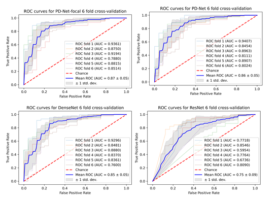
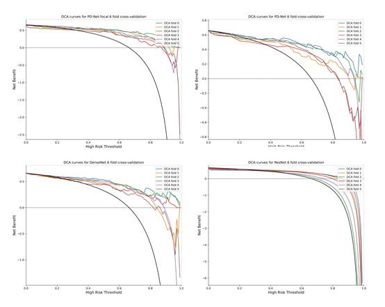
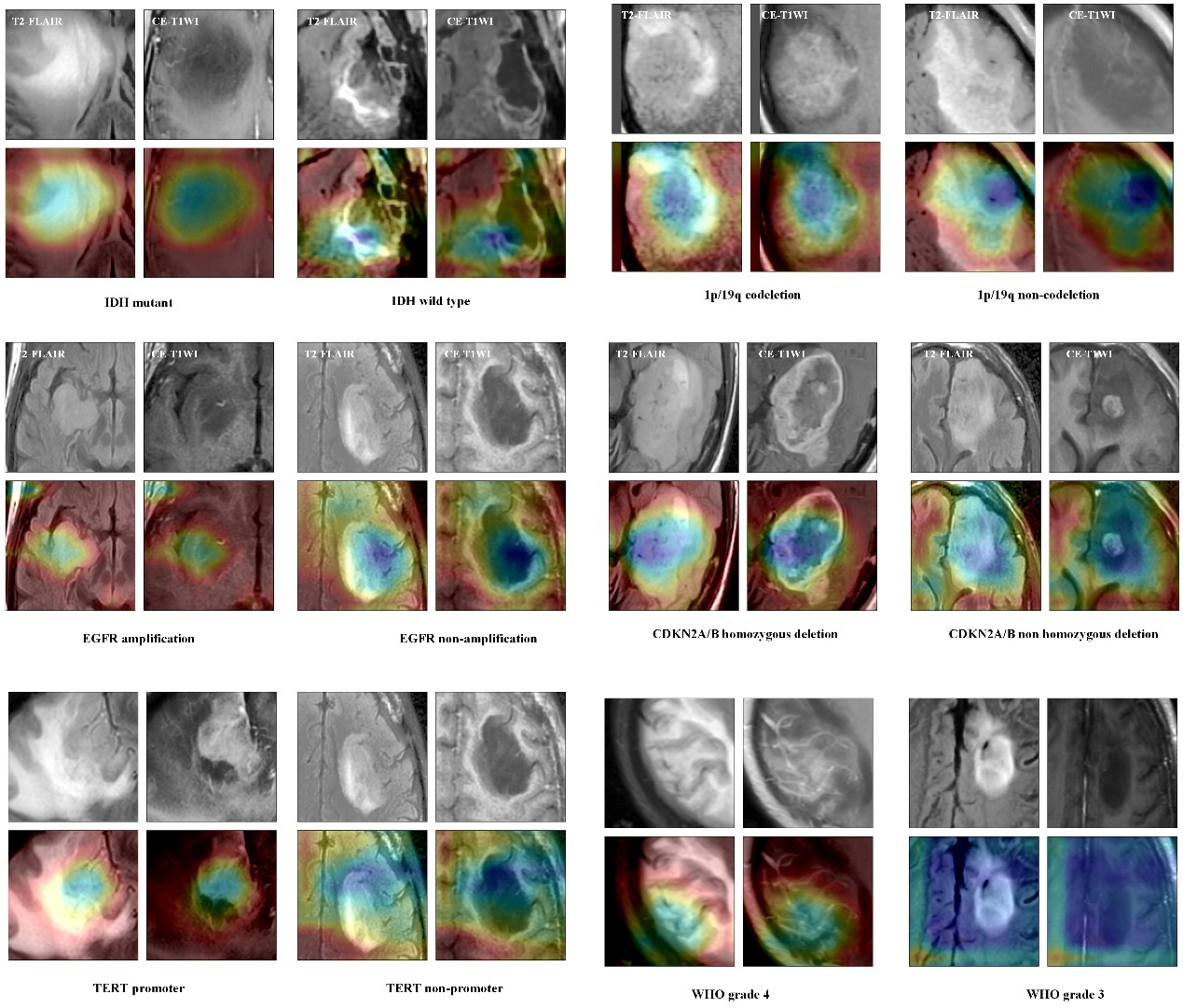
A total of 6 individualized CNNs models (PD-Net-focal) were developed for the adult-type diffuse gliomas binary classification tasks, which learned the genetic and histological features of glioma MR images. The PD-Net-focal models achieved mean cross-validation in IDH, 1p/19q, CDKN2A/B, EGFR, TERT and histological grading, with ACC of 86.82%,84.25%, 84.31%, 86.72%, 86.36% and 84.25%, and with AUC of 91.23%, 91.46%, 93.42%, 93.21%, 90.41% and 89.34%, respectively. The CNNs-based DTDP demonstrated promising diagnostic performance, with ACC of 86.67%. Fig. 2 shows the confusion matrix of the proposed decision tree diagnosis model. The ROC curves for each cross-validation fold for PD-Net-focal, PD-Net, DenseNet, and ResNet are provided in Fig.3. The DCA curves for each cross-validation fold for PD-Net-focal, PD-Net, DenseNet, and ResNet are provided in Fig.4. Provided representative GradCAM images for different genes status in Fig. 5.Discussion
The different models trained on each dataset were integrated as decision tree nodes, and 60 patients with 2760 MR images were independently tested using the DTDP. The preliminary results seem to be promising, showing 86.67% accuracy for classifying diffuse gliomas into 5 categories on the independent testing dataset. The prediction results of different models from 6-fold cross-validation were integrated using a vote-based ensemble method and used as decision nodes to construct the DTDP. This can aggregate the prediction results of different models, improve the performance and stability of the model, and reduce errors caused by data distribution.Conclusion
The DTDP was capable of completing multiple classification tasks and realized the automatic classification and comprehensive diagnosis of adult-type diffuse gliomas, which effectively helped neuroradiologists.Acknowledgements
We wish to thank the help given by Mr. Shanxiong Chen in Statistics analysis and we thank Dr. Yongmei LI for their advice on experimental design.References
1. Clark K, Vendt B, Smith K, et,al. The Cancer Imaging Archive (TCIA): maintaining and operating a public information repository. J Digit Imaging. 2013 Dec;26(6):1045-57.
2. Ceccarelli M, Barthel FP, Malta TM, et,al. Molecular Profiling Reveals Biologically Discrete Subsets and Pathways of Progression in Diffuse Glioma. Cell. 2016 Jan 28;164(3):550-63.
3. Lin TY, Goyal P, Girshick R, He K, Dollar P. Focal Loss for Dense Object Detection. IEEE Trans Pattern Anal Mach Intell. 2020 Feb;42(2):318-327.
Figures